ABSTRACT
Background Osteopontin (OPN) is a glycoprotein involved
in Th1 and Th17 differentiation, tissue inflammation and remodelling. We explored the role of serum OPN (sOPN) as a biomarker in patients with giant cell arteritis (GCA).
Methods sOPN was measured by immunoassay in
76 treatment-naïve patients with GCA and 25 age-matched and sex-age-matched controls. In 36 patients, a second measurement was performed after 1 year of glucocorticoid treatment. Baseline clinical and laboratory findings, as well as relapses and glucocorticoid requirements during follow-up, were prospectively recorded. sOPN and C reactive protein (CRP) were measured in 32 additional patients in remission treated with glucocorticoids or tocilizumab (interleukin 6 (IL-6) receptor antagonist). In cultured temporal arteries exposed and unexposed to tocilizumab, OPN mRNA expression and protein production were measured by reverse transcription polymerase chain reaction (RT-PCR) and immunoassay, respectively.
Results sOPN concentration (ng/mL; mean±SD) was
significantly elevated in patients with active disease (116.75±65.61) compared with controls (41.10±22.65; p<0.001). A significant decline in sOPN was observed in paired samples as patients entered disease remission (active disease 102.45±57.72, remission 46.47±23.49; p<0.001). sOPN correlated with serum IL-6 (r=0.55; p<0.001). Baseline sOPN concentrations were significantly higher in relapsing versus non-relapsing patients (relapsers 129.08±74.24, non-relapsers 90.63±41.02; p=0.03). OPN mRNA expression and protein production in cultured arteries were not significantly modified by tocilizumab. In tocilizumab-treated patients, CRP became undetectable, whereas sOPN was similar in patients in tocilizumab-maintained (51.91±36.25) or glucocorticoid-maintained remission (50.65±23.59; p=0.49).
Conclusions sOPN is a marker of disease activity and a
predictor of relapse in GCA. Since OPN is not exclusively IL-6-dependent, sOPN might be a suitable disease activity biomarker in tocilizumab-treated patients.
IntRoduCtIon
Giant cell arteritis (GCA) is an inflamma-tory disease of large-sized and medium-sized arteries with a chronic and relapsing course.1 2 About 43%–64% of patients
expe-rience recurrences3–5 and require long-term
glucocorticoid treatment with substantial toxicity.4 6 7 For years, attempts to identify
glucocorticoid-sparing agents have not been
ORIGINAL ARTICLe
Serum osteopontin: a biomarker
of disease activity and predictor of
relapsing course in patients with
giant cell arteritis. Potential clinical
usefulness in tocilizumab-treated
patients
Sergio Prieto-González,1 Nekane Terrades-García,1 Marc Corbera-Bellalta,1
Ester Planas-Rigol,1 Chie Miyabe,2 Marco A Alba,1 Ariel Ponce,1
Itziar Tavera-Bahillo,1 Giuseppe Murgia,1 Georgina Espígol-Frigolé,1
Javier Marco-Hernández,1 José Hernández-Rodríguez,1 Ana García-Martínez,3
Sebastian H Unizony,2 Maria C Cid1
to cite: Prieto-González S, Terrades-García N, Corbera-Bellalta M, et al. Serum osteopontin: a biomarker of disease activity and predictor of relapsing course in patients with giant cell arteritis. Potential clinical usefulness in tocilizumab-treated patients. RMD Open 2017;3:e000570. doi:10.1136/ rmdopen-2017-000570
►Prepublication history and additional material for this paper are available online. To view these files, please visit the journal online (http:// dx. doi. org/ 10. 1136/ rmdopen- 2017- 000570).
SP-G and NT-G contributed equally.
Received 4 September 2017 Revised 23 October 2017 Accepted 24 November 2017
For numbered affiliations see end of article.
Correspondence to
Dr Sergio Prieto-González; sprieto@ clinic. ub. es and Dr Maria C Cid; mccid@ clinic. ub. es
Key messages
► To date, serum osteopontin (OPN) concentrations have not been explored in patients with giant cell arteritis (GCA).
► Baseline serum OPN concentration is significantly elevated in patients with active GCA compared with controls and patients in remission, and significantly higher in relapsing versus non-relapsing patients. ► In cultured GCA arteries, OPN mRNA expression and
protein production are not significantly modified by short-term exposure to tocilizumab.
► While in tocilizumab-treated patients C reactive protein becomes undetectable, serum OPN is similar in patients in tocilizumab-maintained or glucocorticoid-maintained remission.
► Serum OPN might be a suitable disease activity biomarker in tocilizumab-treated patients with GCA. This needs to be explored in larger studies. on May 7, 2020 by guest. Protected by copyright.
http://rmdopen.bmj.com/
clearly successful,8–10 but a new treatment paradigm
based on the inhibition of interleukin 6 (IL-6) signalling is emerging in the field of GCA, supported by two recent randomised controlled trials with the IL-6 receptor neutralising antibody tocilizumab.11 12
The inflammatory markers erythrocyte sedimentation rate (ESR) and C reactive protein (CRP) are widely used in clinical practice to monitor disease activity in patients with GCA.3–5 8–10 However, IL-6 receptor blockade with
tocilizumab abrogates the hepatic synthesis of acute-phase reactants and renders CRP and ESR measure-ment unreliable for the purpose of monitoring disease activity.11 12 For these reasons, there is an urgent need for
novel biomarkers that reflect overall disease activity in the era of tocilizumab treatment for GCA.
Osteopontin (OPN) is a multifunctional intracellular and secreted glycoprotein that functions as a matrix protein or as a soluble mediator.13 14 It is expressed by
a variety of cells involved in immune and inflammatory responses, including dendritic cells, T and B lymphocytes, macrophages, neutrophils and eosinophils. OPN partic-ipates in innate and adaptive immune responses.13–15 It
is highly induced after T lymphocyte activation, stimu-lates Th1 and Th17 differentiation and inhibits Th2-me-diated responses. It also promotes B cell differentiation and immunoglobulin production. OPN is not expressed by circulating monocytes, but it is highly upregulated during macrophage differentiation.16 OPN has integrin
and CD44 binding sequences and supports lymphocyte and monocyte migration and survival.14–16 In addition, OPN enhances endothelial and vascular smooth muscle cell (VSMC) migration, contributing to angiogenesis and vascular remodelling.17 According to these functions,
OPN is highly expressed at the sites of inflammation and tissue injury and reflects concomitant activation of different pathways relevant to immune and inflammatory responses that participate in the pathogenesis of GCA.18 19
Based on its production by activated macrophages, OPN expression was investigated by immunohistochem-istry in a variety of granulomatous diseases. In this survey, increased tissue expression of OPN was observed in two temporal artery biopsies from patients with GCA.20
Moreover, increased circulating soluble OPN has been shown in several inflammatory diseases of blood vessels, including Behçet’s disease and anti-neutrophil cyto-plasmic antibodies (ANCA)-associated vasculitis.21 22 Elevated tissue and serum concentrations of OPN have been demonstrated in patients with other vascular condi-tions, such as non-vasculitic thoracic or abdominal aortic aneurysms.23 24 To date, serum OPN (sOPN)
concentra-tions have not been explored in patients with GCA. In this study, we investigated the value of sOPN as biomarker of disease activity and risk of disease relapse in patients with GCA. In addition, we explored the temporal artery expression of OPN in the context of IL-6 receptor blockade. Finally, we compared the serum concentra-tions of OPN and CRP in patients in remission receiving
tocilizumab (with or without low-dose glucocorticoids) or glucocorticoids alone.
MateRIals and Methods Patients
The study group consisted of 76 biopsy-proven patients with GCA, selected from two reported cohorts.25–27 The
first cohort consisted of patients with GCA prospectively imaged in order to detect large vessel involvement at diagnosis (cohort 1).25 The second cohort incorporated
patients who were cross-sectionally evaluated to detect aortic dilatation during follow-up (cohort 2).26 27 Patient
inclusion in the present study was based on the availa-bility of serum samples collected at the time of diagnosis, before the initiation of glucocorticoid treatment. Cohort 1 included 42 patients and cohort 2 included 34.
Cranial symptoms (headache, jaw claudication, scalp tenderness), systemic manifestations (weight loss, fever), polymyalgia rheumatica and disease-related cranial isch-aemic complications were recorded at the time of diag-nosis. CRP, haemoglobin concentration and ESR were determined by laboratory standardised systems.
Patients were followed for at least 2 years (mean 187 weeks, range 114–360), and time to first disease relapse, number of disease relapses, time to reach a stable (at least for 3 months) prednisone maintenance dosage <10 mg/day or <5 mg /day, as well as time to prednisone discontinuation and cumulative prednisone dose were recorded. We used a consensus definition of relapse established in the context of international multicentre clinical trials.9 10 Relapses were defined as reappearance
of GCA manifestations, usually accompanied by elevation of acute-phase reactants, that required treatment adjust-ment. Disease-related symptoms considered were poly-myalgia rheumatica, cranial symptoms (headache, scalp tenderness, jaw claudication, cranial ischaemic compli-cations), systemic manifestations (anaemia, fever and/ or weight loss) or symptomatic large vessel involvement (extremity claudication). Cranial ischaemic manifesta-tions included amaurosis fugax, GCA-related visual loss, diplopia, transient ischaemic attacks or stroke. Isolated increases in ESR or CRP were not considered relapses unless the above symptoms occurred after close follow-up.
In 36 patients from the discovery cohort, a second sample was obtained after approximately 1 year (402±48 days) of glucocorticoid treatment following a uniform, previously reported tapering protocol.4 28 At the time of
the second sample collection, patients were receiving a median daily prednisone dose of 5 mg/day (range 2.5–10 mg/day), and all patients were in clinical remis-sion defined by the absence of disease-related manifesta-tions and the presence of ESR and CRP levels within the normal range.
The control group consisted of two sets of 13 (A) and 12 (B) healthy individuals with no chronic inflamma-tory diseases and matched for age and gender. sOPN was measured in 17 additional patients with GCA in
on May 7, 2020 by guest. Protected by copyright.
prednisone-maintained remission. Seven patients were receiving ≥20 mg/day (high dose) and 10 were
receiving ≤10 mg/day (low-dose). sOPN was subjected to
cross-sectional comparison with that from 15 patients in remission treated with tocilizumab (4–8 mg/kg/month) (as monotherapy or together with low-dose prednisone) to explore the effect of tocilizumab on sOPN concen-tration. CRP was simultaneously measured in the same samples for comparison purposes. The clinical character-istics of these additional 32 patients have been previously described.29
soPn, CRP and Il-6 measurement
Serum samples were stored at −80°C until use. sOPN, IL-6 and CRP in sera were determined by immunoassay (Quantikine R&D Systems, Minneapolis, Minnesota, USA). OPN was also detected in the supernatant fluid from cultured temporal arteries.
Culture of temporal arteries from patients with GCa and controls
Temporal artery fragments from 16 patients with GCA and normal temporal arteries from 15 controls (patients with suspected GCA who eventually were diagnosed with other conditions) were cut into 0.8–1 mm sections and were embedded in Matrigel (Corning Matrigel Basement Membrane Matrix, Life Sciences, Tewksbury, Massachu-setts, USA) to ensure prolonged survival as described30 31
with or without tocilizumab (RoActemra, purchased from Hoffman-La Roche, Basel, Switzerland) or an IgG isotype control (Sigma, Ayrshire, UK) both at 10 µg/mL. Each condition was tested in two to three replicate wells. Temporal artery biopsies were obtained for diagnostic purposes and, since appropriate diagnosis was the first priority, all conditions could not be tested in all samples due to the small size of tissue spared for research. After 5-day culture, sections were homogenised and frozen in TRIzol reagent (Life Technologies, Carlsbad, California, USA) for RNA extraction.
Rna extraction and real-time reverse transcription polymerase chain reaction (Rt-PCR)
Total RNA was obtained from 11 cultured arteries from six patients with GCA and five controls, following the instructions of the manufacturer, and reverse-tran-scribed to cDNA using Archive Kit (Applied Biosys-tems, Life Technologies) in a final volume of 100 µL, employing random hexamer priming. OPN and signal transducer and activator of transcription 3 (STAT3) expression, as well as expression of the housekeeping gene GUSb, was investigated using specific predeveloped TaqMan probes (Hs00959010_m1, Hs00374280_m1 and Hs99999908_m1, respectively) from Applied Biosystems (TaqMan Gene Expression Assays). Fluorescence was detected using ViiA 7 Real-Time PCR System and results were analysed with the QuantStudio Real-Time PCR V.1.1 software (both from Applied Biosystems). Gene expres-sion was normalised to the expresexpres-sion of the endogenous
control GUSb using comparative threshold cicles (ΔCt) method. mRNA concentration was expressed in relative units with respect to GUSb.
statistical analysis
Mann-Whitney U test and Student’s t-test, when appli-cable, were used for quantitative independent or paired data. Correlations were calculated using Spearman’s test. Receiver-operator characteristic (ROC) curves were applied to sOPN concentrations to calculate cut-offs with best sensitivity and specificity. Time (weeks) to first relapse and time required to achieving a maintenance prednisone dose <10 mg/day or <5 mg/day were deter-mined by Kaplan-Meier survival method and compared using the log-rank test. Statistical significance was defined as P<0.05. Calculations were performed with the IBM SPSS Statistics V.20.0.
Results
soPn concentrations are increased in patients with active GCa
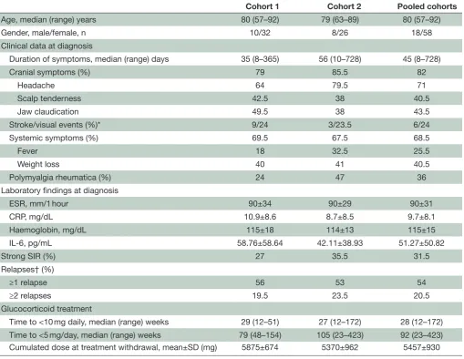
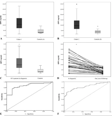
Table 1 summarises clinical and laboratory data from cohort 1 and cohort 2 of patients with GCA. sOPN concentrations (ng/mL; mean±SD) were significantly elevated in patients with active GCA compared with controls in both cohorts: GCA 119.59±70.36 vs controls 42.82±24.58 (p<0.001) in cohort 1, and GCA 113.21±69.56 vs controls 39.24±21.28 (p<0.001) in cohort 2 (figure 1A,B); as well as in the pooled cohorts of patients with active GCA (116.75±69.61) compared with pooled healthy controls (41.10±22.65; p<0.001) (figure 1C). In the 36 patients who underwent a second sample collection at the time of disease remission, a significant decline in sOPN concentrations was observed (active disease at baseline 102.45±57.72 vs remission 46.47±23.49; p<0.001) (figure 1D). Using ROC analysis, an sOPN cut-off of 59.79 ng/dL resulted in a sensitivity and specificity of 80% and 84%, respectively, for patients with active GCA compared with healthy controls (area under the curve (AUC) 0.862, 95% CI 0.788 to 0.937; p<0.001) (figure 1E). Moreover, a sOPN cut-off of 67.28 ng/ dL resulted in a sensitivity and specificity of 77% and 78%, respectively, to detect disease activity when analysing active patients and those in remission (AUC 0.836, 95% CI 0.764 to 0.907; p<0.001) (figure 1F). No significant differences in sOPN concentrations were found between patients in remission (48.78±23.97) and healthy controls (41.10±22.65; p=0.213).
Patients with GCA with active disease and systemic symptoms demonstrated significantly higher sOPN concentrations than those with no systemic symp-toms (118.45±61.70 vs 82.70±57.50; p=0.028) (table 2). Contrarily, and as previously described for serum IL-6,32
sOPN concentrations were significantly lower in patients with cranial ischaemic complications compared with patients without these complications (79.91±57.90 vs 117.29±61.32; p=0.028) (table 2). There were no differ-ences in baseline sOPN levels between patients with or
on May 7, 2020 by guest. Protected by copyright.
http://rmdopen.bmj.com/
Table 1 Baseline clinical, laboratory and follow-up data of patients with giant cell arteritis
Cohort 1 Cohort 2 Pooled cohorts
Age, median (range) years 80 (57–92) 79 (63–89) 80 (57–92)
Gender, male/female, n 10/32 8/26 18/58
Clinical data at diagnosis
Duration of symptoms, median (range) days 35 (8–365) 56 (10–728) 45 (8–728)
Cranial symptoms (%) 79 85.5 82
Headache 64 79.5 71
Scalp tenderness 42.5 38 40.5
Jaw claudication 49.5 38 43.5
Stroke/visual events (%)* 9/24 3/23.5 6/24
Systemic symptoms (%) 69.5 67.5 68.5
Fever 18 32.5 25.5
Weight loss 40 41 40.5
Polymyalgia rheumatica (%) 24 47 36
Laboratory findings at diagnosis
ESR, mm/1 hour 90±34 90±29 90±31
CRP, mg/dL 10.9±8.6 8.7±8.5 9.7±8.1
Haemoglobin, mg/dL 115±18 114±13 115±15
IL-6, pg/mL 58.76±58.64 42.11±38.93 51.27±50.82
Strong SIR (%) 27 35.5 31.5
Relapses† (%)
≥1 relapse 56 53 54
≥2 relapses 19.5 23.5 20.5
Glucocorticoid treatment
Time to <10 mg daily, median (range) weeks 29 (12–51) 27 (12–172) 28 (12–172) Time to <5 mg/day, median (range) weeks 79 (48–154) 105 (23–423) 92 (23–423) Cumulated dose at treatment withdrawal, mean±SD (mg) 5875±674 5370±962 5457±930
Strong SIR was defined as the presence of ≥3 of the following: ESR ≥85 mm/hour, haemoglobin <110 g/L, fever >37°C and weight loss >3 kg as described.25
*Visual events include diplopia and visual loss due to anterior ischaemic optic neuropathy.
†Relapses were clinically defined as depicted in the Materials and methods section and, except for three patients, were always accompanied by a rebound in ESR and CRP.
CRP, C reactive protein; ESR, erythrocyte sedimentation rate; IL-6, interleukin 6; SIR, systemic inflammatory response.
without vascular imaging compatible with large vessel inflammation (table 2). In addition, there was no correla-tion between baseline sOPN concentracorrela-tions and subse-quent aortic dilatation (table 2).
soPn correlates with acute-phase reactants and circulating Il-6
At the time of GCA diagnosis, sOPN showed a positive correlation with serum IL-6 (r=0.55; p<0.001) and with the IL-6-dependent acute-phase reactants ESR (r=0.32; p=0.009) and CRP (r=0.42; p<0.001). In addition, base-line sOPN negatively correlated with haemoglobin concentrations (r=−0.34; p=0.005).
Baseline soPn concentration predicts disease relapse and glucocorticoid use
Baseline sOPN was significantly higher in patients who developed disease relapses during follow-up (relapsers
129.08±74.24 vs non-relapsers 90.63±41.02 ng/mL; p=0.03). Within the group of relapsers, patients with more than one disease relapse demonstrated significantly higher sOPN levels (194.00±77.02) than those with only one relapse (98.52±50.72; p=0.007) (figure 2A).
Furthermore, the percentage of patients in remission over time was significantly lower in patients with base-line sOPN concentrations superior to the mean value (p=0.03) (figure 2B).
The proportion of patients requiring a prednisone maintenance dose ≥10 mg/day over time was significantly
higher among patients with sOPN levels above the mean sOPN concentration at the time of diagnosis (p=0.036) (figure 2C).
Moreover, there was a positive correlation between the baseline sOPN and the time to achieve a stable glucocor-ticoid dose <10 mg/day (r=0.43, p=0.02) or <5 mg/day
on May 7, 2020 by guest. Protected by copyright.
Figure 1 Serum osteopontin (sOPN) concentrations in patients with giant cell arteritis and healthy controls. (A) sOPN concentrations in active patients with giant cell arteritis (GCA) from the cohort 1 and in controls (subgroup A). (B) sOPN concentrations in active patients with GCA from the cohort 2 and controls (subgroup B). (C) sOPN concentrations in the pooled cohorts of patients with active GCA and controls. Box-plot in (A), (B) and (C) represent median, 25%–75% percentile and range. (D) sOPN concentrations in patients with GCA at diagnosis and when in remission by paired comparison. (E) Receiver-operator characteristic (ROC) analysis of sOPN concentrations in patients with active GCA versus controls. (F) ROC curve of patients with active GCA versus patients in remission.
Table 2 Serum OPN concentrations according to clinical/imaging data in the pooled cohort of patients with giant cell arteritis
OPN concentrations (ng/mL) (mean±SD)
Presence Absence p
Cranial symptoms 104.78±59.60 118.56±75.16 0.491
Systemic symptoms 118.45±61.70 82.70±57.50 0.028
Ischaemic symptoms 79.91±57.90 117.29±61.32 0.028
Strong SIR 132.56±77.56 97.46±53.40 0.039
PMR 118.17±74.27 101.15±54.45 0.287
LVV* 109.44±61.88 115.55±69.45 0.799
Aortic dilation† 111.58±46.74 105.56±70.96 0.754
*Evaluated in 42 patients. †Evaluated in 34 patients.
LVV, large vessel vasculitis; OPN, osteopontin; PMR, polymyalgia rheumatica; SIR, systemic inflammatory response.
on May 7, 2020 by guest. Protected by copyright.
http://rmdopen.bmj.com/
[image:5.595.46.553.580.710.2]Figure 2 Baseline serum osteopontin in patients with giant cell arteritis as predictor of relapses and duration of
glucocorticosteroid treatment. (A) Baseline sOPN concentrations in patients with no subsequent relapses, with 1 relapse and with ≥2 relapses. (B) Percentage of patients in remission over time according to baseline sOPN (≥mean sOPN vs <mean sOPN). (C) Percentage of patients requiring a daily maintenance prednisone dose ≥ 10 mg over time according to baseline sOPN (≥ mean sOPN v s < mean sOPN).
(r=0.48, p=0.03), and between baseline sOPN and the cumulated prednisone dose at the time of prednisone discontinuation (r=0.37; p=0.01).
In contrast to the sOPN findings, no significant differences were found in baseline IL-6 (49.76±41.52 vs 28.00±30.86 pg/mL; p=0.237), ESR (83.88±31.10 vs 95.28±20.38 mm/1 hour; p=0.188), CRP (9.77±8.37 vs 6.70±3.91 mg/dL; p=0.117) or haemoglobin (116.56±14.33 vs 113.11±12.15 mg/dL; p=0.412) concen-trations between relapsing patients and patients who achieved sustained clinical remission (online supple-mentary table S1). As with sOPN, the proportion of patients who relapsed over time was significantly higher in patients with CRP concentrations above the mean level than in patients with lower concentrations (p=0.022), but no differences were found according to IL-6, ESR or haemoglobin values (online supplementary figure S1). Together, these data underline the predictive value of baseline sOPN in terms of disease relapse and long-term glucocorticoid requirements in patients receiving gluco-corticoid monotherapy.
oPn expression and secretion in cultured GCa arteries are not suppressed by short-term Il-6 receptor blockade
IL-6 receptor signalling results in activation and expres-sion of transcription factor STAT-3.32 We have previously
shown that cultured temporal arteries have constitutive IL-6 and STAT3 expression and activation.30 31 As
previ-ously shown, STAT3 expression was higher in cultured control arteries than in cultured GCA involved arteries, possibly reflecting loss of STAT-3 producing VSMCs31
(figure 3A). Short-term treatment with the anti-IL-6 receptor monoclonal antibody tocilizumab for 5 days, at concentrations able to reduce STAT3 expression (figure 3A), did not modify OPN mRNA expression or protein secretion in cultured GCA arteries (figure 3B,C). These findings support that, in spite of the correlation
between serum IL-6 and sOPN, OPN production in GCA arteries is not exclusively dependent on IL-6 signalling.
soPn is reduced but not abrogated in patients with GCa in remission treated with tocilizumab
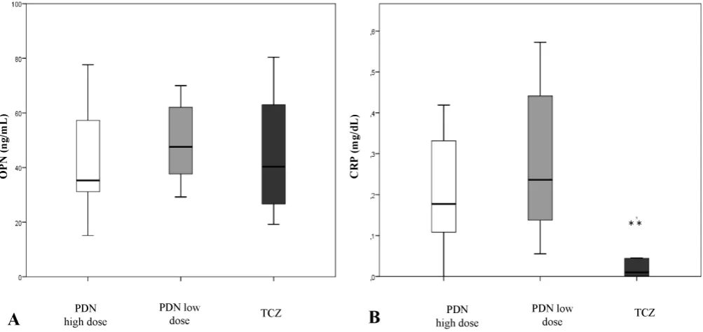
Based on our results on cultured arteries, we measured sOPN and serum CRP concentrations in a subset of patients with GCA who were in remission maintained with glucocorticoids or tocilizumab (alone or in combi-nation with low-dose prednisone) to see whether sOPN could be a useful biomarker in patients receiving IL-6 blockade therapy. As shown in figure 4, serum CRP was significantly lower and sometimes undetectable in tocili-zumab-treated patients (0.06±0.16 mg/dL) compared with prednisone only-treated patients at high dose (0.25±0.24 mg/dL; p=0.017) or low dose (0.28±0.19 mg/dL; p<0.001). In contrast, sOPN was detected in all cases without significant differences between patients under tocilizumab (51.91±36.25) or glucocorticoid only treatment either at high dose (43.55±21.36; p=0.861) or low dose (55.62±24.87; p=0.24). These results suggest that, unlike CRP, sOPN might be an interesting disease activity biomarker to be explored in tocilizumab-treated patients.
dIsCussIon
In this study, we demonstrated that sOPN was signifi-cantly elevated in patients with active GCA compared with age-matched and sex-matched healthy individuals but returned to normal when patients achieved remis-sion with glucocorticoid treatment. In addition, patients with the highest sOPN concentrations at diagnosis expe-rienced more relapses and cumulated higher glucocorti-coid doses. Previous studies have indicated that an intense systemic inflammatory response assessed by a composite combination of clinical and biological data is associated
on May 7, 2020 by guest. Protected by copyright.
Figure 3 Effect of tocilizumab (TCZ) on osteopontin (OPN) and STAT3 expression in cultured temporal arteries from patients with giant cell arteritis (GCA) and controls. (A) STAT3 mRNA expression in relative units (RU) by cultured temporal arteries from negative controls (n=5) and from patients with GCA untreated (n=6), treated with TCZ at 10 µg/mL (n=6) or treated with non-immune human control IgG at 10 µg/mL (n=4). *p=0.030 vs negative biopsies, #p=0.046 vs untreated GCA biopsies (paired comparison). (B) OPN mRNA expression in RU by cultured temporal arteries from negative controls and from patients with GCA untreated, treated with TCZ at 10 µg/mL or treated with non-immune human control IgG at 10 µg/mL as in (A). (C) OPN concentrations in the supernatants of cultured temporal arteries from negative controls (n=15) and from patients with GCA untreated (n=14), treated with TCZ at 10 µg/mL (n=15) or treated with non-immune human control IgG at 10 µg/mL (n=16). **p<0.001 vs negative control samples. Sensitivity of the immunoassay (minimal detectable concentration) was 0.011 ng/mL.
Figure 4 Serum osteopontin (sOPN) and C reactive protein (CRP) in patients with giant cell arteritis (GCA) in remission according to treatment. (A) sOPN in patients with GCA in remission with high-dose prednisone (PDN) (≥20 mg/day), low-dose prednisone (≤10 mg/day) or with tocilizumab (TCZ). (B) Serum CRP concentration in patients with GCA in remission with high-dose prednisone (≥20 mg/day), low-high-dose prednisone (≤10 mg/day) or with TCZ. **p=0.017 vs high-high-dose prednisone group and p<0.001 vs low-dose prednisone group. Sensitivities (minimal detectable concentration) of OPN and CRP immunoassay are 0.011 ng/mL and 0.010 ng/mL, respectively.
with recurrent disease.4 5 33 Accordingly, in the present
study, sOPN was significantly higher in patients with strong systemic inflammatory response than in patients with weak acute-phase reaction. Interestingly, although sOPN at baseline correlated with ESR, CRP and IL-6, and negatively with haemoglobin concentration, sOPN
performed better than individual detection of ESR, CRP, haemoglobin or IL-6 as predictor of relapsing course. sOPN is increased in a variety of conditions conveying tissue injury and remodelling, including malignan-cies, infection and other systemic vasculitis.21 22 34
Therefore, sOPN may not be a suitable diagnostic
on May 7, 2020 by guest. Protected by copyright.
http://rmdopen.bmj.com/
[image:7.595.48.550.436.673.2]biomarker. However, if confirmed in larger studies, our data indicate that sOPN may be a useful biomarker of disease activity and predictor of relapsing disease and glucocorticoid requirements.
Regarding additional clinical data, patients with cranial ischaemic complications had significantly lower sOPN concentrations. A similar pattern has been previously shown for serum IL-6,35 and, in fact, in the present study
sOPN and sIL-6 concentrations significantly correlated, suggesting coordinated regulation. As hypothesised for IL-6, angiogenic activity of OPN might compensate for ischaemia at distal sites.35 36
Although glucocorticoids are very efficient in inducing remission in the majority of patients, glucocorticoid tapering results in relapse in a substantial number of individuals. No clear alternatives to prolonged use of glucocorticoids were available to treat patients with GCA until very recently.8–12 37 A phase II and a phase III trial
have demonstrated the efficacy and safety of blocking IL-6 receptor with tocilizumab in maintaining remission and sparing glucocorticoid in GCA.11 12 However,
neutral-ising IL-6 receptor blocks the synthesis of acute-phase response and leads to a transient rebound in serum IL-6, making difficult the assessment of disease activity in individuals treated with tocilizumab.38 OPN synthesis
is activated through different pathways. Its promoter region has binding sites for transcription factors relevant to immune and inflammatory responses such as nuclear factor kB (NFkB), activator protein 1 (AP-1) and STAT3, which also regulate IL-6 expression.39 Although OPN can
be induced by IL-6, it is not exclusively IL-6-dependent. It can be also induced by IL-1β, tumour necrosis factor-α
and interferon-γ, among others.16 Our preliminary results
indicate that short-term treatment exposure of ex vivo cultured GCA arteries to tocilizumab does not have a selective impact on OPN expression in GCA lesions. Consistently, while tocilizumab virtually abolished serum CRP, sOPN was similar in patients in glucocorticoid-main-tained or tocilizumab-mainglucocorticoid-main-tained remission. All together, these data suggest that sOPN could be a useful biomarker of disease activity for tocilizumab-treated patients.
Our data on baseline sOPN were obtained from two patient cohorts subjected to imaging.25–27 As with serum
IL-6, sOPN did not seem to be associated with anatom-ical disease extension in these patients since there was no relationship between sOPN and computed tomography angiography (CTA) detection or extension of large-vessel inflammation at diagnosis. Moreover, as shown for serum IL-6,26 27 sOPN at diagnosis did not predict
subse-quent development of aortic dilatation in our patients. Our study has important strengths: it is performed in a sizeable and unique cohort of patients with treat-ment-naïve, biopsy-proven GCA, subjected to stan-dardised clinical evaluation, imaging and treatment. However, detection of sOPN in patients in tocilizum-ab-induced vs glucocorticoid only-induced remission was cross-sectional, and the performance of sOPN as a marker of disease activity in these patients could not be
specifically assessed, since no baseline samples or samples during relapse were available. The absence of baseline samples from patients treated with tocilizumab also prevents assessment of sOPN as predictor of response to tocilizumab as demonstrated in rheumatoid arthritis.40
This would be an interesting point to be investigated in future studies.
In summary, our data suggest that OPN might be a biomarker of disease activity in patients with GCA, and our preliminary data suggest that it might be particularly useful for patients treated with tocilizumab. Its perfor-mance in this setting deserves to be explored in larger prospective, longitudinal cohorts or in sera collected during clinical trials.
author affiliations
1Department of Autoimmune Diseases, Vasculitis Research Unit, University of
Barcelona, Institut d'Investigacions Biomèdiques August Pi i Sunyer (IDIBAPS), CRB-CeLLeX, Barcelona, Spain
2Division of Rheumatology, Allergy and Immunology, Vasculitis and
Glomerulonephritis Center, Massachusetts General Hospital, Harvard Medical School, Boston, Massachusetts, USA
3Department of emergency Medicine, Hospital Clínic, University of Barcelona,
IDIBAPS, CRB-CeLLeX, Barcelona, Spain
acknowledgements The authors thank Anna Jordán and Carmen Ligero for their valuable help in getting blood samples from the patients.
Contributors MCC had full access to all of the data in the study and takes responsibility for the integrity of the data and the accuracy of the data analysis. Study design: SP-G, NT-G, MCC. Acquisition of data: SP-G, NT-G, MC-B, CM, eP-R, AG-M, Ge-F, IT-B, AP, JM-H, MAA, JH-R, SHU, MCC. Analysis and interpretation of data: G, Ge-F, MAA, MC-B, eP-R, JH-R, SHU, MCC. Manuscript preparation: SP-G, NT-SP-G, SHU, MCC. Statistical analysis: SP-SP-G, MAA, NT-SP-G, MCC. All authors read, made improvements and approved the manuscript.
Funding Supported by Ministerio de economía y Competitividad (SAF 14/57708-R and SAF17/82275-R), Marató TV3 (201507), Instituto de Salud Carlos III
(PIe13/00033) and Fondo europeo de Desarrollo Regional (FeDeR, una manera de hacer europa). SP-G and IT-B were supported by a research award from Hospital Clínic, Ge-F by Instituto de Salud Carlos III (PI 15/00092), and MAA by Consejo Nacional de Ciencia y Tecnología (CONACyT), Mexico, and by Agencia de Gestió d’Ajuts Universitaris i de Recerca (AGAUR), Generalitat de Catalunya.
Competing interests Ge-F, SP-G, JH-R, SHU and MCC have participated in the GiACTA trial sponsored by Hoffmann-La Roche. MCC has received consultation fees from Hoffman-La Roche.
ethics approval The study was approved by the ethics committee of Hospital Clínic (Barcelona, Spain) and the Massachusetts General Hospital (Boston, Massachussets, USA).
Provenance and peer review Not commissioned; externally peer reviewed.
data sharing statement No additional data are available.
open access This is an Open Access article distributed in accordance with the Creative Commons Attribution Non Commercial (CC BY-NC 4.0) license, which permits others to distribute, remix, adapt, build upon this work non-commercially, and license their derivative works on different terms, provided the original work is properly cited and the use is non-commercial. See: http:// creativecommons. org/ licenses/ by- nc/ 4. 0/
© Article author(s) (or their employer(s) unless otherwise stated in the text of the article) 2017. All rights reserved. No commercial use is permitted unless otherwise expressly granted.
RefeRences
1. Jennette JC, Falk RJ, Bacon PA, et al. Revised international chapel hill consensus conference nomenclature of vasculitides. Arthritis Rheum 2012;2013:1–11.
2. Hoffman GS. Giant cell arteritis. Ann Intern Med 2016;165:65–80. 3. Martinez-Lado L, Calviño-Díaz C, Piñeiro A, et al. Relapses and
recurrences in giant cell arteritis: a population-based study of
on May 7, 2020 by guest. Protected by copyright.
patients with biopsy-proven disease from northwestern Spain. Medicine 2011;90:186–93.
4. Alba MA, García-Martínez A, Prieto-González S, et al. Relapses in patients with giant cell arteritis: prevalence, characteristics, and associated clinical findings in a longitudinally followed cohort of 106 patients. Medicine 2014;93:194–201.
5. Restuccia G, Boiardi L, Cavazza A, et al. Flares in biopsy-proven giant cell arteritis in northern italy: characteristics and predictors in a long-term follow-up study. Medicine 2016;95:e3524.
6. Andersson R, Malmvall BE, Bengtsson BA. Long-term corticosteroid treatment in giant cell arteritis. Acta Med Scand 1986;220:465–9. 7. Proven A, Gabriel SE, Orces C, et al. Glucocorticoid therapy in
giant cell arteritis: duration and adverse outcomes. Arthritis Rheum 2003;49:703–8.
8. Mahr AD, Jover JA, Spiera RF, et al. Adjunctive methotrexate for treatment of giant cell arteritis: an individual patient data meta-analysis. Arthritis Rheum 2007;56:2789–97.
9. Hoffman GS, Cid MC, Hellmann DB, et al. A multicenter, randomized, double-blind, placebo-controlled trial of adjuvant methotrexate treatment for giant cell arteritis. Arthritis Rheum 2002;46:1309–18.
10. Hoffman GS, Cid MC, Rendt-Zagar KE, et al. Infliximab for maintenance of glucocorticosteroid-induced remission of giant cell arteritis: a randomized trial. Ann Intern Med 2007;146:621–30. 11. Villiger PM, Adler S, Kuchen S, et al. Tocilizumab for induction
and maintenance of remission in giant cell arteritis: a phase 2, randomised, double-blind, placebo-controlled trial. Lancet 2016;387:1921–7.
12. Stone JH, Tuckwell K, Dimonaco S, et al. Trial of tocilizumab in giant-cell arteritis. N Engl J Med 2017;377:317–28.
13. Denhardt DT, Noda M, O'Regan AW, et al. Osteopontin as a means to cope with environmental insults: regulation of inflammation, tissue remodeling, and cell survival. J Clin Invest 2001;107:1055–61. 14. Uede T. Osteopontin, intrinsic tissue regulator of intractable
inflammatory diseases. Pathol Int 2011;61:265–80.
15. Clemente N, Raineri D, Cappellano G, et al. Osteopontin bridging innate and adaptive immunity in autoimmune diseases. J Immunol Res 2016;2016:1–15.
16. Krause SW, Rehli M, Kreutz M, et al. Differential screening identifies genetic markers of monocyte to macrophage maturation. J Leukoc Biol 1996;60:540–5.
17. Isoda K, Nishikawa K, Kamezawa Y, et al. Osteopontin plays an important role in the development of medial thickening and neointimal formation. Circ Res 2002;91:77–82.
18. Planas Rigol E, Corbera Bellalta M, et al. Giant-cell arteritis: immunopathogenic mechanisms involved in vascular inflammation and remodeling. J Vasc 2016;2:1–7.
19. Samson M, Corbera-Bellalta M, Audia S, et al. Recent advances in our understanding of giant cell arteritis pathogenesis. Autoimmun Rev 2017;16:833–44.
20. Carlson I, Tognazzi K, Manseau EJ, et al. Osteopontin is strongly expressed by histiocytes in granulomas of diverse etiology. Lab Invest 1997;77:103–8.
21. Bassyouni IH, El-Wakd MM, Bassyouni RH. Soluble levels of osteopontin in patients with Behcet’s disease: association with disease activity and vascular involvement. J Clin Immunol 2013;33:361–7.
22. Lorenzen J, Lovric S, Krämer R, et al. Osteopontin in antineutrophil cytoplasmic autoantibody-associated vasculitis: relation to disease activity, organ manifestation and immunosuppressive therapy. Ann Rheum Dis 2010;69:1169–71.
23. Hellenthal FA, Buurman WA, Wodzig WK, et al. Biomarkers of abdominal aortic aneurysm progression. Part 2: inflammation. Nat Rev Cardiol 2009;6:543–52.
24. Huusko T, Salonurmi T, Taskinen P, et al. Elevated messenger RNA expression and plasma protein levels of osteopontin and matrix
metalloproteinase types 2 and 9 in patients with ascending aortic aneurysms. J Thorac Cardiovasc Surg 2013;145:1117–23. 25. Prieto-González S, Arguis P, García-Martínez A, et al. Large vessel
involvement in biopsy-proven giant cell arteritis: prospective study in 40 newly diagnosed patients using CT angiography. Ann Rheum Dis 2012;71:1170–6.
26. García-Martínez A, Hernández-Rodríguez J, Arguis P, et al. Development of aortic aneurysm/dilatation during the followup of patients with giant cell arteritis: a cross-sectional screening of fifty-four prospectively followed patients. Arthritis Rheum
2008;59:422–30.
27. García-Martínez A, Arguis P, Prieto-González S, et al. Prospective long term follow-up of a cohort of patients with giant cell arteritis screened for aortic structural damage (aneurysm or dilatation). Ann Rheum Dis 2014;73:1826–32.
28. Prieto-González S, García-Martínez A, Tavera-Bahillo I, et al. Effect of glucocorticoid treatment on computed tomography angiography detected large-vessel inflammation in giant-cell arteritis. A prospective, longitudinal study. Medicine 2015;94:e486.
29. Miyabe C, Miyabe Y, Strle K, et al. An expanded population of pathogenic regulatory T cells in giant cell arteritis is abrogated by IL-6 blockade therapy. Ann Rheum Dis 2017;76:898–905. 30. Corbera-Bellalta M, García-Martínez A, Lozano E, et al. Changes
in biomarkers after therapeutic intervention in temporal arteries cultured in Matrigel: a new model for preclinical studies in giant-cell arteritis. Ann Rheum Dis 2014;73:616–23.
31. Corbera-Bellalta M, Planas-Rigol E, Lozano E, et al. Blocking interferon γ reduces expression of chemokines CXCL9, CXCL10 and CXCL11 and decreases macrophage infiltration in ex vivo cultured arteries from patients with giant cell arteritis. Ann Rheum Dis 2016;75:1177–86.
32. Rincon M. Interleukin-6: from an inflammatory marker to a target for inflammatory diseases. Trends Immunol 2012;33:571–7.
33. Hernández-Rodríguez J, García-Martínez A, Casademont J, et al. A strong initial systemic inflammatory response is associated with higher corticosteroid requirements and longer duration of therapy in patients with giant-cell arteritis. Arthritis Rheum
2002;47:29–35.
34. Scatena M, Liaw L, Giachelli CM. Osteopontin: a multifunctional molecule regulating chronic inflammation and vascular disease. Arterioscler Thromb Vasc Biol 2007;27:2302–9.
35. Hernández-Rodríguez J, Segarra M, Vilardell C, et al. Elevated production of interleukin-6 is associated with a lower incidence of disease-related ischemic events in patients with giant-cell arteritis: angiogenic activity of interleukin-6 as a potential protective mechanism. Circulation 2003;107:2428–34.
36. Ramchandani D, Weber GF. Interactions between osteopontin and vascular endothelial growth factor: Implications for cancer. Biochim Biophys Acta 2015;1855:202–22.
37. Langford CA, Cuthbertson D, Ytterberg SR, et al. A randomized, double-blind trial of abatacept (CTLA-4Ig) for the treatment of giant cell arteritis. Arthritis Rheumatol 2017;69:837–45.
38. Nishimoto N, Terao K, Mima T, et al. Mechanisms and pathologic significances in increase in serum interleukin-6 (IL-6) and soluble IL-6 receptor after administration of an anti-IL-6 receptor antibody, tocilizumab, in patients with rheumatoid arthritis and Castleman disease. Blood 2008;112:3959–64.
39. Zhao W, Wang L, Zhang M, et al. NF-κB- and AP-1-mediated DNA looping regulates osteopontin transcription in endotoxin-stimulated murine macrophages. J Immunol 2011;186:3173–9.
40. Izumi K, Kaneko Y, Hashizume M, et al. Baseline serum osteopontin levels predict the clinical effectiveness of tocilizumab but not infliximab in biologic-naïve patients with rheumatoid arthritis: a single-center prospective study at 1 year (the Keio first-bio cohort study). PLoS One 2015;10:e0145468.
on May 7, 2020 by guest. Protected by copyright.
http://rmdopen.bmj.com/