organic papers
Acta Cryst.(2005). E61, o2389–o2390 doi:10.1107/S1600536805020751 Liet al. C
17H14N2O20.5H~2~O
o2389
Acta Crystallographica Section E
Structure Reports
Online
ISSN 1600-5368
N
-Phenyl-2-(quinolin-8-yloxy)acetamide
hemihydrate
Xue-Mei Li, Yong-Hong Wen, Mao-Jie Li and
Shu-Sheng Zhang*
College of Chemistry and Molecular
Engineering, Qingdao University of Science and Technology, 266042 Qingdao, Shandong, People’s Republic of China
Correspondence e-mail: [email protected]
Key indicators
Single-crystal X-ray study T= 293 K
Mean(C–C) = 0.003 A˚ Rfactor = 0.049 wRfactor = 0.126
Data-to-parameter ratio = 10.8
For details of how these key indicators were automatically derived from the article, see http://journals.iucr.org/e.
#2005 International Union of Crystallography Printed in Great Britain – all rights reserved
In the title compound, C17H14N2O20.5H2O, all bond lengths
and angles are within normal ranges. The dihedral angle formed by the phenyl ring with the quinoline moiety is 27.30 (9). The crystal packing is stabilized by intermolecular
N—H O, C—H O and O—H N hydrogen bonds involv-ing the solvent water molecule.
Comment
Recently, we have reported the structure of an amide-type acyclic compound with an 8-hydroxyquinolinate skeleton, namely N,N-diphenyl-2-(quinolin-8-yloxy)acetamide mono-hydrate, (II) (Wen et al., 2005). In order to investigate the effect of the substituent groups of the acyclic compounds on the coordination selectivity and extractability for metal ions, we have synthesized and carried out the structure determi-nation of the title compound, (I).
All bond lengths and angles in (I) (Table 1) are within normal ranges (Allenet al., 1987) and comparable with those in the related compound (II). The dihedral angle formed by the phenyl ring with the quinoline moiety is 27.30 (9). There
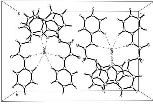
are two intramolecular hydrogen bonds,viz. N2—H1N2 O1 and C17—H17 O2, forming a five- and six-membered ring, respectively. The crystal packing is stabilized by inter-molecular N2—H1N2 O1W, C13—H13 O1Wand O1W— H1W1 N1 hydrogen bonds (Table 2 and Fig. 2) involving the solvent water molecule.
Experimental
2-Chloro-N-phenylacetamide was prepared by the reaction of phenylamine and chloroacetyl chloride in the presence of triethyl-amine, according to the literature method of Wenet al.(2004). To a solution of 8-hydroxyquinoline (1.45 g, 10 mmol) in acetone (40 ml) were added 2-chloro-N-phenylacetamide (1.69 g, 10 mmol), K2CO3
(1.52 g, 11 mmol) and KI (0.5 g), and the resulting mixture was stirred at 333 K for 5 h. After cooling to room temperature, the mixture was washed three times with water and then filtered. Colorless single crystals suitable for an X-ray diffraction study were obtained by slow evaporation of a petroleum ether–ethyl acetate (1:2, v/v) solution over a period of 3 d.
Crystal data
C17H14N2O20.5H2O Mr= 287.31 Monoclinic,C2=c a= 11.093 (5) A˚
b= 12.944 (6) A˚
c= 19.584 (9) A˚ = 91.855 (9)
V= 2811 (2) A˚3
Z= 8
Dx= 1.358 Mg m3 MoKradiation Cell parameters from 1582
reflections = 2.4–21.7
= 0.09 mm1 T= 293 (2) K Block, colorless 0.390.170.12 mm
Data collection
Siemens SMART 1000 CCD area-detector diffractometer !scans
Absorption correction: multi-scan (SADABS; Sheldrick, 1996)
Tmin= 0.965,Tmax= 0.989
7758 measured reflections
2766 independent reflections 1883 reflections withI> 2(I)
Rint= 0.029
max= 26.1 h=13!10
k=14!15
l=24!23
Refinement
Refinement onF2 R[F2> 2(F2)] = 0.049
wR(F2) = 0.126 S= 1.03 2766 reflections 255 parameters
All H-atom parameters refined
w= 1/[2(F
o2) + (0.062P)2
+ 0.2335P]
whereP= (Fo2+ 2Fc2)/3
(/)max< 0.001 max= 0.15 e A˚
3 min=0.17 e A˚
3
Table 1
Selected bond lengths (A˚ ).
O1—C8 1.370 (2) O1—C10 1.424 (2) O2—C11 1.224 (2)
N2—C11 1.342 (2) N2—C12 1.415 (2)
Table 2
Hydrogen-bond geometry (A˚ ,).
D—H A D—H H A D A D—H A
O1W—H1W1 N1i 0.91 (2) 1.99 (3) 2.853 (3) 157 (2) N2—H1N2 O1ii
0.87 (2) 2.35 (2) 2.722 (2) 106 (2) N2—H1N2 O1Wii
0.87 (2) 2.26 (2) 3.109 (2) 170 (2) C13—H13 O1Wii
0.95 (2) 2.55 (2) 3.359 (3) 143 (2) C17—H17 O2ii
0.96 (2) 2.16 (2) 2.824 (3) 125 (2)
Symmetry codes: (i)x;y;zþ1 2; (ii)x;y;z.
All H atoms were located in difference Fourier maps and refined freely. The C—H distances are in the range 0.93 (2)–1.01 (2) A˚ .
Data collection:SMART(Siemens, 1996); cell refinement:SAINT
(Siemens, 1996); data reduction: SAINT; program(s) used to solve structure: SHELXTL (Sheldrick, 1997); program(s) used to refine structure:SHELXTL; molecular graphics:SHELXTL; software used to prepare material for publication: SHELXTL,PARST(Nardelli, 1995) andPLATON(Spek, 2003).
This project was supported by the Program for New Century Excellent Talents in University (No. NCET-04–0649), and the Project of Educational Administration of Shandong Province (No. J04B12).
References
Allen, F. H., Kennard, O., Watson, D. G., Brammer, L., Orpen, A. G. & Taylor, R. (1987).J. Chem. Soc. Perkin Trans. 2, S1–19.
Nardelli, M. (1995).J. Appl. Cryst.28, 659.
Sheldrick, G. M. (1996).SADABS. University of Go¨ttingen, Germany. Sheldrick, G. M. (1997).SHELXTL. Version 5.1. Bruker AXS, Inc., Madison,
Wisconsin, USA.
Siemens (1996).SMARTandSAINT. Siemens Analytical X-ray Instruments Inc., Madison, Wisconsin, USA.
Spek, A. L. (2003).J. Appl. Cryst.36, 7–13.
Wen, Y.-H., Zhang, S.-S., Li, M.-J. & Li, X.-M. (2005).Acta Cryst.E61, o1807– o1809.
[image:2.610.311.564.73.190.2]Wen, Y.-H., Zhang, S.-S., Liang, J. & Li, X.-M. (2004).Acta Cryst.E60, o1702– o1703.
Figure 1
[image:2.610.317.564.238.407.2]The structure of the compound (I), showing 50% probability displace-ment ellipsoids and the atom-numbering scheme.
Figure 2
supporting information
sup-1 Acta Cryst. (2005). E61, o2389–o2390
supporting information
Acta Cryst. (2005). E61, o2389–o2390 [https://doi.org/10.1107/S1600536805020751]
N
-Phenyl-2-(quinolin-8-yloxy)acetamide hemihydrate
Xue-Mei Li, Yong-Hong Wen, Mao-Jie Li and Shu-Sheng Zhang
N-Phenyl-2-(quinolin-8-yloxy)acetamide hemihydrate
Crystal data C17H14N2O2·0.5H2O
Mr = 287.31
Monoclinic, C2/c a = 11.093 (5) Å b = 12.944 (6) Å c = 19.584 (9) Å β = 91.855 (9)° V = 2811 (2) Å3
Z = 8
F(000) = 1208 Dx = 1.358 Mg m−3
Mo Kα radiation, λ = 0.71073 Å Cell parameters from 1582 reflections θ = 2.4–21.7°
µ = 0.09 mm−1
T = 293 K Block, colourless 0.39 × 0.17 × 0.12 mm
Data collection
Siemens SMART 1000 CCD area-detector diffractometer
Radiation source: fine-focus sealed tube Graphite monochromator
Detector resolution: 8.33 pixels mm-1
ω scans
Absorption correction: multi-scan (SADABS; Sheldrick, 1996) Tmin = 0.965, Tmax = 0.989
7758 measured reflections 2766 independent reflections 1883 reflections with I > 2σ(I) Rint = 0.029
θmax = 26.1°, θmin = 2.1°
h = −13→10 k = −14→15 l = −24→23
Refinement Refinement on F2
Least-squares matrix: full R[F2 > 2σ(F2)] = 0.049
wR(F2) = 0.126
S = 1.03 2766 reflections 255 parameters 0 restraints
Primary atom site location: structure-invariant direct methods
Secondary atom site location: difference Fourier map
Hydrogen site location: inferred from neighbouring sites
All H-atom parameters refined w = 1/[σ2(F
o2) + (0.062P)2 + 0.2335P]
where P = (Fo2 + 2Fc2)/3
(Δ/σ)max < 0.001
Δρmax = 0.15 e Å−3
Δρmin = −0.17 e Å−3
Special details
Refinement. Refinement of F2 against ALL reflections. The weighted R-factor wR and goodness of fit S are based on F2,
conventional R-factors R are based on F, with F set to zero for negative F2. The threshold expression of F2 > σ(F2) is used
only for calculating R-factors(gt) etc. and is not relevant to the choice of reflections for refinement. R-factors based on F2
are statistically about twice as large as those based on F, and R- factors based on ALL data will be even larger.
Fractional atomic coordinates and isotropic or equivalent isotropic displacement parameters (Å2)
x y z Uiso*/Ueq
supporting information
sup-3 Acta Cryst. (2005). E61, o2389–o2390
Atomic displacement parameters (Å2)
U11 U22 U33 U12 U13 U23
O1W 0.0732 (15) 0.0369 (11) 0.0646 (13) 0.000 −0.0082 (11) 0.000 O1 0.0768 (9) 0.0396 (8) 0.0415 (8) 0.0090 (6) −0.0024 (6) −0.0013 (6) O2 0.1259 (14) 0.0625 (10) 0.0487 (9) 0.0171 (9) −0.0192 (9) −0.0108 (7) N1 0.0552 (10) 0.0464 (10) 0.0447 (9) 0.0025 (7) −0.0021 (7) 0.0030 (7) N2 0.0666 (11) 0.0401 (9) 0.0406 (9) 0.0002 (7) −0.0017 (8) −0.0043 (7) C1 0.0629 (13) 0.0612 (14) 0.0458 (12) 0.0024 (10) −0.0007 (10) 0.0019 (11) C2 0.0729 (15) 0.0686 (16) 0.0499 (13) 0.0014 (11) −0.0002 (11) −0.0140 (12) C3 0.0642 (14) 0.0509 (14) 0.0659 (15) 0.0014 (11) −0.0032 (11) −0.0148 (11) C4 0.0491 (11) 0.0430 (11) 0.0565 (12) −0.0008 (8) −0.0010 (9) −0.0064 (9) C5 0.0745 (15) 0.0364 (12) 0.0733 (15) 0.0038 (10) −0.0024 (11) −0.0012 (11) C6 0.0909 (17) 0.0398 (13) 0.0655 (15) 0.0045 (11) −0.0020 (12) 0.0116 (11) C7 0.0765 (15) 0.0460 (12) 0.0496 (12) 0.0056 (10) −0.0015 (10) 0.0058 (10) C8 0.0536 (11) 0.0355 (10) 0.0471 (11) 0.0038 (8) −0.0017 (8) 0.0000 (8) C9 0.0414 (10) 0.0388 (11) 0.0489 (11) 0.0008 (8) −0.0008 (8) 0.0002 (8) C10 0.0782 (16) 0.0505 (13) 0.0415 (12) 0.0068 (11) −0.0001 (11) −0.0009 (9) C11 0.0680 (13) 0.0457 (12) 0.0423 (11) 0.0031 (9) 0.0014 (9) −0.0022 (9) C12 0.0522 (12) 0.0416 (11) 0.0464 (11) −0.0026 (8) 0.0049 (9) −0.0008 (8) C13 0.0634 (14) 0.0488 (13) 0.0623 (14) −0.0010 (10) −0.0088 (11) −0.0055 (10) C14 0.0664 (15) 0.0524 (14) 0.0790 (17) 0.0042 (11) −0.0118 (12) 0.0054 (12) C15 0.0751 (16) 0.0419 (14) 0.0863 (18) 0.0022 (11) −0.0003 (13) −0.0006 (12) C16 0.119 (2) 0.0452 (14) 0.0716 (17) 0.0017 (13) −0.0083 (15) −0.0136 (12) C17 0.111 (2) 0.0483 (13) 0.0518 (13) 0.0063 (12) −0.0120 (13) −0.0073 (11)
Geometric parameters (Å, º)
C8—O1—C10 115.56 (14) N1—C9—C8 119.51 (16) C1—N1—C9 117.50 (17) C4—C9—C8 118.25 (17) C11—N2—C12 127.00 (17) O1—C10—C11 112.61 (16) C11—N2—H1N2 119.1 (13) O1—C10—H10B 107.7 (14) C12—N2—H1N2 113.8 (13) C11—C10—H10B 108.9 (14) N1—C1—C2 123.9 (2) O1—C10—H10A 108.9 (13) N1—C1—H1 114.0 (12) C11—C10—H10A 108.3 (14) C2—C1—H1 122.1 (12) H10B—C10—H10A 110.4 (19) C3—C2—C1 119.1 (2) O2—C11—N2 124.71 (19) C3—C2—H2 122.4 (15) O2—C11—C10 116.74 (18) C1—C2—H2 118.4 (15) N2—C11—C10 118.51 (18) C2—C3—C4 120.0 (2) C17—C12—C13 118.9 (2) C2—C3—H3 121.0 (14) C17—C12—N2 123.59 (18) C4—C3—H3 119.0 (14) C13—C12—N2 117.49 (17) C3—C4—C5 122.94 (19) C14—C13—C12 120.2 (2) C3—C4—C9 117.24 (19) C14—C13—H13 120.0 (12) C5—C4—C9 119.82 (19) C12—C13—H13 119.7 (12) C6—C5—C4 120.2 (2) C15—C14—C13 120.7 (2) C6—C5—H5 121.2 (12) C15—C14—H14 121.5 (14) C4—C5—H5 118.7 (12) C13—C14—H14 117.8 (14) C5—C6—C7 121.0 (2) C16—C15—C14 118.8 (2) C5—C6—H6 119.5 (13) C16—C15—H15 121.2 (13) C7—C6—H6 119.5 (13) C14—C15—H15 119.9 (13) C8—C7—C6 120.6 (2) C15—C16—C17 121.7 (2) C8—C7—H7 122.3 (12) C15—C16—H16 118.1 (16) C6—C7—H7 117.1 (12) C17—C16—H16 120.1 (16) C7—C8—O1 124.43 (18) C16—C17—C12 119.7 (2) C7—C8—C9 120.16 (18) C16—C17—H17 122.6 (15) O1—C8—C9 115.41 (15) C12—C17—H17 117.6 (15) N1—C9—C4 122.23 (17)
supporting information
sup-5 Acta Cryst. (2005). E61, o2389–o2390
C3—C4—C9—C8 179.79 (17) C13—C12—C17—C16 0.0 (3) C5—C4—C9—C8 −1.0 (3) N2—C12—C17—C16 178.6 (2)
Hydrogen-bond geometry (Å, º)
D—H···A D—H H···A D···A D—H···A O1W—H1W1···N1i 0.91 (2) 1.99 (3) 2.853 (3) 157 (2)
N2—H1N2···O1 0.87 (2) 2.35 (2) 2.722 (2) 106 (2) N2—H1N2···O1W 0.87 (2) 2.26 (2) 3.109 (2) 170 (2) C13—H13···O1W 0.95 (2) 2.55 (2) 3.359 (3) 143 (2) C17—H17···O2 0.96 (2) 2.16 (2) 2.824 (3) 125 (2)