organic papers
Acta Cryst.(2006). E62, o81–o82 doi:10.1107/S1600536805039711 Wanget al. C
19H19NO4
o81
Acta Crystallographica Section E Structure Reports
Online
ISSN 1600-5368
Cheilanthifoline
Xiao-Ling Wang,a* Zong-Xiao Lia
and Guo-Wei Qinb
a
Department of Chemistry, Baoji College of Arts and Science, Baoji 721007, People’s Republic of China, andbInstitute of Materia Medica,
Shanghai Institute for Biological Sciences, Chinese Academy of Sciences, Shanghai 201203, People’s Republic of China
Correspondence e-mail: [email protected]
Key indicators
Single-crystal X-ray study
T= 296 K
Mean(C–C) = 0.004 A˚
Rfactor = 0.036
wRfactor = 0.089 Data-to-parameter ratio = 7.5
For details of how these key indicators were automatically derived from the article, see http://journals.iucr.org/e.
#2006 International Union of Crystallography Printed in Great Britain – all rights reserved
The title compound, 6,6a,11,14-tetrahydro-8-hydroxy-9-methoxy-12H-benzo[a]-1,3-benzodioxolo[4,5-g]quinolizine, C19H19NO4, a protoberberine-type alkaloid, was isolated from
the roots of the plant Sinomenium acutum. Both the
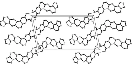
piperidine rings adopt half-chair conformations. The mol-ecules exist in a dimeric form through O—H O hydrogen bonding between the hydroxy group and methoxy O atom.
Comment
Sinomenium acutum is distributed mainly in hilly regions of
southwest, northwest and southeast China. The roots and stems of the plant are used as folk medicine to cure rheuma-tism, dropsy and dermatophytosis. A number of alkaloids with different kinds of skeletons have been isolated from the plant (Jiangsu New Medical College, 1985; Chen et al., 1991; Moriyasuet al., 1993, 1994). In the course of our systematic search for bioactive substances from Chinese traditional herb
medicines, we have studied the roots of S. acutum and
obtained the title compound, (I). Compound (I) was first isolated in some species of Corydalis and identified on the basis of its mass, NMR, IR and UV spectra (Blasket al., 1981; Haisov & Slavk, 1973). We report here the crystal structure of (I).
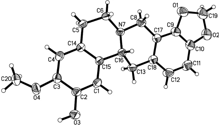
The benzo[1,3]dioxole ring system is essentially planar (Fig. 1). Both the piperidine rings adopt half-chair confor-mations. The methoxy group attached at atom C3 is twisted away from the benzene ring with a torsion angle C20—O4— C3—C4 of 25.2 (4). In the crystal packing, intermolecular hydrogen bonding between the hydroxy group and the methoxy O atom of an adjacent molecule leads to the formation of dimers (Fig. 2). In addition, a C—H inter-action is observed (Table 1).
Experimental
Sinomenine is produced from the powder of the roots ofS. acutumby the Baoji Yongjia Plant Medicine Extracting Limited Company,
Baoji, People’s Republic of China. It is obtained from the benzene extract of the powder in a vacuum (Chenet al., 1995). The remaining benzene mother liquor (3 kg), after the extraction of sinomenine, was obtained from the company. It was subjected to repeated chroma-tography on a silica gel column, and eluted with petroleum ether/ acetone (from 20:1 to 3:1) to afford compound (I) (0.2 g). Single crystals of (I) were obtained after repeated recrystallization from acetone.
Crystal data
C19H19NO4
Mr= 325.35
Monoclinic,C2
a= 13.314 (2) A˚
b= 5.151 (1) A˚
c= 23.585 (3) A˚
= 100.39 (1)
V= 1590.9 (4) A˚3
Z= 4
Dx= 1.358 Mg m
3
MoKradiation Cell parameters from 39
reflections
= 2.6–11.5 = 0.10 mm1
T= 296 (2) K Rhombohedron, yellow 0.480.340.14 mm
Data collection
SiemensP4 diffractometer
!scans
Absorption correction: none 1816 measured reflections 1669 independent reflections 1331 reflections withI> 2(I)
Rint= 0.013
max= 25.5
h= 0!16
k= 0!6
l=28!28 3 standard reflections
every 97 reflections intensity decay: 3.9%
Refinement
Refinement onF2 R[F2> 2(F2)] = 0.036
wR(F2) = 0.089
S= 0.99 1669 reflections 223 parameters
H atoms treated by a mixture of independent and constrained refinement
w= 1/[2
(Fo2) + (0.051P)2]
whereP= (Fo2+ 2Fc2)/3
(/)max= 0.001 max= 0.14 e A˚3 min=0.14 e A˚3
Extinction correction:SHELXTL
Extinction coefficient: 0.0144 (14)
Table 1
Hydrogen-bond geometry (A˚ ,).
D—H A D—H H A D A D—H A
O3—H3O O4 0.91 (4) 2.29 (3) 2.688 (3) 106 (2) O3—H3O O4i
0.91 (4) 1.98 (4) 2.870 (3) 163 (3) C20—H20C Cg1ii
0.96 2.62 3.460 (3) 146
Symmetry codes: (i)xþ1;y;z; (ii)x;y1;z. Cg1 is the centroid of the C1–C4/ C14 ring.
The hydroxy H atom was located in a difference Fourier map and refined freely. The other H atoms were placed in idealized positions and constrained to ride on their parent atoms, with C—H distances in the range 0.93–0.98 A˚ , and withUiso(H) = 1.2Ueq(C). In the absence
of significant anomalous scattering, Friedel pairs were merged prior to the final refinement.
Data collection: XSCANS (Siemens, 1994); cell refinement: XSCANS; data reduction:SHELXTL(Sheldrick, 1997b); program(s) used to solve structure: SHELXS97(Sheldrick, 1997a); program(s) used to refine structure: SHELXL97(Sheldrick, 1997a); molecular graphics:SHELXTL; software used to prepare material for publi-cation:SHELXTL.
The authors thank the Phytochemistry Key Laboratory of Shaanxi province for research grants (Nos. 02js40 and 05js43). The authors also thank Professor Kai-Bei Yu, Chengdu Institute of Organic Chemistry, Chinese Academy of Sciences, for diffraction measurements.
References
Blask, G., Hussain, S. F. & Shamma, M. (1981).J. Nat. Prod.44, 475–477. Chen, C., Sun, L. J. & Xu, H. Q. (1995). Chinese Patent No. 1125724. Chen, Y. Y., Qiu, C. C. & Shen, L. (1991).Beijing Yike Daxue Xuebao,23, 235–
237. (In Chinese.)
Haisov, K. & Slavk, J. (1973).Collect. Czech. Chem. Commun.38, 2307–2311. Jiangsu New Medical College (1985).The Dictionary of Chinese Medicine,
pp. 1234-1236. Shanghai: Shanghai Science and Technology Press. Moriyasu, M., Ichimaru, M. & Nishiyama, Y. (1993).Bunseki Kagaku,42, 659–
665. (In Japanese.)
Moriyasu, M., Ichimaru, M. & Nishiyama, Y. (1994).Nat. Med. 48, 287– 290.
Sheldrick, G. M. (1997a). SHELXS97 and SHELXL97. University of Go¨ttingen, Germany.
Sheldrick, G. M. (1997b).SHELXTL.Version 5.0. Siemens Analytical X-ray Instruments Inc., Madison, Wisconsin, USA.
Siemens (1994). XSCANS. Siemens Analytical X-ray Instruments Inc., Madison, Wisconsin, USA.
Figure 2
[image:2.610.314.568.72.217.2] [image:2.610.316.566.269.397.2]The crystal packing of (I), showing O—H O hydrogen-bonded (dashed lines) dimers. H atoms not involved in the interactions shown have been omitted.
Figure 1
supporting information
sup-1
Acta Cryst. (2006). E62, o81–o82
supporting information
Acta Cryst. (2006). E62, o81–o82 [doi:10.1107/S1600536805039711]
Cheilanthifoline
Xiao-Ling Wang, Zong-Xiao Li and Guo-Wei Qin
S1. Comment
Sinomenium acutum is distributed mainly in hilly regions of southwest, northwest and southeast China. The roots and
stems of the plant are used as folk medicine to cure rheumatism, dropsy and dermatophytosis. A number of alkaloids with
different kinds of skeletons have been isolated from the plant (Jiangsu New Medical College, 1985; Chen et al., 1991;
Moriyasu et al., 1993, 1994). In the course of our systematic search for bioactive substances from Chinese traditional
herb medicines, we have studied the roots of S. acutum and obtained the title compound, (I). Compound (I) was first
isolated in some species of Corydalis and identified on the basis of its mass and its NMR, IR and UV spectra (Blask et
al., 1981; Haisov & Slavk, 1973). We report here the crystal structure of (I).
The benzo[1,3]dioxole ring system is essentially planar (Fig. 1). Both the piperidine rings adopt half-chair
conformations. The methoxy group attached at atom C3 is twisted away from the benzene ring with a torsion angle C20
—O4—C3—C4 of 25.2 (4)°. In the crystal packing, intermolecular hydrogen bonding between the hydroxy group and
the methoxy O atom of an adjacent molecule leads to the formation of dimers (Fig. 2). In addition, a C—H···π interaction
is observed (Table 1).
S2. Experimental
Sinomenine is produced from the powder of the roots of S. acutum by the Baoji Yongjia Plant Medicine Extracting
Limited Company, Baoji, People's Republic of China. It is obtained from the benzene extract of the powder in a vacuum
(Chen et al., 1995). The remaining benzene mother liquor (3 kg), after the extraction of sinomenine, was obtained from
the company. It was subjected to repeated chromatography on a silica gel column, and eluted with petroleum
ether/acetone (from 20:1 to 3:1) to afford compound (I) (0.2 g). Single crystals of (I) were obtained after repeated
recrystallization from acetone.
S3. Refinement
The hydroxy H atom was located in a difference Fourier map and refined freely. The other H atoms were placed in
idealized positions and constrained to ride on their parent atoms, with C—H distances in the range 0.93–0.98 Å, and with
Uiso(H) = 1.2Ueq(C). In the absence of significant anomalous scattering, Friedel pairs were merged prior to the final
Figure 1
The structure of (I), showing 40% probability displacement ellipsoids and the atom-numbering scheme.
Figure 2
The crystal packing of (I), showing O—H···O hydrogen-bonded (dashed lines) dimers. H atoms not involved in the
interactions shown have been omitted.
6,6a,11,14-Tetrahydro-8-hydroxy-9-methoxy-12H-benzo[a]- 1,3-benzodioxolo[4,5-g]quinolizine
Crystal data
C19H19NO4 Mr = 325.35
Monoclinic, C2 Hall symbol: C 2y a = 13.314 (2) Å b = 5.151 (1) Å c = 23.585 (3) Å β = 100.39 (1)° V = 1590.9 (4) Å3 Z = 4
F(000) = 688 Dx = 1.358 Mg m−3
Mo Kα radiation, λ = 0.71073 Å Cell parameters from 39 reflections θ = 2.6–11.5°
µ = 0.10 mm−1 T = 296 K
[image:4.610.126.481.312.492.2]supporting information
sup-3
Acta Cryst. (2006). E62, o81–o82
Data collection
Siemens P4 diffractometer
Radiation source: normal-focus sealed tube Graphite monochromator
ω scans
1816 measured reflections 1669 independent reflections 1331 reflections with I > 2σ(I)
Rint = 0.013
θmax = 25.5°, θmin = 1.8° h = 0→16
k = 0→6 l = −28→28
3 standard reflections every 97 reflections intensity decay: 3.9%
Refinement
Refinement on F2
Least-squares matrix: full R[F2 > 2σ(F2)] = 0.036 wR(F2) = 0.089 S = 0.99 1669 reflections 223 parameters 1 restraint
Primary atom site location: structure-invariant direct methods
Secondary atom site location: difference Fourier map
Hydrogen site location: inferred from neighbouring sites
H atoms treated by a mixture of independent and constrained refinement
w = 1/[σ2(F
o2) + (0.051P)2]
where P = (Fo2 + 2Fc2)/3
(Δ/σ)max = 0.001
Δρmax = 0.14 e Å−3
Δρmin = −0.14 e Å−3
Extinction correction: SHELXTL, Fc*=kFc[1+0.001xFc2λ3/sin(2θ)]-1/4
Extinction coefficient: 0.0144 (14)
Special details
Geometry. All e.s.d.'s (except the e.s.d. in the dihedral angle between two l.s. planes) are estimated using the full covariance matrix. The cell e.s.d.'s are taken into account individually in the estimation of e.s.d.'s in distances, angles and torsion angles; correlations between e.s.d.'s in cell parameters are only used when they are defined by crystal symmetry. An approximate (isotropic) treatment of cell e.s.d.'s is used for estimating e.s.d.'s involving l.s. planes.
Refinement. Refinement of F2 against ALL reflections. The weighted R-factor wR and goodness of fit S are based on F2,
conventional R-factors R are based on F, with F set to zero for negative F2. The threshold expression of F2 > σ(F2) is used
only for calculating R-factors(gt) etc. and is not relevant to the choice of reflections for refinement. R-factors based on F2
are statistically about twice as large as those based on F, and R- factors based on ALL data will be even larger.
Fractional atomic coordinates and isotropic or equivalent isotropic displacement parameters (Å2)
x y z Uiso*/Ueq
O1 0.82863 (15) 1.3199 (5) 0.40355 (8) 0.0684 (8) O2 0.70057 (17) 1.4199 (5) 0.45455 (8) 0.0682 (7) O3 0.45140 (13) 0.3567 (5) 0.07211 (8) 0.0537 (6) O4 0.60253 (14) 0.0927 (4) 0.03625 (7) 0.0509 (6) N7 0.79074 (14) 0.7517 (5) 0.26188 (8) 0.0368 (5) C1 0.56992 (19) 0.5446 (6) 0.14804 (10) 0.0406 (7)
H1 0.5160 0.6316 0.1598 0.049*
C2 0.54997 (18) 0.3825 (6) 0.10126 (10) 0.0388 (7) C3 0.63021 (19) 0.2486 (6) 0.08397 (9) 0.0377 (7) C4 0.72871 (18) 0.2873 (6) 0.11289 (10) 0.0376 (6)
H4 0.7823 0.1991 0.1011 0.045*
C5 0.85767 (18) 0.5095 (6) 0.18788 (11) 0.0388 (7)
H5A 0.8997 0.5252 0.1585 0.047*
H5B 0.8831 0.3646 0.2126 0.047*
H6A 0.9339 0.7713 0.2457 0.050*
H6B 0.8532 0.9049 0.1978 0.050*
C8 0.8125 (2) 0.9656 (7) 0.30289 (11) 0.0477 (8)
H8A 0.8225 1.1232 0.2821 0.057*
H8B 0.8755 0.9292 0.3294 0.057*
C9 0.7416 (2) 1.1829 (7) 0.38215 (10) 0.0457 (8) C10 0.6660 (2) 1.2426 (7) 0.41200 (10) 0.0503 (8) C11 0.5717 (2) 1.1338 (8) 0.39869 (11) 0.0596 (10)
H11 0.5200 1.1757 0.4188 0.072*
C12 0.5563 (2) 0.9556 (8) 0.35317 (11) 0.0573 (9)
H12 0.4928 0.8765 0.3431 0.069*
C13 0.61563 (17) 0.7075 (6) 0.27260 (10) 0.0424 (8)
H13A 0.5451 0.7188 0.2532 0.051*
H13B 0.6279 0.5315 0.2868 0.051*
C14 0.74894 (17) 0.4575 (5) 0.15967 (9) 0.0330 (6) C15 0.66902 (18) 0.5829 (6) 0.17850 (9) 0.0341 (6) C16 0.68580 (16) 0.7671 (6) 0.22975 (9) 0.0352 (6)
H16 0.6727 0.9449 0.2156 0.042*
C17 0.72891 (19) 1.0100 (6) 0.33682 (10) 0.0401 (7) C18 0.63294 (18) 0.8930 (6) 0.32253 (9) 0.0410 (7) C19 0.8035 (3) 1.4720 (9) 0.44975 (13) 0.0719 (11)
H19A 0.8486 1.4280 0.4855 0.086*
H19B 0.8118 1.6551 0.4421 0.086*
C20 0.6676 (2) −0.1137 (6) 0.02803 (11) 0.0493 (7)
H20A 0.7265 −0.0468 0.0147 0.059*
H20B 0.6319 −0.2319 0.0000 0.059*
H20C 0.6889 −0.2034 0.0639 0.059*
H3O 0.443 (2) 0.248 (8) 0.0411 (14) 0.087 (12)*
Atomic displacement parameters (Å2)
U11 U22 U33 U12 U13 U23
supporting information
sup-5
Acta Cryst. (2006). E62, o81–o82
C13 0.0327 (13) 0.059 (2) 0.0354 (13) −0.0049 (14) 0.0062 (11) −0.0048 (15) C14 0.0336 (12) 0.0323 (15) 0.0333 (12) 0.0046 (12) 0.0064 (10) 0.0061 (13) C15 0.0354 (13) 0.0360 (15) 0.0307 (12) 0.0000 (13) 0.0054 (10) −0.0006 (13) C16 0.0317 (13) 0.0380 (16) 0.0353 (12) 0.0019 (13) 0.0047 (10) −0.0002 (13) C17 0.0400 (14) 0.0489 (17) 0.0313 (12) −0.0009 (14) 0.0063 (10) −0.0003 (14) C18 0.0401 (14) 0.0525 (19) 0.0306 (12) 0.0003 (15) 0.0072 (10) 0.0009 (14) C19 0.082 (2) 0.082 (3) 0.0537 (17) −0.017 (2) 0.0199 (16) −0.023 (2) C20 0.0648 (18) 0.0365 (17) 0.0467 (15) 0.0039 (16) 0.0104 (13) −0.0049 (15)
Geometric parameters (Å, º)
O1—C9 1.373 (3) C8—C17 1.500 (3)
O1—C19 1.430 (4) C8—H8A 0.97
O2—C10 1.373 (3) C8—H8B 0.97
O2—C19 1.421 (4) C9—C10 1.363 (4)
O3—C2 1.374 (3) C9—C17 1.378 (4)
O3—H3O 0.91 (4) C10—C11 1.360 (4)
O4—C3 1.378 (3) C11—C12 1.399 (4)
O4—C20 1.407 (3) C11—H11 0.93
N7—C8 1.461 (4) C12—C18 1.391 (3)
N7—C16 1.467 (3) C12—H12 0.93
N7—C6 1.468 (3) C13—C18 1.501 (4)
C1—C2 1.371 (3) C13—C16 1.526 (3)
C1—C15 1.398 (3) C13—H13A 0.97
C1—H1 0.93 C13—H13B 0.97
C2—C3 1.393 (3) C14—C15 1.385 (3)
C3—C4 1.379 (3) C15—C16 1.521 (3)
C4—C14 1.397 (3) C16—H16 0.98
C4—H4 0.93 C17—C18 1.398 (4)
C5—C14 1.504 (3) C19—H19A 0.97
C5—C6 1.511 (4) C19—H19B 0.97
C5—H5A 0.97 C20—H20A 0.96
C5—H5B 0.97 C20—H20B 0.96
C6—H6A 0.97 C20—H20C 0.96
C6—H6B 0.97
C9—O1—C19 105.3 (2) C10—C11—C12 116.7 (3)
C10—O2—C19 105.6 (2) C10—C11—H11 121.7
C2—O3—H3O 116 (2) C12—C11—H11 121.7
C3—O4—C20 118.2 (2) C18—C12—C11 122.1 (3)
C8—N7—C16 111.0 (2) C18—C12—H12 119.0
C8—N7—C6 109.0 (2) C11—C12—H12 119.0
C16—N7—C6 111.64 (18) C18—C13—C16 111.5 (2)
C2—C1—C15 121.8 (2) C18—C13—H13A 109.3
C2—C1—H1 119.1 C16—C13—H13A 109.3
C15—C1—H1 119.1 C18—C13—H13B 109.3
C1—C2—O3 119.4 (2) C16—C13—H13B 109.3
O3—C2—C3 121.3 (2) C15—C14—C4 119.8 (2)
O4—C3—C4 125.4 (2) C15—C14—C5 120.6 (2)
O4—C3—C2 114.9 (2) C4—C14—C5 119.6 (2)
C4—C3—C2 119.6 (2) C14—C15—C1 118.6 (2)
C3—C4—C14 120.8 (2) C14—C15—C16 122.3 (2)
C3—C4—H4 119.6 C1—C15—C16 119.1 (2)
C14—C4—H4 119.6 N7—C16—C15 111.5 (2)
C14—C5—C6 111.1 (2) N7—C16—C13 106.67 (18)
C14—C5—H5A 109.4 C15—C16—C13 112.4 (2)
C6—C5—H5A 109.4 N7—C16—H16 108.7
C14—C5—H5B 109.4 C15—C16—H16 108.7
C6—C5—H5B 109.4 C13—C16—H16 108.7
H5A—C5—H5B 108.0 C9—C17—C18 116.4 (2)
N7—C6—C5 110.0 (2) C9—C17—C8 120.9 (3)
N7—C6—H6A 109.7 C18—C17—C8 122.6 (2)
C5—C6—H6A 109.7 C12—C18—C17 119.9 (3)
N7—C6—H6B 109.7 C12—C18—C13 122.1 (2)
C5—C6—H6B 109.7 C17—C18—C13 117.9 (2)
H6A—C6—H6B 108.2 O2—C19—O1 108.6 (3)
N7—C8—C17 113.0 (2) O2—C19—H19A 110.0
N7—C8—H8A 109.0 O1—C19—H19A 110.0
C17—C8—H8A 109.0 O2—C19—H19B 110.0
N7—C8—H8B 109.0 O1—C19—H19B 110.0
C17—C8—H8B 109.0 H19A—C19—H19B 108.4
H8A—C8—H8B 107.8 O4—C20—H20A 109.5
C10—C9—O1 110.3 (2) O4—C20—H20B 109.5
C10—C9—C17 123.2 (3) H20A—C20—H20B 109.5
O1—C9—C17 126.5 (2) O4—C20—H20C 109.5
C11—C10—C9 121.7 (3) H20A—C20—H20C 109.5
C11—C10—O2 128.1 (3) H20B—C20—H20C 109.5
C9—C10—O2 110.3 (3)
supporting information
sup-7
Acta Cryst. (2006). E62, o81–o82
C19—O1—C9—C17 179.7 (3) O1—C9—C17—C18 −179.9 (3) O1—C9—C10—C11 179.4 (3) C10—C9—C17—C8 175.7 (3) C17—C9—C10—C11 −0.1 (5) O1—C9—C17—C8 −3.7 (4) O1—C9—C10—O2 −0.5 (4) N7—C8—C17—C9 172.2 (3) C17—C9—C10—O2 −179.9 (3) N7—C8—C17—C18 −11.7 (4) C19—O2—C10—C11 −179.4 (4) C11—C12—C18—C17 −0.2 (5) C19—O2—C10—C9 0.5 (4) C11—C12—C18—C13 −178.2 (3) C9—C10—C11—C12 0.6 (5) C9—C17—C18—C12 0.7 (4) O2—C10—C11—C12 −179.6 (3) C8—C17—C18—C12 −175.5 (3) C10—C11—C12—C18 −0.4 (5) C9—C17—C18—C13 178.8 (2) C3—C4—C14—C15 2.1 (4) C8—C17—C18—C13 2.6 (4) C3—C4—C14—C5 −175.9 (2) C16—C13—C18—C12 152.2 (3) C6—C5—C14—C15 −16.5 (3) C16—C13—C18—C17 −25.9 (4) C6—C5—C14—C4 161.5 (2) C10—O2—C19—O1 −0.3 (4) C4—C14—C15—C1 −3.1 (4) C9—O1—C19—O2 0.0 (4)
Hydrogen-bond geometry (Å, º)
D—H···A D—H H···A D···A D—H···A
O3—H3O···O4 0.91 (4) 2.29 (3) 2.688 (3) 106 (2) O3—H3O···O4i 0.91 (4) 1.98 (4) 2.870 (3) 163 (3)
C20—H20C···Cg1ii 0.96 2.62 3.460 (3) 146