Detection of ochratoxin A in aptamer assay using total
internal reflection ellipsometry
AL RUBAYE, Ali, NABOK, Aleksey <http://orcid.org/0000-0002-9078-1757>, CATANANTE, Gaelle, MARTY, Jean-Louis, TAKACS, Ezster and SZEKACS, Andras
Available from Sheffield Hallam University Research Archive (SHURA) at:
http://shura.shu.ac.uk/18924/
This document is the author deposited version. You are advised to consult the publisher's version if you wish to cite from it.
Published version
AL RUBAYE, Ali, NABOK, Aleksey, CATANANTE, Gaelle, MARTY, Jean-Louis, TAKACS, Ezster and SZEKACS, Andras (2018). Detection of ochratoxin A in
aptamer assay using total internal reflection ellipsometry. Sensors and Actuators B: Chemical, 263, 248-251.
Copyright and re-use policy
See http://shura.shu.ac.uk/information.html
1
Detection of ochratoxin A in aptamer assay
using total internal reflection ellipsometry
Ali Al Rubayea*, Alexei Naboka, Gaelle Catananteb, Jean-Louis Martyb, Ezster Takacsc, Andras Szekacsc
a
Sheffield Hallam University, Materials and Engineering Research Institute, UK
b
University of Perpignan, Department of Biochemistry and Molecular Biology, France
c
Agro-Environmental Research Institute, NARIC, Budapest, Hungary.
Abstract
The current work is a continuation of our research targeted the development of novel optical
sensing technologies for detection of mycotoxins. The method of (TIRE) was developed in
the last decade as a combination of spectroscopic ellisometry and SPR and was proved to be a
highly sensitive analytical tool in bio-sensing particularly attractive for detection of low
molecular weight analytes, such as mycotoxins. The use of aptamers as highly specific
artificial molecular receptors to ochratoxin A (OTA) in conjunction with the method Total
Internal Reflection Ellipsometry (TIRE) is reported here for the first time. Our results showed
a possibility of label-free optical detection of OTA down to 0.01 ppb in concentration in
direct assay with specific aptamer. The kinetics of aptamer/OTA binding was studied with
dynamic TIRE spectral measurements and allowed evaluating the affinity constant KD = 1.8.10-8 Mol which is characteristic for highly specific aptamer/OTA binding.
Keywords: Aptamer, Ochratoxin A, Label-free optical biosensor, Total Internal Reflection Ellipsometry, Binding kinetics
2
1. Introduction
The main goal of this work is the development of novel optical sensing technologies for
detection of mycotoxins. One of the most attractive optical biosensing technology developed
in the last decade was the method of total internal reflection ellipsometry (TIRE) which is a
combination of spectroscopic ellipsometry (SE) and surface plasmon resonance (SPR)
(Arwin et al., 2004).
This method appeared to be more sensitive than SPR (Nabok et al., 2008), and thus suitable
for detection of small molecular analytes, including pesticides (atrazine and simazine)
(Nabok et al., 2005) mycotoxins T2 (Nabok et al., 2005; Nabok et al., 2007a), zearalenone
(Nabok et al., 2011a), and aflatoxin B1 (Nabok et al., 2011b), alkyl-phenols (Nabok et al.,
2007b), and microcystine (Al-Ammar et al., 2015).
On the biochemistry side, the specific bio-receptors for the above mentioned toxins were
IgG-based antibodies (Ab). In majority of cases, the direct immunoassay sensing format was
used with the antibodies immobilized electrostatically on the surface of gold via the layers of
polycations (PAH or PEI) and proteins A or G (Nabok et al., 2008; Al-Ammar et al., 2015).
Such immobilization procedure was relatively simple, universal (in respect of using different
substrates, i.e. gold, glass, silicon), and was providing quite strong binding of Ab to the
substrate (second strongest after covalent binding). However the stability of immobilized
antibodies was always in question as well as a possibility of non-specific binding which
resulted in a number of negative control tests to perform. Also, a multi-stage process of Ab
immobilization prolongs the time of analysis.
Aptamers were developed recently as a synthetic alternative to antibodies in bio-sensing
applications. Aptamers are linear bio-polymers with specifically designed sequences of RNA
or DNA oligonucleotides which bind to target molecules of both organic and inorganic
origins (Hamula et al., 2006; Mairal et al., 2008). The technology of aptamers synthesis
improved dramatically in the last few years, so they became and commercially available for a
wide variety of analytes, and in many cases less expensive than antibodies. Aptamers have a
number of advantages over traditional antibodies, mainly in their robustness and simple
immobilization chemistry. Aptamers being relatively small size receptors seem to be
particularly suitable for optical detection of small toxin molecules because of a large relative
increment of thickness (or refractive index) when binding analytes. Aptamers were recently
used successfully in detection mycotoxins, such as ochratoxin A (Rhouati et al., 2013a).
Ochratoxin A (OTA), an object our present study, is a mycotoxin produced by some of
3
known by its carcinogenic, genotoxic and mutagenic actions on human (Varga et al., 1996;
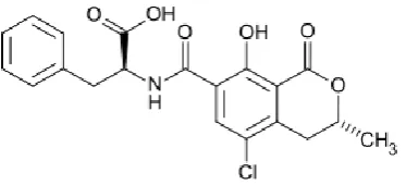
Clark et al., 2006; Phfohl et al., 2007). OTA is a relatively small molecule (see chemical
[image:4.595.206.393.139.224.2]structure in Fig. 1) with the molecular weight of 403.8 Da.
Fig. 1. Chemical structure of ochratoxin A
Traditional biosensing technique such as ELISA is capable of detecting OTA in the
concentration range down to of 5−100 ng/mL (Barna-Vetro et al., 1996). More recent work
(Barthelmebs et al., 2011) achieved the detection limit for OTA of 1 ng/mL using ELISA
aptamer assay. The detection limit for OTA detection in aptamer assay was lowered
substantially down to single ng/ml using other transducing techniques such as
electrochemical, optical and piezo-electrical methods (Yang et al., 2012; Bueno et al., 2016;
Catanante et al., 2016; Mishra et al., 2016). Aptamers can be immobilised on the surface of
gold via functional thiol group on one end (Balamurugan et al.,2008), and may contain either
fluorescent groups or redox labels such as methylene blue on the other end as described by
(Sheng et al., 2011; Ferapontova and Gothelf, 2009) and therefore can be used in optical or
electrochemical biosensors. The chromatographic strip assay method (Wang et al., 2011)
utilising labled aptamer for rapid toxin detection. High sensitivity of toxin detection can be
achived in a sandwich assay format, for example using a pair of aptamer and antibody
specific to toxins (Costantini et al., 2016; Seo et al., 2017). ( Bonel et al., 2011; Rhouati et
al.,2013b ). The sensitivity of electrochemical detection can be boosted using competitive aptasensor assay test for OTA coupled with paramagnetic beads. In this work we report for the first time the label-free optical detection of ochratoxin A in the direct assay with highly
specific aptamers using the method of TIRE.
2. Experimental methodology
2.1. Samples’ preparation and immobilization of aptamers
Standard microscopic glass slides were cleaned in hot piranha solution (3 : 1 mixture of
4
a stream of nitrogen gas. Gold layers of about 25 nm in thickness were evaporated on glass
slides using Edwards E306A metal evaporator unit. A thin (2 to 3 nm) layer of chromium was
evaporated first to improve the adhesion of a gold layer to glass. Such two-stage evaporation
was carried out without breaking the vacuum of no less than 10−6 Torr.
DNA-based aptamers specific to OTA acquired from M/sMicrosynth (Schutzenstrasse,
Balgach, Switzerland) have a following oligonucleotides sequence:
5'-SH-GATCGGGTGTGGGTGGCGTAAAGGGAGCATCGGACA-3'. The aptamer was
functionalized with thiol group (C3-SH) on the 5′ terminal position to obtain a strong and
oriented binding to gold. The immobilization of aptamers on gold surface was carried out by
mixing the original 100 µM aptamer solution with 2 mM of 1,4-Dithiothreitol (DTT) diluted
in 100 mM HEPES buffer (pH 7.4) supplemented with 2mM of MgCl2 in order to remove the protecting group and subsequently release aptamers with the SH end-groups which then bind
to gold (Rhouatiet et al., 2013; Yang et al., 2012). Before immobilization, the liquid aptamer
samples were activated by quick (5 min) heating up to 90°C followed by 5 min cooling to
4oC using thermo-cycling PCR unit (Master cycler personal Eppendorf VWR, Leuven, Belgium). Immobilization was carried out by casting aptamer solution onto gold coated
slides; the immobilization time was 10-12 hours at room temperature in moist atmosphere.
The unreacted oligonucleotide was removed from the gold slides by several rinsing stages
with HEPES/MgCl2 buffer. Then, gold coated glass slides with immobilized aptamers were kept in HEPES/MgCl2 to prevent aptamers from coiling. Interestingly, the samples prepared were quite stable and keep their functionality for few weeks.
2.2. TIRE measurements
The method of total internal reflection ellipsometry (TIRE) and its application for
detection of mycotoxins has been described previously in detail (Nabok et al., 2008; Nabok et
al., 2011b). In the TIRE method being a combination of spectroscopic ellipsometry and SPR,
the spectra of two ellipsometric parameters and were recorded, where and represent,
respectively, the amplitude ratio and phase shift between p- and s- components of polarized
light. The spectrum of resembles the traditional SPR graph, while -spectrum exhibits a
phase drop near the plasmon resonance, position of which is much more sensitive (as
compared to ) to molecular adsorption. That is why, -spectra are typically recorded in
5
The TIRE experimental set-up (shown schematically as inset in Fig. 2) is based on J.A.
Woollam M2000 spectroscopic ellipsometer with the addition of a 68o glass prism (providing the light coupling at total internal reflection conditions) optically connected via index
matching fluid with the gold coated glass slide. The reaction cell with the inlet and outlet
tubes were attached underneath to the gold surface and allowed the injection of the required
chemicals to perform binding reactions.
In our case of gold coated glass slides with immobilized aptamers, the injected solution was
ochratoxin A (OTA) acquired from Microsynth (Switzerland), the original stock solution
(10g/ml) of OTA in acetonitrile was multiply diluted with PBS buffer to obtain the required
concentrations of 0.01, 0.1, 1, 10, 100, and 1000 ng/ml.
Two types of ellipsometric measurements were carried out: (i) dynamic measurements, e.g.
a large number of spectroscopic scans taken during the binding of analytes to receptors which
give the information on the reactions kinetics, and (ii) single spectroscopic scans carried out
in a standard buffer solution after completion of the adsorption (or binding) stage. The latter
measurements are actually used for sensing. Typically, the shift of spectra of (a phase
related ellipsometric parameter), constitutes the TIRE sensor response.
3. Results and discussion
3.1. TIRE detection of OTA in direct assay with specific aptamer
A typical series of TIRE -spectra recorded after injecting OTA of different
concentrations is shown in Fig. 2. As one can see a progressive blue (e.g. to the shorter
wavelengths) spectral shift has developed upon OTA bind binding. This is unusual
observation which can be interpreted by decreasing in the molecular layer thickness (or
refractive index).
The ellipsometry data fitting using a four-layer model (glass-gold-molecular layer-water)
allows evaluating the thickness of the immobilized aptamer layers (the data fitting procedure
was described in detail previously (Nabok et al., 2008; Nabok et al., 2011b)). Similar to our
earlier works, it was assumed that the refractive index of the molecular layer was not much
affected during binding OTA, and thus the effect was associated entirely with changes in the
6
Fig.2. TIRE spectra recorded on aptamer layer (1), and after binding OTA of 0.01ng/ml (2), 1ng/ml (3), and
10ng/ml (4).
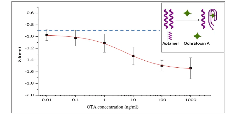
The results of TIRE data fitting are shown in Fig. 3 as the dependence of thickness increment
against the concentration of OTA. As one can see, the thickness increment appeared to be
negative thus corresponding to decrease in the aptamer layer thickness upon binding of OTA.
Such process is well-understood assuming that aptamers are coiling around the OTA target
molecules as illustrated on inset in Fig. 3. The minimal detected concentration of OTA in our
[image:7.595.68.501.474.695.2]experiments was 0.01ng/ml.
Fig. 3. Dependence of the thickness increment of the aptamer layer vs OTA concentration
.
0.01 0.1 1 10 100 1000
-2.0 -1.8 -1.6 -1.4 -1.2 -1.0 -0.8 -0.6
d(
nm
)
C OTA (ng/ml)
δd
(n
m
)
7
Negative control experiments were carried out by injecting pure PBS buffer solution (with no
OTA) into the cell. Such tests were also resulted in a small “blue” spectral shift
corresponding to the thickness decrease of about 0.9 nm. This can be explained by
spontaneous self-coiling of aptamers in BPS buffer (aptamers are usually kept in HEPES
buffer containing MgCl2 salt in order to prevent self-coiling). Such 0.9 nm thickness decrease can be considered as a baseline for detection (shown as a dotted line in Fig. 3). The thickness
increment of about 1 nm corresponding to 0.01 ng/ml concentration of OTA is just above the
baseline. The achieved detection limit of 0.01 ng/ml for OTA is remarkable for direct assay
format, an order of magnitude lower that it was reported earlier for TIRE detection of
mycotoxins in direct immunoassay with antibodies (Nabok et al., 2008; Nabok et al., 2011b).
3.2. OTA-aptamer binding kinetics
The kinetics of OTA binding to aptamers was studied using the data analysis protocol
developed earlier for immune binding reaction and successfully applied for detection of
mycotoxins in direct immunoassay with specific antibodies (Nabok et al., 2007a; Nabok et al.,
2011b). This approach is based on Langmuir adsorption model of binding analytes of
concentration C to molecular receptors with concentration N on the surface. The solution of
differential equation of adsorption for the concentration of analyte molecules (n) adsorbed on
the surface is given as:
1 exp( t/)
k C k
C k N n
d a
a
,
where ka and kd are the rates of adsorption and desorption, and τ is the time constant given as
d aC k
k
1
.
The binding kinetics was therefore studied by recording a number of TIRE spectra during
binding of OTA to aptamers and plotting the resulted time dependences of ∆ (or Ψ) at
particular wavelength, e.g. 600nm, as shown in Fig. 4a. The time constant () was then
8
Fig.4. Typical time dependence of ∆ during binding OTA of 1ng/ml to aptamers (a); evaluation of KA (b).
Such procedure was repeated for all dynamic scans taken during binding OTA of different concentrations, and the results were presented in a graph 1/ against the OTA concentration in Fig. 4b. The values of k𝑎 and k𝑑 can be found as the gradient and intercept, respectively, of the linear dependence 1/𝑘𝑎 𝐶 + 𝑘𝑑in Fig. 4b. Then both the association constant (KA)
and affinity constant (KD) can be found as KA = ka / kd and KD = 1 / KA . The obtained
values of KA = 5.63. 107 (mol -1) and KD = 1.77.10-8 (mol) for aptamers-OTA binding are typical for high affinity reactions and similar to those of antibody-antigen binding (Nabok et al., 2007a; Nabok et al., 2011b).
y = 0.0121Ln(x) + 0.0867
0 0.02 0.04 0.06 0.08 0.1 0.12 0.14 0.16 0.18
0.1 1 10 100 1000
OTA concentration (ng/ml)
1
/t
(
m
in
-1
)
(a)
9
4. Conclusions
The detection of OTA in aptamer assay using TIRE method was successful. The minimal
detected concentration of OTA was 0.01ng/ml which is a remarkable result for direct aptamer
assay format. The association constant KA = 5.63.107 mol-1 and the affinity constant KD =
1.77.10-8 mol found from the binding kinetics study are characteristic for highly specific aptamer-OTA binding. The other advantages of the proposed method are label-free detection
and the use of a simple, quick and cost-effective direct assay format. Further work is focused
on TIRE detection of other mycotoxins (aflatoxin, zearalenone) in direct assay with specific
aptamers and the use of LSPR in nano-structured gold films. Detection of mycotoxins in real
samples extracted from food and feed is also considered in future.
Acknowledgments
This work was funded by NATO SPS program, project NUKR.SFPP 984637.
References
Al-Ammar, R., Nabok, A., Hashim, A., Smith, T., 2015. Microcystin-LR produced by bacterial algae: Optical detection and purification of contaminated substances. Sensors & Actuators B- Chemical, 209, 1070-1076.
Al-Rubaye, A. G., Nabok, A., & Tsargorodska, A., 2017. Spectroscopic ellipsometry study of
gold nanostructures for LSPR bio-sensing applications. Sensing and Bio-Sensing Research,
12, 30-35.
Arwin, H., Poksinski, M. and Johansen, K., 2004. Total internal reflection ellipsometry: principles and applications. Applied Optics, 43(15), 3028-3036.
Balamurugan, S., Obubuafo, A., Soper, S. A., & Spivak, D. A., 2008. Surface immobilization methods for aptamer diagnostic applications. Analytical and bioanalytical chemistry, 390(4), 1009-1021.
Barna-Vetró, I., Solti, L., Téren, J., Gyöngyösi, Á., Szabó, E. and Wölfling, A., 1996. Sensitive ELISA test for determination of ochratoxin A. Journal of Agricultural and Food Chemistry, 44(12), 4071-4074.
10
Bonel, L., Vidal, J. C., Duato, P., & Castillo, J. R.,2011. An electrochemical competitive biosensor for ochratoxin A based on a DNA biotinylated aptamer. Biosensors and Bioelectronics, 26(7), 3254-3259.
Bueno, D., Mishra, R.K., Hayat, A., Catanante, G., Sharma, V., Muñoz, R. and Marty, J.L., 2016. Portable and low cost fluorescence set-up for in-situ screening of Ochratoxin A. Talanta, 159, 395-400.
Catanante, G., Mishra, R.K., Hayat, A. and Marty, J.L., 2016. Sensitive analytical
performance of folding based biosensor using methylene blue tagged aptamers. Talanta, 153, 138-144.
Clark, H.A., Snedeker, S.M., 2006. Ochratoxin A: its cancer risk and potential for exposure. Journal of Toxicology and Environmental Health, Part B:Critical Reviews, 9(3), 265-296.
Costantini, F., Sberna, C., Petrucci, G., Reverberi, M., Domenici, F., Fanelli, C., ... & Caputo, D.,2016. Aptamer-based sandwich assay for on chip detection of Ochratoxin A by an array of amorphous silicon photosensors. Sensors and Actuators B: Chemical, 230, 31-39.
Ferapontova, E. E., & Gothelf, K. V., 2009. Optimization of the Electrochemical RNA‐
Aptamer Based Biosensor for Theophylline by Using a Methylene Blue Redox Label. Electroanalysis, 21(11), 1261-1266.
Hamula, C.L., Guthrie, J.W., Zhang, H., Li, X.F. and Le, X.C., 2006. Selection and analytical applications of aptamers. Trends in Analytical Chemistry, 25(7), 681-691.
Mairal, T., Ozalp, V.C., Lozano, P., Sanchez, Mir, M., Katakis, I., O’Sullivan, C.K., 2008 Aptamers: Molecular tools for analytical applications. Anal. Bioanal. Chem., 390, 989–1007.
Mishra, R.K., Hayat, A., Catanante, G., Istamboulie, G. and Marty, J.L., 2016. Sensitive quantitation of Ochratoxin A in cocoa beans using differential pulse voltammetry based aptasensor. Food chemistry, 192, 799-804.
Nabok, A., Tsargorodskaya, A., Mustafa, M.K., Szekacs, I., Starodub, N.F., Szekacs, A., 2011a. Detection of low molecular weight toxins using optical phase detection techniques, Sensors and Actuators B: Chemical , 154, 232-237.
Nabok, A.V., Mustafa, M.K., Tsargorodskaya, A. and Starodub, N.F., 2011b. Detection of aflatoxin B1 with a label-free ellipsometry immunosensor. BioNanoScience, 1(1-2), 38-45.
Nabok, A., Tsargorodskaya, A., 2008. The method of total internal reflection ellipsometry for thin film characterisation and sensing. Thin Solid Films, 516(24), 8993-9001.
11
Nabok, A., Tsargorodskaya, A., Holloway, A., Starodub, N.F. and Demchenko, A., 2007b. Specific binding of large aggregates of amphiphilic molecules to the respective antibodies. Langmuir, 23(16), 8485-8490.
Nabok, A.V., Tsargorodskaya, A., Hassan, A.K. and Starodub, N.F., 2005. Total internal reflection ellipsometry and SPR detection of low molecular weight environmental toxins. Applied Surface Science, 246(4), 381-386.
Pfohl‐Leszkowicz, A., Manderville, R.A., 2007. Ochratoxin A: An overview on toxicity and carcinogenicity in animals and humans. Molecular nutrition & food research, 51(1), 61-99.
Rhouati, A., Yang, C., Hayat, A. and Marty, J.L., 2013a. Aptamers: A promising tool for ochratoxin A detection in food analysis. Toxins, 5(11), 1988-2008.
Rhouati, A., Hayat, A., Hernandez, D. B., Meraihi, Z., Munoz, R., & Marty, J. L.,2013b. Development of an automated flow-based electrochemical aptasensor for on-line detection of ochratoxin A. Sensors and Actuators B: Chemical, 176, 1160-1166.
Seo, H. B., & Gu, M. B. (2017). Aptamer-based sandwich-type biosensors. Journal of
Biological Engineering, 11(1), 11.
Sheng, L., Ren, J., Miao, Y., Wang, J., & Wang, E.,2011. PVP-coated graphene oxide for selective determination of ochratoxin A via quenching fluorescence of free aptamer.
Biosensors and Bioelectronics, 26(8), 3494-3499.
Varga, J., Kevei, E., Rinyu, E., Téren, J., and Z. Kozakiewicz,1996. Ochratoxin production by Aspergillus species. Applied and Environmental Microbiology, 62, 4461-4464.
Wang, L., Ma, W., Chen, W., Liu, L., Ma, W., Zhu, Y., ... & Xu, C.,2011. An aptamer-based chromatographic strip assay for sensitive toxin semi-quantitative detection. Biosensors and Bioelectronics, 26(6), 3059-3062.