Received 24 April 2015 Accepted 5 May 2015
Edited by M. Weil, Vienna University of Technology, Austria
†
Keywords:neutron powder diffraction; sodium molybdate; sodium tungstate
CCDC references:1063434; 1063433
Supporting information:this article has supporting information at journals.iucr.org/e
Crystal structures of spinel-type Na
2MoO
4and
Na
2WO
4revisited using neutron powder diffraction
A. Dominic Fortes
ISIS Facility, Rutherford Appleton Laboratory, Harwell Science and Innovation Campus, Didcot, Oxfordshire OX11 0QX, England, Department of Earth Sciences, University College London, Gower Street, London WC1E 6BT, England, and Department of Earth and Planetary Sciences, Birkbeck, University of London, Malet Street, London WC1E 7HX, England. *Correspondence e-mail: [email protected]
Time-of-flight neutron powder diffraction data have been collected from Na2MoO4 and Na2WO4 to a resolution of sin ()/ = 1.25 A˚ 1, which is
substantially better than the previous analyses using MoKX-rays, providing roughly triple the number of measured reflections with respect to the previous studies [Okadaet al.(1974).Acta Cryst.B30, 1872–1873; Bramnik & Ehrenberg (2004).Z. Anorg. Allg. Chem.630, 1336–1341]. The unit-cell parameters are in excellent agreement with literature data [Swanson et al. (1962). NBS Monograph No. 25, sect. 1, pp. 46–47] and the structural parameters for the molybdate agree very well with those of Bramnik & Ehrenberg (2004). However, the tungstate structure refinement of Okadaet al.(1974) stands apart as being conspicuously inaccurate, giving significantly longer W—O distances, 1.819 (8) A˚ , and shorter Na—O distances, 2.378 (8) A˚, than are reported here or in other simple tungstates. As such, this work represents an order-of-magnitude improvement in precision for sodium molybdate and an equally substantial improvement in both accuracy and precision for sodium tungstate. Both compounds adopt the spinel structure type. The Na+ions have site symmetry .3m
and are in octahedral coordination while the transition metal atoms have site symmetry 43mand are in tetrahedral coordination.
1. Chemical context
Both Na2MoO4 and Na2WO4 have rich phase diagrams in
pressure and temperature space (Pistorius, 1966). The stable form at room temperature is the -Ag2MoO4 cubic spinel
structure type, space groupFd3m, which has been known for almost a century (Wyckoff, 1922). Among the alkali metal sulfates, chromates, molybdates and tungstates, only Na2MoO4
and Na2WO4 adopt the normal spinel structure at ambient
pressure. Li2MoO4 forms a cubic spinel structure at high
pressure (Liebertz & Rooymans, 1967). Li2WO4 forms a
‘spinel-like’ phase at high pressure (Pistorius, 1975; Horiuchi
et al., 1979). Cubic sodium molybdate and sodium tungstate have been examined intermittently over subsequent decades using a variety of crystallographic techniques (Lindqvist, 1950; Becka & Poljak, 1958; Swanson et al., 1957, 1962; Singh Mudher et al., 2005) and vibrational spectroscopic methods (Busey & Keller, 1964; Preudhomme & Tarte, 1972; Breitinger
et al., 1981; Luz Limaet al., 2010, 2011), or by nuclear magnetic resonance and quadrupole coupling (Lynch & Segel, 1972). However, the extant structural information on both phases is derived from X-ray diffraction data of low to modest preci-sion. The first published structure refinement of Na2MoO4was
only reported recently (Bramnik & Ehrenberg, 2004) from X-ray powder diffraction data measured to sin ()/ = 0.71 A˚ 1; the last structure refinement of Na2WO4 was
reported by Okada et al. (1974) from X-ray single-crystal diffraction data to sin ()/= 0.81 A˚ 1. Both compounds are highly soluble in water, crystallizing at room temperature as orthorhombic dihydrates (space group Pbca, Atovmyan & D’yachenko, 1969; Farrugia, 2007). Below 283.5 K for the molybdate and 279.2 K for the tungstate, crystals grow with ten water molecules per formula unit (Funk, 1900; Cadbury, 1955; Zhilova et al., 2008). The high solubility in water and propensity towards forming hydrogen-bonded hydrates (unlike the heavier alkali metal molybdates and tungstates) suggests that both compounds would be excellent candidates for formation of hydrogen-bonded complexes with water soluble organics, such as amino acids, producing metal– organic crystals with potentially useful optical properties (cf., glycine lithium molybdate; Flecket al., 2006).
In the course of preparing deuterated specimens of the dihydrated and decahydrated forms of Na2MoO4and Na2WO4
for neutron diffraction analysis, the anhydrous phases were synthesised and an opportunity arose to acquire neutron powder diffraction data. The advantage of using a neutron radiation probe is that the scattering lengths of the atoms concerned are fairly similar, coherent scattering lengths being 6.715 fm for Mo, 4.86 fm for W, 3.63 fm for Na and 5.803 fm for O (Sears, 2006). Secondly, with the time-of-flight method, particularly with a very long primary flight path and high-angle backscattering detectors, one can acquire unparalleled resolution at very short flight times (i.e., small d-spacings), ensuring an order of magnitude improvement in parameter precision over the previous studies. In this work, usable data were obtained at a resolution of sin ()/= 1.25 A˚ 1, roughly tripling the number of measured reflections with respect to Okada et al. (1974) and Bramnik & Ehrenberg (2004). This work provides the most accurate and precise foundation on which to build future discussion of the hydrated forms of
Na2MoO4and Na2WO4. Neutron powder diffraction data for
Na2MoO4and Na2WO4are given in Figs. 1 and 2.
2. Structural commentary
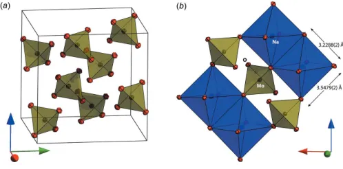
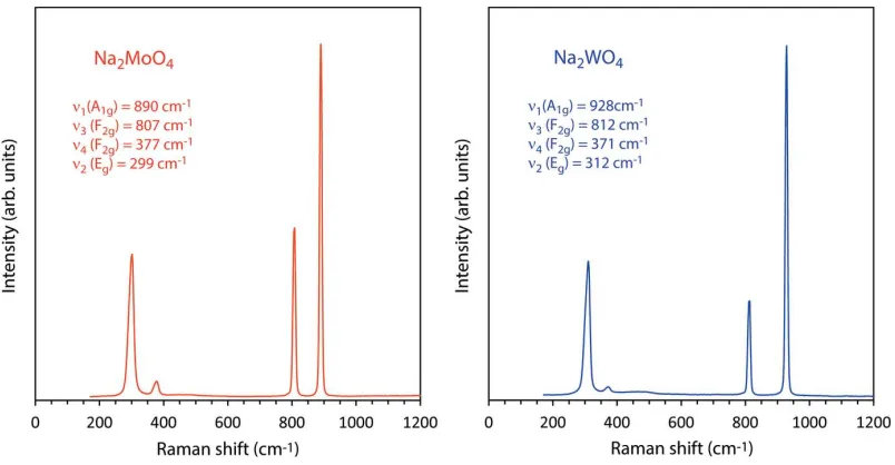
The structure of both compounds is the normal spinel type with Na+ions on the 16csites in octahedral coordination and Mo6+/W6+ ions on 8b sites in tetrahedral coordination. The coordinating oxygen atoms occupy the 32egeneral positions, their location being defined by a single variable parameteru. For ideal cubic close packing, theucoordinate adopts a value of 0.25 although for various spinels is found in the range 0.24 to 0.275. In Na2MoO4 the u parameter has a value of
0.262710 (15) and in Na2WO4it has a value of 0.262246 (15).
The practical consequence of this compared with the ‘ideal’ value ofu= 0.25 is that the shared edges of the NaO6
[image:2.610.69.565.66.257.2]octa-hedra are shorter than the unshared edges (Fig. 3b). In the Figure 1
Neutron powder diffraction data for Na2MoO4; red points are the observations, the green line is the calculated profile and the pink line beneath the diffraction pattern represents Obs Calc. Vertical black tick marks report the expected positions of the Bragg peaks. The inset shows the data measured at very short flight times (i.e., smalld-spacing).
Figure 2
[image:2.610.315.564.570.693.2]Neutron powder diffraction data for Na2WO4; red points are the observations, the green line is the calculated profile and the pink line beneath the diffraction pattern represents Obs Calc. Vertical black tick marks report the expected positions of the Bragg peaks. The inset shows the data measured at very short flight times (i.e., smalld-spacing).
Figure 3
molybdate, these lengths are 3.2288 (2) and 3.5479 (2) A˚ , the ratio being 1.0988 (1); in the tungstate, the lengths of the two inequivalent octahedral edges are 3.2356 (2) A˚ and 3.5441 (2) A˚ , their ratio being 1.0953 (1). The MoO4
2
and WO4
2
tetrahedra have perfectTdsymmetry with Mo—O and
W—O bond lengths of 1.7716 (3) and 1.7830 (2) A˚ , respec-tively. The unit-cell parameters for both compounds are in excellent agreement with those of Swansonet al.(1962) and the structural parameters for the molybdate agree very well with those of Bramnik & Ehrenberg (2004). However, the Na2WO4 structure refinement of Okada et al. (1974) stands
apart as being conspicuously inaccurate, giving significantly longer W—O distances, 1.819 (8) A˚ , and shorter Na—O distances, 2.378 (8) A˚ , than are reported here or in many other simple tungstates. Indeed the ionic radii of four-coordinated Mo6+ and W6+ obtained from analysis of a large range of crystal structures are nearly identical, being 0.41 and 0.42 A˚ , respectively (Shannon, 1976). The values reported here agree very well with the majority of Mo—O and W—O bond lengths in isolated MoO4
2
and WO4 2
tetrahedral oxyanions from a range of alkali metal and alkaline earth compounds tabulated in the literature (e.g., Zachariasen & Plettinger, 1961; Gate-house & Leverett, 1969; Koster et al., 1969; Gu¨rmen et al., 1971; Wandahl & Christensen, 1987; Farrugia, 2007; van den Berg & Juffermans, 1982). As such, this work represents an improvement in accuracy for sodium molybdate and an improvement in both accuracy and precision for sodium tungstate.
3. Synthesis and crystallization
Na2MoO42H2O (Sigma Aldrich M1003, > 99.5%) and
Na2WO42H2O (Sigma Aldrich 14304, > 99.0%) were heated
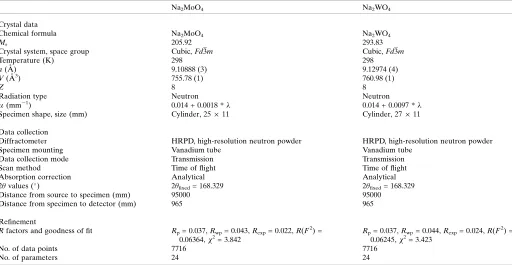
to 673 K in ceramic crucibles for 24 hr. Loss of water was confirmed by Raman spectroscopy; X-ray powder diffraction confirmed the phase identity and purity of the two anhydrous products, Na2MoO4and Na2WO4.
Raman spectra were acquired using a B&WTeki-Raman plus portable spectrometer; this device uses a 532 nm laser (37 mW power at the fiber-optic probe tip) to stimulate Raman scattering, which is measured in the range 170– 4000 cm 1 with a spectral resolution of 3 cm 1. Data were collected in a series of 20 x 9 sec integrations for Na2MoO4and
20 x 7 sec integrations for Na2WO4; after summation, the
background was removed and peaks fitted using Pseudo-Voigt functions in OriginPro (OriginLab, Northampton, MA) (Fig. 4). These data are provided as an electronic supplement in the form of an ASCII file.
4. Refinement
Crystal data, data collection and structure refinement details are summarized in Table 1. For the neutron scattering experiments, each specimen was loaded into a vanadium tube of 11 mm internal diameter to a depth of approximately 25 mm. The exact sample volume and mass were measured in order to determine the number density for correction of the specimen self-shielding. The samples were mounted on the HRPD beamline (Ibberson, 2009) at the ISIS neutron spal-lation source and data were collected in the 10–110 ms time-of-flight window for 2.5 h (Na2MoO4) and 3.5 h (Na2WO4).
Data were corrected for self-shielding, focussed to a common scattering angle and normalized to the incident spectrum by reference to a V:Nb null-scattering standard before being output in a format suitable for Rietveld refinement with
[image:3.610.106.508.72.281.2]GSAS/Expgui(Larsen & Von Dreele, 2000: Toby, 2001). Figure 4
Acknowledgements
The author thanks the STFC ISIS facility for beam-time access and acknowledges financial support from the STFC, grant No. ST/K000934/1.
References
Arnold, O., & 27 co-authors (2014). Nucl. Instrum. Methods Phys. Res. A,764, 156–166.
Atovmyan, L. O. & D’yachenko, O. A. (1969).J. Struct. Chem.10, 416–418.
Becka, L. N. & Poljak, R. J. (1958).Anales Asoc. Quim. Arg.46, 204– 209.
Berg, A. J. van den & Juffermans, C. A. H. (1982).J. Appl. Cryst.15, 114–116.
Bramnik, K. G. & Ehrenberg, H. (2004).Z. Anorg. Allg. Chem.630, 1336–1341.
Breitinger, D. K., Emmert, L. & Kress, W. (1981). Ber. Bunsenges. Phys. Chem.85, 504–505.
Busey, R. H. & Keller, O. L. Jr (1964).J. Chem. Phys.41, 215–225. Cadbury, W. E. Jr (1955).J. Phys. Chem.59, 257–260.
Farrugia, L. J. (2007).Acta Cryst.E63, i142.
Fleck, M., Schwendtner, K. & Hensler, A. (2006). Acta Cryst.C62, m122–m125.
Funk, R. (1900).Ber. Dtsch. Chem. Ges.33, 3696–3703.
Gatehouse, B. M. & Leverett, P. (1969).J. Chem. Soc. A, pp. 849. Gu¨rmen, E. (1971).J. Chem. Phys.55, 1093–1097.
Horiuchi, H., Morimoto, N. & Yamaoka, S. (1979). J. Solid State Chem.30, 129–135.
Ibberson, R. M. (2009).Nucl. Instrum. Methods Phys. Res. A,600, 47– 49.
Koster, A. S., Kools, F. X. N. M. & Rieck, G. D. (1969).Acta Cryst.
B25, 1704–1708.
Larsen, A. C. & Von Dreele, R. B. (2000).General Structure Analysis System (GSAS). Los Alamos National Laboratory Report LAUR 86-748, Los Alamos, New Mexico, USA. http://www.ncnr.NIST.gov/ Xtal/software/GSAS.html.
Liebertz, J. & Rooymans, C. J. M. (1967).Solid State Commun.5, 405– 409.
Lindqvist, I. (1950).Acta Chem. Scand.4, 1066–1074.
Luz Lima, C., Saraiva, G. D., Freire, P. T. C., Maczka, M., Paraguassu, W., de Sousa, F. F. & Mendes Filho, J. (2011).J. Raman Spectrosc.
42, 799–802.
Luz Lima, C., Saraiva, G. D., Souza Filho, A. G., Paraguassu, W., Freire, P. T. C. & Mendes Filho, J. (2010).J. Raman Spectrosc.41, 576–581.
Lynch, G. F. & Segel, S. L. (1972).Can. J. Phys.50, 567–572. Mantid (2013). Manipulation and Analysis Toolkit for Instrument
Data; Mantid Project. http://dx.doi.org/10.5286/SOFTWARE/ MANTID.
Okada, K., Morikawa, H., Marumo, F. & Iwai, S. (1974).Acta Cryst.
B30, 1872–1873.
Pistorius, C. W. F. T. (1966).J. Chem. Phys.44, 4532–4537. Pistorius, C. W. F. T. (1975).J. Solid State Chem.13, 325–329. Preudhomme, J. & Tarte, P. (1972).Spectrochim. Acta Part A,28, 69–
79.
Putz, H. & Brandenburg, K. (2006). DIAMOND. Crystal Impact GbR, Bonn, Germany. http://www.crystalimpact.com/diamond. Sears, V. F. (2006).Neutron News,3, 26–37.
Shannon, R. D. (1976).Acta Cryst.A32, 751–767.
Singh Mudher, K. D., Keskar, M., Krishnan, K. & Venugopal, V. (2005).J. Alloys Compd.396, 275–279.
Swanson, H. E., Gilfrich, N. T. & Cook, M. I. (1957). Natl. Bur. Stand. (US) Circ. 539, Vol. 7, p. 45.
Swanson, H. E., Morris, M. C., Stinchfield, R. P. & Evans, E. H. (1962). NBS Monograph No. 25, sect. 1, pp. 46–47.
[image:4.610.47.559.93.358.2]Toby, B. H. (2001).J. Appl. Cryst.34, 210–213.
Table 1
Experimental details.
Na2MoO4 Na2WO4
Crystal data
Chemical formula Na2MoO4 Na2WO4
Mr 205.92 293.83
Crystal system, space group Cubic,Fd3m Cubic,Fd3m
Temperature (K) 298 298
a(A˚ ) 9.10888 (3) 9.12974 (4)
V(A˚3) 755.78 (1) 760.98 (1)
Z 8 8
Radiation type Neutron Neutron
(mm 1) 0.014 + 0.0018 * 0.014 + 0.0097 *
Specimen shape, size (mm) Cylinder, 2511 Cylinder, 2711 Data collection
Diffractometer HRPD, high-resolution neutron powder HRPD, high-resolution neutron powder Specimen mounting Vanadium tube Vanadium tube
Data collection mode Transmission Transmission Scan method Time of flight Time of flight Absorption correction Analytical Analytical 2values () 2
fixed= 168.329 2fixed= 168.329
Distance from source to specimen (mm) 95000 95000 Distance from specimen to detector (mm) 965 965 Refinement
Rfactors and goodness of fit Rp= 0.037,Rwp= 0.043,Rexp= 0.022,R(F2) =
0.06364,2= 3.842
Rp= 0.037,Rwp= 0.044,Rexp= 0.024,R(F2) =
0.06245,2= 3.423
No. of data points 7716 7716
No. of parameters 24 24
Wandahl, G. & Christensen, A. N. (1987).Acta Chem. Scand. Ser. A,
41, 358–360.
Westrip, S. P. (2010).J. Appl. Cryst.43, 920–925.
Wyckoff, R. W. G. (1922).J. Am. Chem. Soc.44, 1994–1998.
Zachariasen, W. H. & Plettinger, H. A. (1961).Acta Cryst.14, 229– 230.
sup-1
Acta Cryst. (2015). E71, 592-596
supporting information
Acta Cryst. (2015). E71, 592-596 [doi:10.1107/S2056989015008774]
Crystal structures of spinel-type Na
2MoO
4and Na
2WO
4revisited using neutron
powder diffraction
A. Dominic Fortes
Computing details
For both compounds, data collection: HRPD control software; cell refinement: GSAS/Expgui (Larsen & Von Dreele,
2000; Toby, 2001); data reduction: MANTID (Arnold et al., 2014; Mantid, 2013); program(s) used to solve structure:
coordinates taken from a previous refinement. Program(s) used to refine structure: GSAS/Expgui (Larsen & Von Dreele,
2000, Toby, 2001) for Na2MoO4; GSAS/Expgui (Larsen & Von Dreele, 2000; Toby, 2001) for Na2WO4. For both
compounds, molecular graphics: DIAMOND (Putz & Brandenburg, 2006); software used to prepare material for
publication: publCIF (Westrip, 2010).
(Na2MoO4) Disodium molybdenum(VI) oxide
Crystal data
Na2MoO4
Mr = 205.92 Cubic, Fd3m
Hall symbol: -F 4vw 2vw 3
a = 9.10888 (3) Å
V = 755.78 (1) Å3
Z = 8
Dx = 3.619 Mg m−3
Melting point: 961 K Neutron radiation
µ = 0.01+ 0.0018 * λ mm−1
T = 298 K white
cylinder, 25 × 11 mm
Specimen preparation: Prepared at 673 K and 100 kPa
Data collection
HRPD, High resolution neutron powder diffractometer
Radiation source: ISIS Facility, Neutron spallation source
Specimen mounting: vanadium tube Data collection mode: transmission Scan method: time of flight
Absorption correction: analytical
Data were corrected for self shielding using σscatt = 29.198 barns and σab(λ) = 3.541 barns at 1.798 Å during the normalization procedure. The linear absorption coefficient is wavelength dependent and is calculated as: µ = 0.014 + 0.0018 * λ (mm-1).
Tmin = 1.000, Tmax = 1.000 2θfixed = 168.329
sup-2
Acta Cryst. (2015). E71, 592-596 Refinement
Least-squares matrix: full
Rp = 0.037
Rwp = 0.043
Rexp = 0.022
R(F2) = 0.06364
χ2 = 3.842 7716 data points
Excluded region(s): Data at d-spacings smaller than 0.4 Å were excluded since the counting statistics became progressively poorer at very short flight times due to the lower neutron flux at the shortest wavelengths.
Profile function: TOF profile function #3 (21 terms). Profile coefficients for exp pseudovoigt convolution [Von Dreele, 1990 (unpublished)] (α) = 0.1919, (β0) = 0.025953, (β1) = 0.005213, (σ0) = 0, (σ1) = 196.3, (σ2) = 23.5, (γ0) = 0, (γ1) = 14.91, (γ2) = 0, (γ2s) = 0, (γ1e) = 0, (γ2e) = 0, (εi) = 0, (εa) = 0, (εA) = 0, (γ11) = 0, (γ22) = 0, (γ33) = 0, (γ12) = 0, (γ13) = 0, (γ23) = 0. Peak tails ignored where intensity <0.0005x peak. Aniso. broadening axis 0.0 0.0 1.0
24 parameters 0 restraints 0 constraints (Δ/σ)max = 0.03
Background function: GSAS Background function #1 (10 terms). Shifted Chebyshev function of 1st kind 1: 1.18715, 2:
-7.466630x10⁻³, 3:8.117230x10⁻², 4: -5.411800x10⁻², 5: -1.714140x10⁻², 6: -1.882400x10⁻², 7: -1.930110x10⁻², 8: -6.255180x10⁻³, 9: 6.598230x10⁻³, 10: 8.478560x10⁻³
Fractional atomic coordinates and isotropic or equivalent isotropic displacement parameters (Å2)
x y z Uiso*/Ueq
O 0.262710 (16) 0.262710 (16) 0.262710 (16) 0.01182
Mo 0.375 0.375 0.375 0.00740
Na 0.0 0.0 0.0 0.01381
Atomic displacement parameters (Å2)
U11 U22 U33 U12 U13 U23
O 0.01182 (6) 0.01182 (6) 0.01182 (6) −0.00156 (6) −0.00156 (6) −0.00156 (6)
Mo 0.00740 (8) 0.00740 (8) 0.00740 (8) 0.0 0.0 0.0
Na 0.01381 (11) 0.01381 (11) 0.01381 (11) −0.00068 (12) −0.00068 (12) −0.00068 (12)
Geometric parameters (Å, º)
Mo—O 1.7716 (3) Naiv—Ovii 2.3986 (2)
Mo—Oi 1.7716 (3) Naiv—Oviii 2.3986 (2)
Mo—Oii 1.7716 (3) Naiv—Oix 2.3986 (2)
Mo—Oiii 1.7716 (3) Na—Naiv 3.2205 (1)
Naiv—O 2.3986 (2) Mo—Naiv 3.7763 (1)
Naiv—Ov 2.3986 (2) O—Ovi 3.2288 (2)
Naiv—Ovi 2.3986 (2) O—Ov 3.5479 (4)
O—Mo—Oi 109.4712 (1) O—Naiv—Ov 95.393 (6)
O—Mo—Oii 109.4712 (1) O—Naiv—Oix 180.000 (1)
sup-3
Acta Cryst. (2015). E71, 592-596
Oi—Mo—Oii 109.4712 (1) O—Naiv—Ovii 95.393 (6)
Oi—Mo—Oiii 109.4712 (1) Ov—Naiv—Ovi 180.000 (1)
Oii—Mo—Oiii 109.4712 (1) Mo—O—Naiv 129.178 (5)
O—Naiv—Ovi 84.607 (6)
Symmetry codes: (i) −x+3/4, y, −z+3/4; (ii) −x+3/4, −y+3/4, z; (iii) x, −y+3/4, −z+3/4; (iv) −x+1/4, −y+1/4, z; (v) x, −y+1/4, −z+1/4; (vi) −y+1/2, x+1/4,
z−1/4; (vii) −x+1/4, y, −z+1/4; (viii) y+1/4, −x+1/2, z−1/4; (ix) −y+1/2, −x+1/2, −z.
(Na2WO4) Disodium tungsten(VI) oxide
Crystal data
Na2WO4
Mr = 293.83 Cubic, Fd3m
Hall symbol: -F 4vw 2vw 3
a = 9.12974 (4) Å
V = 760.98 (1) Å3
Z = 8
Dx = 5.129 Mg m−3
Melting point: 969 K Neutron radiation
µ = 0.01+ 0.0097 * λ mm−1
T = 298 K white
cylinder, 27 × 11 mm
Specimen preparation: Prepared at 673 K and 100 kPa
Data collection
HRPD, High resolution neutron powder diffractometer
Radiation source: ISIS Facility, Neutron spallation source
Specimen mounting: vanadium tube Data collection mode: transmission Scan method: time of flight
Absorption correction: analytical
Data were corrected for self shielding using σscatt = 28.088 barns and σab(λ) = 19.361 barns at 1.798 Å during the normalisation procedure. The linear absorption coefficient is wavelength dependent and is calculated as: µ = 0.014 + 0.0097 * λ [mm-1]
Tmin = 1.000, Tmax = 1.000 2θfixed = 168.329
Distance from source to specimen: 95000 mm Distance from specimen to detector: 965 mm
Refinement
Least-squares matrix: full
Rp = 0.037
Rwp = 0.044
Rexp = 0.024
R(F2) = 0.06245
χ2 = 3.423 7716 data points
Excluded region(s): Data at d-spacings smaller than 0.4 Å were excluded since the counting statistics became progressively poorer at very short flight times due to the lower neutron flux at the shortest wavelengths.
Profile function: TOF profile function #3 (21 terms). Profile coefficients for exp pseudovoigt convolution [Von Dreele, 1990 (unpublished)] (α) = 0.1603, (β0) = 0.026115, (β1) = 0.004558, (σ0) = 0, (σ1) = 237.2, (σ2) = 45.0, (γ0) = 0, (γ1) = 14.21, (γ2) = 0, (γ2s) = 0, (γ1e) = 0, (γ2e) = 0, (εi) = 0, (εa) = 0, (εA) = 0, (γ11) = 0, (γ22) = 0, (γ33) = 0, (γ12) = 0, (γ13) = 0, (γ23) = 0. Peak tails ignored where intensity <0.0005x peak. Aniso. broadening axis 0.0 0.0 1.0
24 parameters 0 restraints 0 constraints (Δ/σ)max = 0.01
sup-4
Acta Cryst. (2015). E71, 592-596
Fractional atomic coordinates and isotropic or equivalent isotropic displacement parameters (Å2)
x y z Uiso*/Ueq
O 0.262246 (15) 0.262246 (15) 0.262246 (15) 0.01312
W 0.375 0.375 0.375 0.00903
Na 0.0 0.0 0.0 0.01538
Atomic displacement parameters (Å2)
U11 U22 U33 U12 U13 U23
O 0.01312 (6) 0.01312 (6) 0.01312 (6) −0.00161 (5) −0.00161 (5) −0.00161 (5)
W 0.00903 (11) 0.00903 (11) 0.00903 (11) 0.0 0.0 0.0
Na 0.01538 (11) 0.01538 (11) 0.01538 (11) −0.00045 (12) −0.00045 (12) −0.00045 (12)
Geometric parameters (Å, º)
W—O 1.7830 (2) Naiv—Ovii 2.3995 (1)
W—Oi 1.7830 (2) Naiv—Oviii 2.3995 (1)
W—Oii 1.7830 (2) Naiv—Oix 2.3995 (1)
W—Oiii 1.7830 (2) Na—Naiv 3.2279 (1)
Naiv—O 2.3995 (1) W—Naiv 3.7850 (1)
Naiv—Ov 2.3995 (1) O—Ovi 3.2356 (2)
Naiv—Ovi 2.3995 (1) O—Ov 3.5441 (4)
O—W—Oi 109.4712 (3) O—Naiv—Ov 95.211 (6)
O—W—Oii 109.4712 (3) O—Naiv—Oix 180.000 (1)
O—W—Oiii 109.4712 (3) O—Naiv—Oviii 84.789 (6)
Oi—W—Oii 109.4712 (3) O—Naiv—Ovii 95.211 (6)
Oi—W—Oiii 109.4712 (3) Ov—Naiv—Ovi 180.000 (1)
Oii—W—Oiii 109.4712 (3) W—O—Naiv 129.043 (4)
O—Naiv—Ovi 84.789 (6)
Symmetry codes: (i) −x+3/4, y, −z+3/4; (ii) −x+3/4, −y+3/4, z; (iii) x, −y+3/4, −z+3/4; (iv) −x+1/4, −y+1/4, z; (v) x, −y+1/4, −z+1/4; (vi) −y+1/2, x+1/4,