332
https://doi.org/10.1107/S2056989018002116 Acta Cryst.(2018). E74, 332–336research communications
Received 5 December 2017 Accepted 4 February 2018
Edited by G. Smith, Queensland University of Technology, Australia
Keywords:crystal structure; aromatic aldehydes; E-aldoximes; hydrogen bonding.
CCDC references:1821856; 1821855
Supporting information:this article has supporting information at journals.iucr.org/e
Crystal structures of (
E
)-1-naphthaldehyde oxime
and (
E
)-phenanthrene-9-carbaldehyde oxime
Jamal Lasri,a* Katherine Chulvib* and Naser Eltaher Eltayeba
aDepartment of Chemistry, Rabigh College of Science and Arts, PO Box 344, King Abdulaziz University, Jeddah, Saudi
Arabia, andbFB 1.3 Strukturanalytik, Bundesanstalt fu¨r Materialforschung und -pru¨fung (BAM), Berlin, Germany. *Correspondence e-mail: [email protected], [email protected]
The aldoximes C11H9NO (I) and C15H11NO (II), synthesized inca90% yield, by
treatment of 1-naphthaldehyde or phenanthrene-9-carbaldehyde, respectively, with hydroxylamine hydrochloride and sodium carbonate, have been character-ized by IR, 1H, 13C and DEPT-135 NMR spectroscopies, and also by single-crystal X-ray diffraction analysis. The molecules of (I) and (II) are conformationally similar, with the aldoxime substituent groups lying outside the planes of the naphthalene or phenanthrene rings, forming dihedral angles with them of 23.9 (4) and 27.9 (6), respectively. The crystal structures of both (I) and (II) are similar with a single intermolecular O—H N hydrogen-bonding interaction, giving rise to the formation of one-dimensional polymeric chains extending along the 21(b) screw axes in each.
1. Chemical context
Oxime compounds have found many applications; for example in the medical field, they are used as antidotes for nerve agents (Kassa, 2002). Oximes are also used as intermediates in the industrial production of caprolactam, a precursor to Nylon 6 (Ritzet al., 2012). Oximes, HO—N CR1R2, are also valuable
and simple reagents containing the O—N C moiety
(Kukushkin & Pombeiro, 1999), which easily adds to nitrile ligands, to form a variety of nitrogen-containing productse.g. iminoacylated compounds (Kopylovichet al., 2009; Lasriet al., 2007, 2008), amidines (Kopylovichet al., 2001), carboxamides (Kopylovichet al., 2002), phthalocyanines (Kopylovichet al., 2004), or 1,3,5-triazapentadiene species (Kopylovich et al., 2007). In this work, we report the synthesis and crystal structures of two aldoximes,viz.(E)-1-naphthaldehyde oxime (I) and (E)-phenanthrene-9-carbaldehyde oxime (II), by treatment of 1-naphthaldehyde or phenanthrene-9-carbalde-hyde, respectively, with hydroxylamine hydrochloride and sodium carbonate.
2. Structural commentary
The title compounds (I) and (II) both crystallize in the non-centrosymmetric monoclinic space groupP21withZ= 2, with
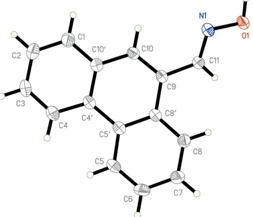
similar unit-cell parameters. The asymmetric unit contents for each are shown in Figs. 1 and 2. Compound (I) comprises a naphthalene unit functionalized with an aldoxime group at position 1. The naphthalene unit is, as expected, essentially
planar but the plane containing the aldoxime atoms lies significantly out of the naphthalene plane [torsion angle N1— C11—C1—C2 = 23.6 (6)] (Table 1). In the case of compound (II), the plane of the aldoxime group lies similarly out-of-plane with the phenanthrene ring system [comparative torsion
angle N1—C11—C9—C10 = 27.6 (4)], corresponding to
dihedral angles between the two planes of 23.9 (4) and 27.9 (5) for (I) and (II), respectively. The aldoxime group shows similar bond lengths for both structures: 1.395 (5) and 1.405 (3) A˚ for O1—N1, 1.273 (5) and 1.268 (3) A˚ for N1— C11, 1.461 (6) and 1.466 (4) A˚ for C1—C11 or C9—C11, for (I) and (II), respectively.
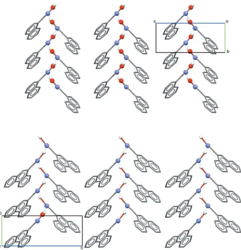
3. Supramolecular features
Similar intermolecular interactions are observed in the crystal structures of both (I) and (II). In each, molecules are linked through a single intermolecular O1—H N1i hydrogen-bonding interaction [Tables 2 and 3 for (I) and (II), respec-tively]. These basic interactions are shown in Fig. 3, defining an oximeC(3) type II motif. It is well known that oximes are able to form different types of hydrogen-bonding motifs (Brutonet al., 2003). In the structures of both (I) and (II), the formation of a one-dimensional polymeric chain arrangement of molecules results, extending along the 21(b) screw axes in
each (Fig. 4).
research communications
Acta Cryst.(2018). E74, 332–336 Lasriet al. C
[image:2.610.45.297.92.152.2]11H9NO and C15H11NO
333
Figure 2The molecular conformation and atom-numbering scheme for (II), with non-H atoms represented as 30% probability ellipsoids.
Table 1
Selected torsion angles () for the aldoxime groups in (I) and (II).
Compound (I) Compound (II)
C1/C9—C11—N1—O1 175.5 (4) 175.3 (2) C2/C10—C1/C9—C11—N1 23.6 (6) 27.6 (4) C80—C1—C11—N1 160.4 (4) – C80
[image:2.610.311.565.171.205.2]—C9—C11—N1 – 156.1 (2)
Figure 1
The molecular conformation and atom-numbering scheme for (I), with non-H atoms represented as 30% probability ellipsoids.
Table 2
Hydrogen-bond geometry (A˚ ,) for (I).
D—H A D—H H A D A D—H A
O1—H1 N1i 0.90 (6) 1.94 (6) 2.834 (5) 177 (6)
Symmetry code: (i)xþ2;y1 2;zþ1. Table 3
Hydrogen-bond geometry (A˚ ,) for (II).
D—H A D—H H A D A D—H A
O1—H N1i 0.88 (3) 1.99 (3) 2.852 (3) 169 (3)
[image:2.610.46.295.281.460.2]Symmetry code: (i)xþ1;yþ1 2;zþ1.
Figure 3
[image:2.610.48.299.507.722.2] [image:2.610.312.563.594.702.2]4. Database survey
Many naphthalene-carbaldehyde oxime derivatives are
present in the Cambridge Structural Database (Version 5.38; Groomet al.2016) but no one crystal structure containing only an aldoxime group in position 1 of the naphthalene ring system has been reported. The most similar structures that can be found are LIVROY/LIVROY01 (Guoet al., 2008; Tarai & Baruah, 2016) with an additional hydroxyl group in position 2 and TIJPOS (Asaadet al., 2005) with a dimethylamino group in position 9. The most important difference between (I) and LIVROY/LIVROY01 are the two hydrogen bonds: one
intramolecular O—H N and another intermolecular O—
H O. As a result of the intramolecular hydrogen-bonding interaction, the aldoxime group in the latter compound is coplanar with the central naphthalene ring with a dihedral angle of 1.21and torsion angles C1—C11—N1—O2 = 179.27,
C3—C1—C11—N1 = 179.91 and C4—C1—C11—N1 =
0.76. However, TIJPOS (Asaadet al., 2005), with just one type of intermolecular hydrogen bond, shows a rotation in the aldoxime group that is more dramatic than in (I) and (II) (Table 1), with a 40.35deviation from the central naphthalene plane.
No examples of structures of phenanthrene-carbaldehyde oxime derivatives are present in the Cambridge Structural Database.
5. Synthesis and crystallization
The aldoximes (E)-1-naphthaldehyde oxime (I) and (E )-phenanthrene-9-carbaldehyde oxime (II) were synthesized, in
ca90% yield, by treatment of 1-naphthaldehyde or phenan-threne-9-carbaldehyde, respectively, with hydroxylamine hydrochloride and sodium carbonate in MeOH at room temperature. To a solution of hydroxylamine hydrochloride (41.6 mg, 0.60 mmol) in MeOH (10 ml) was added sodium carbonate (31.7 mg, 0.30 mmol). The reaction mixture was stirred at room temperature for 5 min. 1-Naphthaldehyde
(85.0 mg, 0.54 mmol) or phenanthrene-9-carbaldehyde
(112.2 mg, 0.54 mmol) was added and the reaction mixture was stirred at room temperature for 12 h. The precipitate formed was then filtered off and the filtrate was evaporatedin vacuo. The crude residue was purified by column chroma-tography on silica (CHCl3as the eluent, 50 ml), followed by
evaporation of the solventin vacuoto give the pure aldoximes [(I), 84 mg, 90% yield and (II), 107 mg, 89% yield] (see reaction scheme).
Single crystals of the aldoximes (I) and (II) suitable for X-ray diffraction were obtained by slow evaporation of their 10 ml CHCl3 solutions at room temperature. Compounds (I)
and (II) were characterized by IR, 1H, 13C and DEPT-135 NMR spectroscopies and also by single crystal X-ray diffrac-tion analysis.
In the IR spectra of (I) and (II), the characteristic bands at wavenumbers 3389 and 3200 cm1(O—H), and 1614 and 1607 cm-1(C N), confirm the formation of the aldoximes (I) and (II), respectively. In the 1H NMR spectra, we observed the absence of the signal of the aldehyde atca10 ppm and a new signal at ca 8.8 ppm due to the imine proton CH N was detected. Moreover, in the13C and DEPT-135 NMR spectra, the signal of the aldehyde atca190 ppm was not observed, and
a new signal at ca 150 ppm due to the oxime carbon
CH NOH was detected, confirming the formation of the
aldoximes (I) and (II).
(E)-1-naphthaldehyde oxime (I)
Yield: 90%. IR (cm1): 3389 (OH), 1614 (C N).1H NMR (CDCl3),: 7.53 (t, JHH 7.5 Hz, 1H, CHaromatic), 7.56 (t, JHH
7.0 Hz, 1H, CHaromatic), 7.61 (t, JHH7.0 Hz, 1H, CHaromatic),
7.82 (d, JHH7.1 Hz, 1H, CHaromatic), 7.93 (t, JHH8.1 Hz, 2H,
334
Lasriet al. C11H9NO and C15H11NO Acta Cryst.(2018). E74, 332–336
[image:3.610.48.295.69.325.2]research communications
Figure 4
CHaromatic), 8.48 (d, JHH8.3 Hz, 1H, CHaromatic), 8.87 (s, 1H,
CH N).13C NMR (CDCl3),: 124.2, 125.4, 126.2, 127.0, 127.1
(CHaromatic), 128.0 (Caromatic), 128.8, 130.6 (CHaromatic), 130.8,
133.8 (Caromatic), 150.0 (CH N). DEPT-135 NMR (CDCl3),:
124.2, 125.4, 126.2, 127.0, 127.1, 128.8, 130.6 (CHaromatic), 150.0
(CH N).
E-phenanthrene-9-carbaldehyde oxime (II)
Yield: 89%. IR (cm1): 3200 (OH), 1607 (C N).1H NMR (CDCl3),: 7.64 (t,JHH7.9 Hz, 1H, CHaromatic), 7.68–7.75 (m,
3H, CHaromatic), 7.94 (d, JHH7.9 Hz, 1H, CHaromatic), 8.04 (s,
1H, CHaromatic), 8.62 (d,JHH7.9 Hz, 1H, CHaromatic), 8.70 (d,
JHH 8.2 Hz, 1H, CHaromatic), 8.77 (d, JHH 8.2 Hz, 1H,
CHaromatic), 8.85 (s, 1H, CH N).13C NMR (CDCl3),: 122.6,
123.1, 125.4 (CHaromatic), 126.8 (Caromatic), 126.9, 127.0, 127.2,
127.9, 129.3 (CHaromatic), 130.7, 131.0, 131.1 (Caromatic), 150.8
(CH N). DEPT-135 NMR (CDCl3), : 122.6, 123.1, 125.4,
126.9, 127.0, 127.2, 127.9, 129.3 (CHaromatic), 150.8 (CH N).
6. Refinement
Crystal data, data collection and structure refinement details are summarized in Table 4. All C-bound H atoms were located in difference-Fourier maps but were subsequently treated as riding with C—H = 0.93 A˚ and withUiso(H) = 1.2Ueq(C). The
H atoms of the OH groups were positioned with idealized geometry and were refined freely in both structures.
Funding information
This project was funded by the Deanship of Scientific Research (DSR) at King Abdulaziz University, Jeddah, under grant No. (G-100–662-37). The authors, therefore, acknowl-edge with thanks the DSR for technical and financial support.
References
Asaad, N., Davies, J. E., Hodgson, D. R. W., Kirby, A. J., van Vliet, L. & Ottavi, L. (2005).J. Phys. Org. Chem.18, 101–109.
Bruker (2016). APEX3, SAINT and SADABS. Bruker AXS Inc., Madison, Wisconsin, USA.
Bruton, E. A., Brammer, L., Pigge, F. C., Aakero¨y, C. B. & Leinen, D. S. (2003).New J. Chem.27, 1084–1094.
Groom, C. R., Bruno, I. J., Lightfoot, M. P. & Ward, S. C. (2016).Acta Cryst.B72, 171–179.
Guo, Z., Li, L., Liu, G. & Dong, J. (2008).Acta Cryst.E64, o568. Kassa, J. (2002).J. Toxicol. Clin. Toxicol.40, 803–816.
Kopylovich, M. N., Haukka, M., Kirillov, A. M., Kukushkin, V. Yu. & Pombeiro, A. J. L. (2007).Chem. Eur. J.13, 786–791.
Kopylovich, M. N., Kukushkin, V. Yu., Guedes da Silva, M. F. C., Haukka, M., Frau´sto da Silva, J. J. R. & Pombeiro, A. J. L. (2001).J. Chem. Soc. Perkin Trans. 1, pp. 1569–1573.
Kopylovich, M. N., Kukushkin, V. Yu., Haukka, M., Frau´sto da Silva, J. J. R. & Pombeiro, A. J. L. (2002).Inorg. Chem.41, 4798–4804. Kopylovich, M. N., Kukushkin, V. Yu., Haukka, M., Luzyanin, K. V. &
Pombeiro, A. J. L. (2004).J. Am. Chem. Soc.126, 15040–15041. Kopylovich, M. N., Lasri, J., Guedes da Silva, M. F. C. & Pombeiro,
A. J. L. (2009).Dalton Trans.pp. 3074–3084.
research communications
Acta Cryst.(2018). E74, 332–336 Lasriet al. C
[image:4.610.47.560.93.385.2]11H9NO and C15H11NO
335
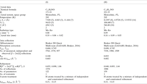
Table 4Experimental details.
(I) (II)
Crystal data
Chemical formula C11H9NO C15H11NO
Mr 171.19 221.25
Crystal system, space group Monoclinic,P21 Monoclinic,P21
Temperature (K) 295 295
a,b,c(A˚ ) 7.928 (5), 4.843 (3), 11.444 (7) 8.2397 (8), 4.9728 (5), 13.9332 (14) (
) 94.03 (5) 106.680 (7)
V(A˚3) 438.3 (5) 546.88 (10)
Z 2 2
Radiation type MoK MoK
(mm1) 0.08 0.09
Crystal size (mm) 0.100.060.02 0.160.090.05
Data collection
Diffractometer Bruker D8 Quest Bruker D8 Quest
Absorption correction Multi-scan (SADABS; Bruker, 2016) Multi-scan (SADABS; Bruker, 2016)
Tmin,Tmax 0.684, 0.745 0.698, 0.745
No. of measured, independent and observed [I> 2(I)] reflections
5762, 1570, 957 7330, 1988, 1509
Rint 0.100 0.053
(sin/)max(A˚
1) 0.603 0.602
Refinement
R[F2> 2(F2)],wR(F2),S 0.055, 0.098, 1.06 0.040, 0.093, 1.04
No. of reflections 1570 1988
No. of parameters 122 159
No. of restraints 1 1
H-atom treatment H atoms treated by a mixture of independent and constrained refinement
H atoms treated by a mixture of independent and constrained refinement
max, min(e A˚
3) 0.15,0.15 0.15,0.15
Kukushkin, V. Yu. & Pombeiro, A. J. L. (1999).Coord. Chem. Rev. 181, 147–175.
Lasri, J., Charmier, M. A. J., da Silva, M. F. C. G. & Pombeiro, A. J. L. (2007).Dalton Trans.pp. 3259–3266.
Lasri, J., Guedes da Silva, M. F. C., Charmier, M. A. J. & Pombeiro, A. J. L. (2008).Eur. J. Inorg. Chem.pp. 3668–3677.
Ritz, J., Fuchs, H., Kieczka, H. & Moran, W. C. (2012).Ullmann’s Encyclopedia of Industrial Chemistry. Weinheim: Wiley-VCH. Sheldrick, G. M. (2015a).Acta Cryst.A71, 3–8.
Sheldrick, G. M. (2015b).Acta Cryst.C71, 3–8.
Tarai, A. & Baruah, J. B. (2016).Cryst. Growth Des.16, 126–135.
336
Lasriet al. C11H9NO and C15H11NO Acta Cryst.(2018). E74, 332–336
supporting information
sup-1 Acta Cryst. (2018). E74, 332-336
supporting information
Acta Cryst. (2018). E74, 332-336 [https://doi.org/10.1107/S2056989018002116]
Crystal structures of (
E
)-1-naphthaldehyde oxime and (
E
)-phenanthrene-9-carbaldehyde oxime
Jamal Lasri, Katherine Chulvi and Naser Eltaher Eltayeb
Computing details
For both structures, data collection: APEX3 (Bruker, 2016); cell refinement: SAINT (Bruker, 2016); data reduction:
SAINT (Bruker, 2016); program(s) used to solve structure: SHELXT2014 (Sheldrick, 2015a); program(s) used to refine
structure: SHELXL2014 (Sheldrick, 2015b); molecular graphics: SHELXL2014 (Sheldrick, 2015b).
(E)-1-Naphthaldehyde oxime (I)
Crystal data
C11H9NO
Mr = 171.19
Monoclinic, P21
a = 7.928 (5) Å
b = 4.843 (3) Å
c = 11.444 (7) Å
β = 94.03 (5)°
V = 438.3 (5) Å3
Z = 2
F(000) = 180
Dx = 1.297 Mg m−3
Mo Kα radiation, λ = 0.71073 Å Cell parameters from 1367 reflections
θ = 2.6–22.6°
µ = 0.08 mm−1
T = 295 K Block, colourless 0.10 × 0.06 × 0.02 mm
Data collection
Bruker D8 Quest diffractometer
φ and ω scans
Absorption correction: multi-scan (SADABS; Bruker, 2016)
Tmin = 0.684, Tmax = 0.745
5762 measured reflections
1570 independent reflections 957 reflections with I > 2σ(I)
Rint = 0.100
θmax = 25.4°, θmin = 2.6°
h = −9→9
k = −5→5
l = −13→13
Refinement
Refinement on F2
Least-squares matrix: full
R[F2 > 2σ(F2)] = 0.055
wR(F2) = 0.098
S = 1.06 1570 reflections 122 parameters 1 restraint
Hydrogen site location: mixed
H atoms treated by a mixture of independent and constrained refinement
w = 1/[σ2(F
o2) + (0.0164P)2 + 0.1408P]
where P = (Fo2 + 2Fc2)/3
(Δ/σ)max < 0.001
Δρmax = 0.15 e Å−3
supporting information
sup-2 Acta Cryst. (2018). E74, 332-336
Special details
Geometry. All esds (except the esd in the dihedral angle between two l.s. planes) are estimated using the full covariance matrix. The cell esds are taken into account individually in the estimation of esds in distances, angles and torsion angles; correlations between esds in cell parameters are only used when they are defined by crystal symmetry. An approximate (isotropic) treatment of cell esds is used for estimating esds involving l.s. planes.
Fractional atomic coordinates and isotropic or equivalent isotropic displacement parameters (Å2)
x y z Uiso*/Ueq
O1 0.8254 (4) −0.0401 (7) 0.5056 (3) 0.0458 (9)
N1 0.8708 (5) 0.1715 (9) 0.4312 (3) 0.0418 (11)
C1 0.7559 (5) 0.4959 (10) 0.2895 (4) 0.0318 (12)
C2 0.8998 (6) 0.5156 (11) 0.2295 (4) 0.0452 (14)
H2 0.9903 0.3987 0.2494 0.054*
C3 0.9129 (7) 0.7072 (14) 0.1393 (5) 0.0569 (16)
H3 1.0128 0.7207 0.1017 0.068*
C4 0.7801 (7) 0.8737 (11) 0.1066 (4) 0.0499 (15)
H4 0.7888 0.9970 0.0450 0.060*
C4′ 0.6296 (6) 0.8624 (10) 0.1645 (4) 0.0377 (12)
C5 0.4924 (6) 1.0383 (10) 0.1338 (4) 0.0468 (15)
H5 0.5009 1.1626 0.0726 0.056*
C6 0.3485 (7) 1.0323 (12) 0.1907 (5) 0.0570 (17)
H6 0.2585 1.1479 0.1677 0.068*
C7 0.3370 (6) 0.8491 (11) 0.2846 (4) 0.0478 (15)
H7 0.2393 0.8461 0.3250 0.057*
C8 0.4663 (5) 0.6760 (11) 0.3176 (4) 0.0387 (13)
H8 0.4557 0.5558 0.3800 0.046*
C8′ 0.6172 (5) 0.6760 (10) 0.2582 (4) 0.0285 (11)
C11 0.7413 (6) 0.2826 (9) 0.3785 (4) 0.0350 (12)
H11 0.6343 0.2264 0.3972 0.042*
H1 0.922 (7) −0.129 (13) 0.528 (5) 0.10 (2)*
Atomic displacement parameters (Å2)
U11 U22 U33 U12 U13 U23
O1 0.041 (2) 0.044 (2) 0.051 (2) 0.008 (2) −0.0047 (18) 0.008 (2)
N1 0.040 (2) 0.038 (3) 0.046 (3) 0.001 (2) −0.005 (2) −0.005 (2)
C1 0.033 (3) 0.029 (3) 0.033 (3) −0.007 (3) −0.002 (2) −0.007 (3)
C2 0.035 (3) 0.054 (4) 0.048 (3) −0.005 (3) 0.009 (2) −0.014 (3)
C3 0.051 (3) 0.080 (5) 0.043 (3) −0.018 (4) 0.021 (3) −0.012 (4)
C4 0.064 (4) 0.051 (4) 0.036 (3) −0.017 (3) 0.011 (3) −0.005 (3)
C4′ 0.046 (3) 0.037 (3) 0.029 (3) −0.008 (3) −0.003 (2) −0.007 (3)
C5 0.060 (4) 0.037 (4) 0.041 (3) −0.009 (3) −0.011 (3) 0.008 (3)
C6 0.049 (3) 0.049 (4) 0.070 (4) 0.006 (3) −0.013 (3) 0.008 (4)
C7 0.035 (3) 0.048 (4) 0.060 (4) 0.002 (3) 0.000 (3) 0.009 (3)
C8 0.036 (3) 0.039 (3) 0.040 (3) 0.002 (3) −0.002 (2) 0.008 (3)
C8′ 0.029 (2) 0.027 (3) 0.029 (3) −0.004 (2) −0.0016 (19) −0.008 (3)
supporting information
sup-3 Acta Cryst. (2018). E74, 332-336
Geometric parameters (Å, º)
O1—N1 1.395 (5) C4′—C5 1.407 (6)
O1—H1 0.90 (6) C4′—C8′ 1.410 (6)
N1—C11 1.273 (5) C5—C6 1.353 (6)
C1—C2 1.375 (6) C5—H5 0.9300
C1—C8′ 1.429 (6) C6—C7 1.402 (7)
C1—C11 1.461 (6) C6—H6 0.9300
C2—C3 1.397 (7) C7—C8 1.357 (6)
C2—H2 0.9300 C7—H7 0.9300
C3—C4 1.358 (7) C8—C8′ 1.417 (5)
C3—H3 0.9300 C8—H8 0.9300
C4—C4′ 1.406 (6) C11—H11 0.9300
C4—H4 0.9300
N1—O1—H1 106 (4) C6—C5—H5 119.0
C11—N1—O1 111.5 (4) C4′—C5—H5 119.0
C2—C1—C8′ 118.9 (4) C5—C6—C7 119.0 (5)
C2—C1—C11 120.4 (5) C5—C6—H6 120.5
C8′—C1—C11 120.6 (4) C7—C6—H6 120.5
C1—C2—C3 121.5 (5) C8—C7—C6 121.0 (5)
C1—C2—H2 119.3 C8—C7—H7 119.5
C3—C2—H2 119.3 C6—C7—H7 119.5
C4—C3—C2 120.1 (5) C7—C8—C8′ 120.9 (5)
C4—C3—H3 119.9 C7—C8—H8 119.5
C2—C3—H3 119.9 C8′—C8—H8 119.5
C3—C4—C4′ 120.9 (5) C4′—C8′—C8 118.1 (4)
C3—C4—H4 119.5 C4′—C8′—C1 119.2 (4)
C4′—C4—H4 119.5 C8—C8′—C1 122.7 (4)
C4—C4′—C5 121.7 (5) N1—C11—C1 121.9 (4)
C4—C4′—C8′ 119.3 (5) N1—C11—H11 119.0
C5—C4′—C8′ 118.9 (4) C1—C11—H11 119.0
C6—C5—C4′ 122.0 (5)
C8′—C1—C2—C3 0.1 (7) C5—C4′—C8′—C8 −0.3 (6)
C11—C1—C2—C3 176.2 (5) C4—C4′—C8′—C1 −2.1 (6)
C1—C2—C3—C4 −2.0 (8) C5—C4′—C8′—C1 179.7 (4)
C2—C3—C4—C4′ 1.8 (8) C7—C8—C8′—C4′ 0.5 (7)
C3—C4—C4′—C5 178.3 (5) C7—C8—C8′—C1 −179.6 (4)
C3—C4—C4′—C8′ 0.3 (7) C2—C1—C8′—C4′ 1.9 (6)
C4—C4′—C5—C6 −178.7 (5) C11—C1—C8′—C4′ −174.2 (4)
C8′—C4′—C5—C6 −0.6 (7) C2—C1—C8′—C8 −178.0 (4)
C4′—C5—C6—C7 1.3 (8) C11—C1—C8′—C8 5.9 (6)
C5—C6—C7—C8 −1.2 (8) O1—N1—C11—C1 −175.5 (4)
C6—C7—C8—C8′ 0.2 (7) C2—C1—C11—N1 23.6 (6)
supporting information
sup-4 Acta Cryst. (2018). E74, 332-336
Hydrogen-bond geometry (Å, º)
D—H···A D—H H···A D···A D—H···A
O1—H1···N1i 0.90 (6) 1.94 (6) 2.834 (5) 177 (6)
Symmetry code: (i) −x+2, y−1/2, −z+1.
(E)-Phenanthrene-9-carbaldehyde oxime (II)
Crystal data
C15H11NO
Mr = 221.25
Monoclinic, P21
a = 8.2397 (8) Å
b = 4.9728 (5) Å
c = 13.9332 (14) Å
β = 106.680 (7)°
V = 546.88 (10) Å3
Z = 2
F(000) = 232
Dx = 1.344 Mg m−3
Mo Kα radiation, λ = 0.71073 Å Cell parameters from 2141 reflections
θ = 2.6–24.9°
µ = 0.09 mm−1
T = 295 K Block, colourless 0.16 × 0.09 × 0.05 mm
Data collection
Bruker D8 Quest diffractometer
φ and ω scans
Absorption correction: multi-scan (SADABS; Bruker, 2016)
Tmin = 0.698, Tmax = 0.745
7330 measured reflections
1988 independent reflections 1509 reflections with I > 2σ(I)
Rint = 0.053
θmax = 25.3°, θmin = 2.6°
h = −9→9
k = −5→5
l = −16→16
Refinement
Refinement on F2
Least-squares matrix: full
R[F2 > 2σ(F2)] = 0.040
wR(F2) = 0.093
S = 1.04 1988 reflections 159 parameters 1 restraint
Hydrogen site location: mixed
H atoms treated by a mixture of independent and constrained refinement
w = 1/[σ2(F
o2) + (0.0478P)2]
where P = (Fo2 + 2Fc2)/3
(Δ/σ)max < 0.001
Δρmax = 0.15 e Å−3
Δρmin = −0.15 e Å−3
Extinction correction: SHELXL2016 (Sheldrick, 2015b),
Fc*=kFc[1+0.001xFc2λ3/sin(2θ)]-1/4
Extinction coefficient: 0.058 (11)
Special details
Geometry. All esds (except the esd in the dihedral angle between two l.s. planes) are estimated using the full covariance matrix. The cell esds are taken into account individually in the estimation of esds in distances, angles and torsion angles; correlations between esds in cell parameters are only used when they are defined by crystal symmetry. An approximate (isotropic) treatment of cell esds is used for estimating esds involving l.s. planes.
Fractional atomic coordinates and isotropic or equivalent isotropic displacement parameters (Å2)
x y z Uiso*/Ueq
O1 0.3294 (3) 1.0407 (5) 0.49418 (15) 0.0446 (6)
N1 0.4084 (3) 0.8293 (5) 0.55737 (16) 0.0371 (6)
supporting information
sup-5 Acta Cryst. (2018). E74, 332-336
H11 0.197024 0.785637 0.585614 0.041*
C9 0.3660 (3) 0.5157 (6) 0.67732 (18) 0.0322 (7)
C10 0.5314 (4) 0.5027 (7) 0.7316 (2) 0.0366 (7)
H10 0.607395 0.622514 0.716727 0.044*
C10′ 0.5935 (3) 0.3140 (6) 0.81023 (19) 0.0349 (7)
C1 0.7655 (4) 0.3096 (7) 0.8651 (2) 0.0472 (8)
H1 0.840812 0.427685 0.848577 0.057*
C2 0.8232 (4) 0.1335 (8) 0.9426 (2) 0.0510 (9)
H2 0.937339 0.131717 0.978623 0.061*
C3 0.7116 (4) −0.0423 (7) 0.9676 (2) 0.0481 (9)
H3 0.751583 −0.162219 1.020293 0.058*
C4 0.5438 (4) −0.0424 (6) 0.9159 (2) 0.0445 (8)
H4 0.470941 −0.162230 0.933886 0.053*
C4′ 0.4792 (4) 0.1366 (6) 0.83561 (19) 0.0333 (7)
C5′ 0.3013 (4) 0.1442 (6) 0.77844 (19) 0.0334 (7)
C5 0.1811 (4) −0.0332 (6) 0.7975 (2) 0.0431 (8)
H5 0.215809 −0.156417 0.849626 0.052*
C6 0.0153 (4) −0.0300 (7) 0.7419 (3) 0.0499 (9)
H6 −0.061247 −0.149898 0.756119 0.060*
C7 −0.0389 (4) 0.1524 (7) 0.6641 (2) 0.0499 (9)
H7 −0.151904 0.154288 0.626056 0.060*
C8 0.0728 (4) 0.3292 (7) 0.6429 (2) 0.0424 (8)
H8 0.034520 0.451063 0.590667 0.051*
C8′ 0.2453 (3) 0.3306 (6) 0.69893 (19) 0.0317 (7)
H 0.413 (4) 1.109 (7) 0.475 (2) 0.061 (12)*
Atomic displacement parameters (Å2)
U11 U22 U33 U12 U13 U23
O1 0.0474 (13) 0.0426 (14) 0.0448 (12) 0.0035 (11) 0.0145 (10) 0.0153 (11)
N1 0.0446 (14) 0.0344 (14) 0.0343 (13) 0.0040 (13) 0.0142 (11) 0.0020 (12)
C11 0.0375 (16) 0.0301 (17) 0.0356 (15) 0.0031 (14) 0.0127 (13) −0.0001 (13)
C9 0.0408 (16) 0.0295 (16) 0.0284 (14) 0.0016 (16) 0.0131 (12) −0.0015 (14)
C10 0.0416 (16) 0.0334 (17) 0.0352 (14) −0.0037 (15) 0.0116 (12) 0.0008 (15)
C10′ 0.0427 (16) 0.0307 (16) 0.0313 (14) 0.0026 (15) 0.0106 (12) −0.0056 (14)
C1 0.0453 (18) 0.046 (2) 0.0475 (18) 0.0005 (19) 0.0093 (14) 0.0020 (17)
C2 0.0461 (19) 0.053 (2) 0.0475 (19) 0.0078 (18) 0.0024 (15) −0.0014 (17)
C3 0.060 (2) 0.044 (2) 0.0371 (17) 0.0147 (18) 0.0089 (15) 0.0060 (15)
C4 0.0555 (19) 0.041 (2) 0.0390 (17) 0.0035 (17) 0.0168 (14) 0.0040 (15)
C4′ 0.0464 (17) 0.0265 (15) 0.0294 (14) 0.0033 (15) 0.0149 (13) −0.0035 (13)
C5′ 0.0432 (16) 0.0297 (15) 0.0306 (14) 0.0021 (15) 0.0157 (12) −0.0040 (14)
C5 0.0552 (19) 0.036 (2) 0.0441 (18) −0.0002 (17) 0.0247 (15) 0.0039 (15)
C6 0.0458 (19) 0.047 (2) 0.065 (2) −0.0071 (18) 0.0282 (16) 0.0019 (18)
C7 0.0401 (18) 0.051 (2) 0.059 (2) −0.0031 (18) 0.0148 (15) 0.0046 (19)
C8 0.0440 (18) 0.0402 (18) 0.0413 (17) 0.0011 (17) 0.0096 (14) 0.0018 (16)
supporting information
sup-6 Acta Cryst. (2018). E74, 332-336
Geometric parameters (Å, º)
O1—N1 1.405 (3) C3—C4 1.363 (4)
O1—H 0.88 (3) C3—H3 0.9300
N1—C11 1.268 (3) C4—C4′ 1.409 (4)
C11—C9 1.466 (4) C4—H4 0.9300
C11—H11 0.9300 C4′—C5′ 1.454 (4)
C9—C10 1.357 (3) C5′—C5 1.408 (4)
C9—C8′ 1.448 (4) C5′—C8′ 1.416 (4)
C10—C10′ 1.422 (4) C5—C6 1.364 (4)
C10—H10 0.9300 C5—H5 0.9300
C10′—C1 1.405 (4) C6—C7 1.385 (4)
C10′—C4′ 1.408 (4) C6—H6 0.9300
C1—C2 1.365 (4) C7—C8 1.365 (4)
C1—H1 0.9300 C7—H7 0.9300
C2—C3 1.384 (5) C8—C8′ 1.412 (4)
C2—H2 0.9300 C8—H8 0.9300
N1—O1—H 102 (2) C3—C4—H4 119.5
C11—N1—O1 110.9 (2) C4′—C4—H4 119.5
N1—C11—C9 121.2 (3) C10′—C4′—C4 117.9 (2)
N1—C11—H11 119.4 C10′—C4′—C5′ 119.2 (2)
C9—C11—H11 119.4 C4—C4′—C5′ 122.9 (3)
C10—C9—C8′ 119.6 (2) C5—C5′—C8′ 118.0 (2)
C10—C9—C11 120.0 (3) C5—C5′—C4′ 122.2 (3)
C8′—C9—C11 120.4 (2) C8′—C5′—C4′ 119.8 (2)
C9—C10—C10′ 122.9 (3) C6—C5—C5′ 122.0 (3)
C9—C10—H10 118.6 C6—C5—H5 119.0
C10′—C10—H10 118.6 C5′—C5—H5 119.0
C1—C10′—C4′ 119.8 (3) C5—C6—C7 119.9 (3)
C1—C10′—C10 120.9 (3) C5—C6—H6 120.1
C4′—C10′—C10 119.2 (2) C7—C6—H6 120.1
C2—C1—C10′ 120.6 (3) C8—C7—C6 120.3 (3)
C2—C1—H1 119.7 C8—C7—H7 119.8
C10′—C1—H1 119.7 C6—C7—H7 119.8
C1—C2—C3 119.9 (3) C7—C8—C8′ 121.2 (3)
C1—C2—H2 120.1 C7—C8—H8 119.4
C3—C2—H2 120.1 C8′—C8—H8 119.4
C4—C3—C2 120.9 (3) C8—C8′—C5′ 118.6 (3)
C4—C3—H3 119.5 C8—C8′—C9 122.0 (3)
C2—C3—H3 119.5 C5′—C8′—C9 119.3 (2)
C3—C4—C4′ 120.9 (3)
O1—N1—C11—C9 −175.3 (2) C4—C4′—C5′—C5 −1.9 (4)
N1—C11—C9—C10 27.6 (4) C10′—C4′—C5′—C8′ −0.5 (4)
N1—C11—C9—C8′ −156.1 (2) C4—C4′—C5′—C8′ 179.7 (3)
C8′—C9—C10—C10′ −0.2 (4) C8′—C5′—C5—C6 0.2 (4)
supporting information
sup-7 Acta Cryst. (2018). E74, 332-336
C9—C10—C10′—C1 −179.3 (3) C5′—C5—C6—C7 −0.1 (5)
C9—C10—C10′—C4′ −1.9 (4) C5—C6—C7—C8 −0.1 (5)
C4′—C10′—C1—C2 0.4 (5) C6—C7—C8—C8′ 0.3 (5)
C10—C10′—C1—C2 177.9 (3) C7—C8—C8′—C5′ −0.2 (4)
C10′—C1—C2—C3 0.0 (5) C7—C8—C8′—C9 179.7 (3)
C1—C2—C3—C4 −0.2 (5) C5—C5′—C8′—C8 −0.1 (4)
C2—C3—C4—C4′ 0.0 (5) C4′—C5′—C8′—C8 178.4 (3)
C1—C10′—C4′—C4 −0.5 (4) C5—C5′—C8′—C9 −179.9 (2)
C10—C10′—C4′—C4 −178.1 (3) C4′—C5′—C8′—C9 −1.5 (4)
C1—C10′—C4′—C5′ 179.7 (3) C10—C9—C8′—C8 −178.0 (3)
C10—C10′—C4′—C5′ 2.2 (4) C11—C9—C8′—C8 5.7 (4)
C3—C4—C4′—C10′ 0.3 (4) C10—C9—C8′—C5′ 1.9 (4)
C3—C4—C4′—C5′ −179.9 (3) C11—C9—C8′—C5′ −174.4 (2)
C10′—C4′—C5′—C5 177.8 (3)
Hydrogen-bond geometry (Å, º)
D—H···A D—H H···A D···A D—H···A
O1—H···N1i 0.88 (3) 1.99 (3) 2.852 (3) 169 (3)