Thesis by Jesse D. Bloom
In Partial Fulfillment of the Requirements for the Degree of
Doctor of Philosophy
California Institute of Technology Pasadena, California
2007
c
2007
Acknowledgements
Graduate school has been a tremendously intellectually exciting time. At the top of the list of reasons why is my advisor, Frances Arnold. If you had told me five years ago that I would have spent my time in graduate school doing research in a Chemical Engineering lab, I would have thought you were crazy! But Frances has integrated the study of proteins, evolution, and engineering together into the fascinating area of evolutionary protein engineering, and I feel lucky to have stumbled into a lab pursuing research in such a fertile area. Frances has encouraged my theoretical/computational bent while reminding me to stay grounded in experiments, and she has encouraged my interests in some of the more obscure areas of evolution while keeping me in touch with the practical concerns of engineering. Above all, she has been tremendously encouraging, and has pushed me to extend myself into new areas. Because Frances is constantly challenging me to defend my ideas and think about what I am doing, I will be extremely well prepared to eventually make the transition to being a professor myself. When I do, one of my goals will be to emulate her skills as a manager and mentor.
The second person who has profoundly shaped my graduate path is Christoph Adami. When I arrived at Caltech, I knew almost nothing about evolution. It was Chris’s Evolution and Biocomplexity class that opened my eyes to this entire field. Chris has some of the most creative and (I hope he won’t mind me saying this) crazy ideas in science, and it is great to talk to someone who is constantly bridging entire scientific disciplines. The last few years at Caltech I have drifted out of Chris’s lab and into the wet lab, but he remains a huge influence. He is one of the friendliest people I know, and it is wonderful to always be able to stop by his lab and chat for a few hours.
time to go over things carefully with me. He is really the one who has taught me how to do theory, walking me through the process of transferring an idea into a mathematical equation. Like Chris, Claus is a biologist who is an expert in areas that most biologists know little about, and I am grateful to have picked up a little of his expertise. In general I have been spoiled by Claus, because any time I have a question I simply send him an e-mail and back comes a carefully reasoned answer. I hope that this situation might continue for decades, as I’m sure that Claus will remain both a collaborator and a mentor.
Claus was responsible for introducing me to Alpan Raval, who might as well be a wizard when it comes to math. I still remember the first time that I drove to Claremont to talk to Alpan about a mathematical problem that Claus and I had been puzzling over for days. Alpan went to the blackboard and essentially solved the problem in real time. Since then I’ve gotten the opportunity to work with Alpan on several more problems, and it is always the same story: He comes up with elegant approaches that never even would have occurred to me. The selfish side of me hopes that he at least stays at Keck for a few more years so I can keep stopping by to have him solve my math problems.
I also owe a debt of gratitude to my undergraduate advisor Susan Lindquist and her postdoc Gunter Kramer. They helped me get started on a project related to role of molecular chaperones in protein evolution, which I still hope to finish at some point. They have been patient in explaining to me the intricacies of a new field. In addition, Sue remains a valuable mentor who has been very supportive. She is a phenomenally creative scientist, and I am still just beginning to appreciate the significance of some of the things I learned as an undergraduate in her lab many years ago.
I would also like to thank the other members of my thesis committee, Steve Mayo, Doug Rees, and Paul Sternberg. Steve has given me a number of pieces of important advice, and even let me mess around with the ORBIT program. Doug has a very deep perspective on protein thermodynamics, and I’ve benefited a lot from my conversations with him.
bright future in science since he is already coming up with outstanding original ideas. David and Sy both did very productive SURF projects which directly helped with publications, and Ophelia was upbeat, enthusiastic, and creative; she will definitely prosper in her own PhD at Caltech.
People in the Arnold lab have been great coworkers. At the top of the list is Chris Otey, who spent countless hours sharing his accumulated knowledge about how to do P450 experiments. Chris’s generosity with his time was essential in getting my work up and running. Matt Peters also played a huge role in getting me started on my P450 work, and a decade from now when he is CEO of the next Chevron (will he be a Republican then?), I will tell people that we used to spend hours rating the merits of the 2004 Democratic presidential candidates. Chris Snow is a relatively recent addition to the Arnold lab, but he has already taught me a lot about computer modeling and helped me with some pretty slick molecular graphics. Allan Drummond and I led somewhat parallel careers in the Adami and Arnold labs, and we had countless great discussion and brainstorming sessions. His ideas and input were always stimulating. Philip Romero spent his first quarter working with me and helping with ideas that turned into Chapter 6 of this thesis. And Marco Landwehr was an entertaining and helpful office-mate who (as he will always remind me) synthesized all of that 12-para-nitrophenoxydodecanoic acid.
There are lots of other people in the Arnold lab who helped me at various times, among them Jorge Rodriquez, Rudi Fasan, Mike Chen, Jeff Endelman, Joff Silberg, Michelle Meyer, Marco Landwehr, Andrew Sawayama, Geethani Bandara, and Cheryl Nakashima. I am grateful to all of these people and everyone else in the Arnold lab for simply making my day-to-day existence so enjoyable.
I also had lots of important scientific interactions with graduate students from other labs. In particular, Christina Vizcarra and Eric Zollars from the Mayo lab were always fun to chat with. Titus Brown gave me lots of good advice, the best of which was to take the time to learn Python.
Sierra Madre, and made Los Angeles a very livable place. This livability was helped by many other friends, especially Dan Feldman, Pete Huskey, Mary Laura Lind, Daven Henze, Amy Eastwood, Alan Hampton, Janie Shelton, and Philip Gable. I’d also like to thank my running coach Evae Silva and all of Team Run With Us for giving me something other than research to think about, but also being understanding when the commitments of finishing my PhD caused a (hopefully temporary) decline in my appearances at practice.
Abstract
Contents
Acknowledgements iii
Abstract vii
1 Introduction 1
2 Thermodynamic Prediction of Protein Neutrality 6
2.1 Abstract . . . 6
2.2 Introduction . . . 6
2.3 Results . . . 7
2.4 Discussion . . . 15
2.5 Methods . . . 17
2.6 Acknowledgements . . . 21
3 Protein Stability Promotes Evolvability 22 3.1 Abstract . . . 22
3.2 Introduction . . . 22
3.3 Results . . . 23
3.4 Discussion . . . 30
3.5 Methods . . . 33
3.6 Acknowledgments . . . 52
4 Thermodynamics of Neutral Protein Evolution 53 4.1 Abstract . . . 53
4.2 Introduction . . . 54
4.4 Discussion . . . 68
4.5 Materials and Methods . . . 72
4.6 Appendix . . . 74
4.7 Acknowledgements . . . 76
5 Evolution Favors Mutational Robustness in Sufficiently Large Populations 77 5.1 Abstract . . . 77
5.2 Background . . . 78
5.3 Results and Discussion . . . 79
5.4 Conclusions . . . 87
5.5 Methods . . . 89
5.6 Mathematical Appendix . . . 99
5.7 Acknowledgments . . . 115
6 Neutral Genetic Drift Can Aid Functional Protein Evolution 116 6.1 Abstract . . . 116
6.2 Background . . . 117
6.3 Results and Discussion . . . 118
6.4 Conclusions . . . 128
6.5 Methods . . . 130
6.6 Acknowledgements . . . 136
Chapter 1
Introduction
Proteins are the molecular workhorses of biology, carrying out a tremendous range of essen-tial biochemical functions. The existence of proteins that are exquisitely tuned to perform such diverse tasks is a testament to the creative power of natural evolution. It is also a source of admiration among bioengineers, who seek to mimic evolution by tailoring proteins for a wide variety of medical and industrial applications. An understanding of the processes by which proteins evolve to perform new functions is therefore of interest to both biologists and engineers.
One of the most fascinating overarching questions about protein evolution was posed over 40 years ago by the great chemist Linus Pauling and his postdoctoral fellow Emile Zuckerkandl in research they performed at the California Institute of Technology. Work-ing at the time when it was first becomWork-ing feasible to obtain the amino acid sequences of proteins, Pauling and Zuckerkandl assembled the sequences of hemoglobin proteins from a range of different species. They compared the protein sequences with an eye towards determining the molecular changes that underpinned the evolutionary divergence of these species. Their analysis showed that the hemoglobin sequences had accumulated many mu-tations since each pair of species had diverged. But although it was already well known (in part from Pauling’s earlier work on sickle-cell anemia [1, 2]) that even a single amino acid mutation could dramatically alter a protein’s function, the number of accumulated muta-tions in hemoglobin seemed more reflective of the amount of elapsed evolutionary time than any measure of functional alteration. Summarizing their research, Pauling and Zuckerkandl wrote [3],
reason to expect that the extent of functional change in a polypeptide chain is proportional to the number of amino acid substitutions in the chain. Many such substitutions may lead to relatively little functional change, whereas at other times the replacement of one single amino acid residue by another may lead to a radical functional change. Of course, the two aspects are not unrelated, since the functional effect of a given single substitution will frequently depend on the
presence or absence of a number of other substitutions.
This last italicized sentence (emphasis added) highlights what is one of the most important issues both for understanding the natural evolution of proteins and for modifying these molecules in engineering applications. In the absence of a dependence of the effect of one mutation on the presence of other mutations, then the evolution or engineering of a new or improved protein property can be viewed as a simple hill-climbing exercise, with each successive beneficial mutation moving a protein further up the path towards some desired objective. But if the impact of a mutation depends upon whether other mutations are present, then the situation becomes much more complicated. In the particular case em-phasized by Pauling and Zuckerkandl, whether a mutation is beneficial depends on the presence of other mutations that themselves have no substantial effect. This type of depen-dence means that evolutionary optimization cannot occur by natural selection for beneficial mutations alone, since selectively favored “uphill” steps may only be possible after several “sideways” steps caused by the random occurrence and spread of mutations that are not favored by selection. In the title of this thesis, selectively neutral mutations that can en-able future beneficial mutations are described as causing changes in “hidden dimensions” in protein evolution. This phrase is a reference to the fact that the effects of these mutations are hidden to direct selection for protein function, but nonetheless have important effects that are revealed by later beneficial mutations.
modeling and experiments to show that one such property is protein stability. The basic idea is that a protein’s stability is under selection only insofar as it must achieve some minimal threshold in order to allow the protein to reliably fold and perform its biological function. A protein has substantial latitude to further increase its stability above the threshold, but provides no direct fitness benefit by doing so. However, if a protein does achieve extra stability beyond the minimal required threshold, it can then tolerate a subsequent destabilizing mutation without any adverse consequences. In these terms, the “interaction” between several mutations can be highly generic, since it is simply due to their cumulative effect on protein stability. The situation is analogous to hanging several small weights from a thread. If the thread is strong enough to support one weight, but snaps under the force of two, then in a sense the effect of one weight on the thread depends on its “interaction” with the other weight. But if one recognizes that the thread is simply responding to the cumulative downward force, then it is clear that the “interaction” between the weights can be understood simply in terms of the sum of the downward forces they impose. Chapters 2 and 3 show that this is an apt analogy for describing the effects of mutations on two different enzymes, TEM1 β-lactamase and cytochrome P450 BM3. In particular, they demonstrate that mutations that make a protein more stable improve its tolerance for subsequent mutations, in effect drawing a link between the biophysical property of protein stability and the evolutionary property of mutational robustness.
Chapter 3 shows that high stability also increases a protein’s “evolvability,” which is defined as the probability that a random mutation improves the protein’s performance in one or more specified functions. Stability promotes evolvability by the same basic mechanism by which it increases mutational robustness. Mutations that improve some desired aspect of a protein’s biochemical function are usually detrimental to its stability. High stability allows a protein to withstand the destabilizing effects of these functionally beneficial mutations without any adverse consequences, thereby increasing its overall evolvability. In Chapter 3 I show how an understanding of this hidden dimension of stability can be exploited to engineer cytochrome P450 proteins with new enzymatic activities.
equations of population genetics with detailed information about the specific biophysical constraints that govern protein evolution. Chapter 4 also predicts that the amount of sta-bility and mutational robustness possessed by natural proteins should depend on the size of the population in which they evolve (specifically, on whether this population is mostly monomorphic or highly polymorphic). Chapter 5 confirms this prediction with a series of epic laboratory evolution experiments on cytochrome P450 proteins. This chapter provides the first experimental evidence of how a population’s mutational robustness can depend on its size.
Finally, Chapter 6 turns to a slightly different mechanism by which selectively neutral mutations can aid in the evolution of proteins. In this chapter, I describe measurements performed on a set of proteins that have all evolved under the same constant selection criterion. I show that while the function of these proteins that was under selection has been preserved above the selection threshold, a number of other “promiscuous” functions have changed substantially. This provides a second mechanism by which selectively neutral mutations can aid in functional evolution: they can create diversity in properties that are not currently under selection, poising the proteins to readily evolve new functions should selection “ask new questions” at some point in the future.
Overall, this thesis establishes two important mechanisms by which selectively neutral mutations can aid in future functional evolution. In the first mechanism, a neutral mutation increases a protein’s stability, thereby improving its tolerance for subsequent mutations, some of which may confer new or improved functions. In the second mechanism, neutral mutations enhance a promiscuous protein function, allowing the protein to more easily undergo adaptive evolution should a change in selection pressures make the promiscuous function beneficial at some point in the future. Both of these mechanisms couple selectively neutral and functionally important mutations, and so confirm Pauling and Zuckerkandl’s contention that these two modes of sequence change are crucially linked during evolution.
Caveat about protein stability
Chapter 2
Thermodynamic Prediction of
Protein Neutrality
A version of this chapter has been published as [7].
2.1
Abstract
We present a simple theory that uses thermodynamic parameters to predict the probability that a protein will retain function after one or more random amino acid substitutions. Our theory predicts that for large numbers of substitutions, the probability that a protein retains function will decline exponentially with the number of substitutions, with the severity of this decline determined by the protein’s structure. Our theory also predicts that a protein can gain extra robustness to the first few substitutions by increasing its thermodynamic stability. We validate our theory with simulations on lattice protein models and by showing that it quantitatively predicts previously published experimental measurements on subtilisin and our own measurements on variants of TEM1 β-lactamase. Our work unifies observations about the clustering of functional proteins in sequence space, and provides a basis for interpreting the response of proteins to substitutions in protein engineering applications.
2.2
Introduction
experimen-tal studies have identified a bewildering array of factors that affect a protein’s tolerance to substitutions. Simulations have highlighted the contributions of both protein stabil-ity and structure by showing that more stable proteins have a higher fraction of folded mutants [8, 9, 10, 11], and that some structures are encoded by more sequences than oth-ers [12, 13, 14]. Experiments have demonstrated that proteins can be extremely tolerant of a single substitution; for example, 84% of single-residue mutants of T4 lysozyme [15] and 65% of single-residue mutants of lac repressor [16] mutants were scored as functional. For multiple substitutions, the fraction of functional proteins decreases roughly exponentially with the number of substitutions, although the severity of this decline varies among pro-teins [17, 18, 19]. Protein mutagenesis experiments have also underscored the contribution of protein stability to mutational tolerance by finding “global suppressor” substitutions that buffer a protein against otherwise deleterious substitutions by increasing its stability [20, 21]. We unify these diverse experimental and computational results into a simple frame-work for predicting a protein’s tolerance for substitutions. A fundamental measure of this tolerance is the fraction of proteins retaining biochemical function after a single random sub-stitution, often called the neutrality [22]. We extend this concept to multiple substitutions by defining them-neutrality as the fraction of functional proteins among all sequences that differ from the wildtype sequence atmresidues. We show that a protein’sm-neutrality can be accurately predicted from measurable thermodynamic parameters, and that these pre-dictions capture the contributions of both stability and structure to determining a protein’s tolerance for substitutions.
2.3
Results
2.3.1 Thermodynamic Framework for Predicting Neutrality
functionally disruptive substitutions disrupt the structure rather than specifically affecting functional residues [20, 23, 24].
The native structure is thermodynamically stable [25], with typical free energies of folding (∆Gf) between −5 and−15 kcal/mol [26]. A mutant sequence will still fold to the wildtype structure so long as its stability in that structure meets some threshold. We call the extra stability of the native structure beyond this minimal threshold ∆Gextraf , and note that functional proteins always have ∆Gextra
f ≤0. A protein’s m-neutrality is therefore the fraction of sequences with msubstitutions that still meet the stability threshold.
A substitution causes a stability change of
∆∆G= ∆Gmut
f −∆Gwtf
where ∆Gwtf and ∆Gmutf are the wildtype and mutant protein stabilities. Substitutions tend to be destabilizing: although there are no large collections of ∆∆Gmeasurements for truly random substitutions, in a likely biased collection of more than 2,000 measured ∆∆G
values for single-residue substitutions [27], the mean is 0.9 kcal/mol and the values at the 10th and 90th percentiles are −1.0 and 3.2.
The thermodynamic effects of most substitutions are independent and additive [28, 29], meaning that if the stability changes due to two different single substitutions are ∆∆Ga
and ∆∆Gb, then the stability change due to both substitutions is ∆∆Ga+ ∆∆Gb. With this additivity assumption, if we know the probability distribution p1 ∆∆G1 that a
sin-gle random substitution causes a stability change ∆∆G1, we can compute the probability
pm(∆∆Gm) that msubstitutions will cause a stability change of ∆∆Gm.
Denote the m-neutrality as Pf(m). When we approximate Pf(m) as the fraction of proteins that continue to meet the stability threshold afterm substitutions, we have
Pf(m) =
−∆Gextraf Z
−∞
pm(∆∆Gm) d (∆∆Gm).
a b
N —W—M— S
| |
W M— F — S
| |
C C C —Q
| | | |
F F F I
| |
K — L — Y —G
0.1 0.3 I fr ac ti on of su b st it u ti on s
S — K
| |
S — F W—K
| |
V M— L I
| | | |
K—C C C C
| | |
P—C— C — A A
0.1 0.3
0 3 6
III
K— L R
| | |
V M—V
|
H— F F —K
| | |
I —W I A
| |
D—M I —A
| |
K —G
II
K—K
| |
H F — K
| |
K— F F — D
|
C V — F —Q
| | |
P M W—K
| |
Q— P — D
0 3 6
IV
0 2 4 6
1 0.1 0.01 Pf ( m ) I II III IV
[image:18.612.117.533.79.258.2]∆∆Gmut number of substitutions
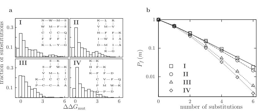
Figure 2.1: Lattice proteins with different structures but the same stability (∆Gf =−1.0) converge to different exponential declines in m-neutrality. (a) The distributions of ∆∆G
for all 380 single amino acid substitutions to the inset lattice proteins. (b) The measured (symbols) and predicted (lines)m-neutralities for the four proteins. A sequence is functional if it folds to the original native structure with ∆Gf <0.0.
2.3.2 Lattice Proteins Support Predictions
We used lattice protein simulations to test our m-neutrality predictions. Lattice proteins are highly simplified models of proteins that provide a useful tool for studying protein folding [30, 31, 32, 33] and evolution [34, 35] (some example lattice proteins are shown in Figure 2.1). Like real proteins, our lattice proteins exist in a vast conformational space (4.2×10⁷ possible conformations), yet we can exactly compute ∆G
f in just fractions of a second. We considered a mutant lattice protein to be functional if it still stably folded to the wildtype structure, and so these simulations directly tested our theory under the assumption that the retention of fold is equivalent to the retention of structure. We carried out our simulations by evolving lattice proteins with various stabilities and structures, and then computing their m-neutralities by sampling mutants with random amino acid substitutions.
simple exponential of the form
Pf(m)∝νm
where ν varies among proteins and gives the severity of the exponential decline of m -neutrality withm. The underlying reason for this exponential decline is clear: after several substitutions, the distribution of ∆Gf among the remaining functional sequences reaches a steady state, and so each new substitution pushes the same fraction of proteins beyond the stability threshold. Interestingly, although Pf(m= 1) is similar for all of the proteins in Figure 2.1, the factors that give rise to the different ν parameters are present in the distribution of single mutant ∆∆Gvalues, since it is used to predict the m-neutralities for all values ofm.
Figure 2.2 shows the m-neutralities of proteins with the same structure but different stabilities. After several substitutions, all of the proteins converge to the same value of ν, suggesting that ν is a generic property of a protein’s structure and does not depend on its particular sequence or stability. On the other hand, the response of a protein to the first few substitutions depends strongly on its stability, with more stable proteins exhibiting higher initial m-neutrality. The high initial m-neutrality of stable proteins is readily rationalized in terms of the thermodynamic model: substitutions tend to disrupt a protein’s structure by pushing its stability below the minimal threshold, but proteins with an extra stability cushion are buffered against the first few substitutions. Proteins that sit on the very margin of the minimal stability threshold exhibit lower 1-neutrality than is predicted by an expo-nential decline because these proteins are less stable than the average functional protein, and so surviving sequences will tend to be more stable than the wildtype sequence and so exhibit a higher tolerance for the next substitution.
2.3.3 Real Proteins Support Predictions
a
number of substitutions
Pf
(
m
)
∆Gf= 0.0 ∆Gf=−1.0 ∆Gf=−2.0
0 2 4 6
1
0.1
0.01
0.001
b
Pf
(
m
)
0.8
0.5
0.2
-1.5 -0.5 -1.5 -0.5 Pf(m= 1) ν
[image:20.612.217.433.206.458.2]∆Gf
1
0.1
0 4 8 12
TEM1 WT subtilisin
fr
ac
ti
on
fu
n
ct
io
n
al
library average DNA mutations
Figure 2.3: Theoretical predictions match measured neutralities in mutant libraries of sub-tilisin (dashed lines) and TEM1 β-lactamase (solid lines) genes. Thick lines show predic-tions made using the PoPMuSiC potential and thin lines show predicpredic-tions made using the FOLDEF potential. The measurements are from Table 2.2, normalized by the values from the control unmutated library.
creating many sequences with specific numbers of amino acid substitutions is extremely difficult, but it is relatively easy to use error-prone PCR to make libraries of mutant genes with randomly distributed nucleotide mutations. The fraction of functional proteins F
encoded by the genes in a mutant gene library can easily be related to the distribution of ∆∆G values for single amino acid substitutions by
F =
∞ X mnt=0
f(mnt)×Pˆf(mnt)
wheref(mnt) is the fraction of genes withmnt nucleotide mutations in the mutant library
and ˆPf(mnt) is the fraction of functional proteins encoded by genes with mnt nucleotide
mutations, computed as for Pf(m) except that we now use ˆp1 ∆∆G1, the probability
that a single nucleotide mutation made according to the particular protocol used causes a stability change of ∆∆G1.
Base pairs sequenced 22,800 Total mutations 172 Mutation frequency (%) 0.75 ±0.06 Mutations per gene 6.5±0.5 Mutation types (%)
A→T, T→A 22
A→C, T→G 9
A→G, T→C 42 G→A, C →T 20
G→C, C→G 1
G→T, C→A 3
[image:22.612.226.421.85.252.2]frameshift 3
Table 2.1: Mutation frequencies for TEM1 β-lactamase mutagenesis determined by se-quencing 20 unselected clones each from the wildtype and M182T error-prone PCR round 5 libraries. Standard errors are calculated assuming Poisson counting statistics.
β-lactamase, and screened genes from these libraries in Escherichia coli for their ability to confer resistance to the antibiotic ampicillin. Table 2.1 shows the frequency of nucleotide mutations introduced by our mutagenesis protocol, while Table 2.2 shows how the fraction of genes in the library that conferred ampicillin resistance decreased with increasing average numbers of nucleotide mutations.
In order to test the ability of our theory to predict the fractions of functional proteins in these libraries, we also needed a method for calculating the ∆∆Gvalues for single amino acid substitutions. We used two existing computational potentials, the database-derived PoPMuSiC potential of Gilis and Rooman [37, 38] and the empirical FOLDEF potential of Serrano and coworkers [39], and corrected for the fact that some amino acid substitutions are more likely than others. The only remaining unknown parameter is the extra stability of the protein, ∆Gextraf , which cannot be directly measured because we do not know the minimal stability required for the protein to function. However, ∆Gextra
f only influences the initial behavior of them-neutrality curve and does not affect the exponential decline parameter ν
(as shown in Figure 2.2), so we can fit ∆Gextra
f to the experimental measurements and still test the ability of the theory to predict the severity of the decline in m-neutrality.
Figure 2.3 shows that the measured fractions functional for subtilisin and wildtype TEM1 β-lactamase are in good agreement with the predictions made with the PoPMuSiC and FOLDEF potentials. Subtilisin and TEM1 β-lactamase exhibit similar declines in
0 3 6
1
0.5
0.2 TEM1 WT TEM1 M182T
fr
ac
ti
on
fu
n
ct
io
n
al
library average DNA mutations
Figure 2.4: The more stable M182T variant of TEM1 β-lactamase (dashed lines) exhibits enhanced neutrality relative to wildtype (solid lines) as predicted by the theory. Thick lines show predictions made using the PoPMuSiC potential and thin lines show predictions made using the FOLDEF potential. The measurements are from Table 2.2, normalized by the values from the control unmutated library.
Round hmnti WT M182T
0 0.0± 0.0 0.76 ±0.03 0.74 ±0.04 1 1.3± 0.2 0.59 ±0.03 0.68 ±0.03 2 2.6± 0.3 0.47 ±0.03 0.54 ±0.02 3 3.9± 0.4 0.28 ±0.02 0.45 ±0.04 4 5.2± 0.4 0.18 ±0.01 0.28 ±0.01 5 6.5± 0.5 0.13 ±0.01 0.20 ±0.02
classification as αβ 3-layer (αβα) sandwiches [40]), and our lattice protein simulations suggest that proteins with the same structure should have the same exponential decline inm -neutrality. Computations on proteins with markedly different structures predicted different declines in m-neutralities (data not shown), but no experimental data is yet available for these proteins.
The second major prediction of our theory is that among proteins with the same struc-ture, more stable variants will exhibit higher initial m-neutralities, but converge to same exponential decline parameter ν. Our measurements on wildtype and the M182T variant of TEM1 β-lactamase allowed us to test this prediction, since the M182T variant is 2.7 kcal/mol more stable than wildtype [36], yet should have the same structure since it differs by only a single amino acid substitution. Figure 2.4 shows the measured fractions func-tional for wildtype and the M182T variant, as well as the theoretical predictions made using both the PoPMuSiC and FOLDEF potentials. The M182T variant exhibits enhanced initial
m-neutrality as predicted by the theory, and once again the predictions made with both potentials are in good agreement with the experimental measurements.
2.4
Discussion
We have presented a theory for calculating the probability that a protein will remain func-tional after random amino acid substitutions, and have confirmed the main theoretical predictions with simulations and experiments. Our theory naturally separates a protein’s
m-neutrality into components due to structure and stability. The eventual severity of the exponential decline in m-neutrality with the number of substitutions is a property of a protein’s structure. On the other hand, a protein can increase its tolerance for the first few mutations by increasing it stability, in effect allowing it to “take a few hits” before it is pushed into the inevitable structurally determined exponential decline in m-neutrality. This increased tolerance to mutations due to extra stability is probably also the underlying reason for the existence of global suppressor mutations [20, 21] that buffer proteins against otherwise deleterious mutations by increasing their thermodynamic stability.
Mutations are most likely to be non-additive if the mutated residues are in direct contact in a protein’s structure [28, 29]. Since proteins are large, two randomly chosen residues will only rarely contact each other, and so although the additivity assumption is violated for some specific mutations, it is accurate when averaged over all possible mutations. The second assumption is that mutations affect function only through their effects on stabil-ity. This assumption ignores some effects of mutating residues that are directly involved in a protein’s function. Therefore, for proteins with a high fraction of functional residues, our theory only provides an upper bound on m-neutrality. However, our theory’s remark-able success in describing the m-neutralities of both subtilisin and the TEM1β-lactamase variants suggests that this assumption is also accurate for most proteins.
Our theory provides a quantitative rationale for earlier work with lattice proteins on the organization of functional proteins in sequence space. Bornberg-Bauer and Chan [9] proposed that proteins are located in superfunnels in sequence space with the most stable sequence having the most neutral neighbors; others have reported that folded proteins surround highly stable prototype sequences in sequence space [41, 10, 11], and Shakhnovich and coworkers [8] showed that proteins with a large energy gap between the lowest and second lowest energy conformations are stabilized against mutations. We provide a clear explanation: more stable proteins are able to tolerate more of the possible mutations before unfolding, and so a higher fraction of their neighboring sequences fold.
In addition to these stability-based effects, different protein structures have different inherent designabilities, with more sequences folding into some structures than others [12, 42, 43]. Proteins with more designable structures might be expected to show a milder exponential decline in m-neutrality (a larger value of ν), since their structures occupy a larger fraction of sequence space. The structural neutrality given by ν therefore provides a quantitative measure of designability that can be estimated with current computational techniques.
if the protein has a large amount of extra stability, as is seen for the M182T variant of TEM1 β-lactamase. Therefore, it is important to examine whether natural proteins have evolved stability above that required for function in their natural environments in order to provide them with additional robustness [22] to the first few amino acid substitutions.
Our theory also has applications in protein engineering. Directed evolution involves screening libraries of mutant proteins for new or improved functions [44]. Each round of directed evolution typically introduces only one or two amino acid substitutions because the rapid decline in m-neutrality means that larger numbers of substitutions will yield libraries of mostly unfolded proteins. Our model suggests that using highly stable parents for directed evolution should increase the fraction of folded mutants at a given level of substitutions. It also provides a method for predicting which structures will respond better to large numbers of substitutions.
2.5
Methods
2.5.1 Convolution of Mutational Effects
The distribution pm(x) was calculated as
pm(x) = 1 2π
Z ∞
−∞
[F(k)]nexp (−ikx) dk
where
F(k) =
Z ∞
−∞
p1(y) exp (iky) dy
is the characteristic function ofp1(x). For the numerical calculations,p1(x) was constructed
by binning all single mutant ∆∆Gvalues with a bin size of 0.01 and representing it with a list padded by a number of zeroes equal to m−1 times the number of bins. Each element of the fast-Fourier transform (FFT) of this list was raised to the m power, and pm was recovered through an inverse FFT.
2.5.2 Lattice Proteins
of the 41,889,578 conformations corresponding to all length 20 self-avoiding walks [45]. The sum over these conformations was tractable because they correspond to only 910,972 unique contact sets. The energyE(C) of conformationC is
E(C) = N X
i=1
i−2
X j=1
Cij(C)×ǫ(Ai,Aj),
where Cij(C) is one if residues i and j are nearest neighbors in conformation C and zero otherwise, and ǫ(Ai,Aj) is the interaction energy between residue types Ai and Aj, given by [46] (Table 5). The energies are in units of kBT where T = 1.0 for all simulations, corresponding to room temperature.
The stability with which a protein folds to a target conformation Ctis
∆Gf(Ct) =E(Ct) +Tln{Q(T)−exp [−E(Ct)/T]}
whereQ(T) is the partition sum
Q(T) = X
{Ci}
exp [−E(Ci)/T].
We considered a mutant functional if ∆Gf(Ct)<0.0, and nonfunctional otherwise.
To generate lattice proteins, Ct was randomly chosen from the subset of conformations with unique contact sets. For each Ct, an adaptive walk was begun with a random starting sequence, with each step of the walk choosing the first point mutant with a better ∆Gf(Ct). The adaptive walk was terminated after a sequence was found with ∆Gf(Ct)≤ −2.0, 500 steps were taken, or no improved mutants were found. If 200 random walks failed to generate a sequence with ∆Gf(Ct) ≤ −2.0, a new Ct was chosen. A clonal population of the final sequence from the random walk was then subjected to 2.5×10⁵ generations of neutral evolution with a population size of 100 and a per residue mutation rate of 5×10⁻⁵, with all
fitness of one to any sequence that met the target stability and a fitness of zero to any other sequence. After the 2.5×10⁵ generations, we selected the first sequence generated with a stability within 0.025 of the target stability, and used this sequence for the m-neutrality analysis. Lattice protein m-neutralities were computed by sampling all mutants form≤2 or 5×10⁵ randomly generated mutants for m > 2. In Figure 2.2, ν was computed as
p
Pf(m= 6)/Pf(m= 4).
2.5.3 Measured Neutralities of Subtilisin and TEM1 β-lactamase
The measured neutralities for subtilisin were those from population 6B in Table 2 of [17], normalized by the fraction of functional clones in the control library.
The 861 bp genes for wildtype and M182T TEM1 β-lactamase were a kind gift from Brian Shoichet [36] and were subcloned into the pMON 1A2 plasmid [47] with SacI and HindIII using PCR primers 5’-GCGGCGGAGCTC TGAGTATTCAACATTTCCGT
GTCGC-3’ and5’-GCGGCGAAGCTTTTACCAATG
CTTAATCAGTGAGGCAC-3’and sequenced. For the round zero library, unmutated gene was cut directly from the plasmid. Each successive round of error-prone PCR used 3 ng of SacI/HindIII digest of gene from the previous round in 100 µl reactions containing 0.5 µM of each of the above primers, 7 mM MgCl2, 75 µM MnCl2, 200 µM of dATP and dGTP,
500 µM of dTTP and dGTP, 1X Applied Biosystems PCR buffer without MgCl2, and 5 U
of Applied BiosystemTaq DNA polymerase. The PCR conditions were 95oC for 5 minutes, and then 14 cycles of 30 s each at 95oC, 50oC, and 72oC. The number of doublings per round was determined to be approximately ten by quantifying the DNA versus a marker on an agarose gel.
The mutation frequency in the round five library was determined by sequencing the first 570 bp of twenty genes each from the unselected wildtype and M182T plates with the sequencing primer5’-GGTCGATGTTTGATGTTATGGAGC-3’. No biases in the locations of mutations were observed. The wildtype and M182T genes were mutated under identical conditions, and the sequencing found the same mutation frequency in the round five library for both (0.77 ± 0.08% for wildtype and 0.74 ± 0.08% for M182T). For better statistics, the sequencing results for both libraries were combined to give the data in Table 2.1. The per round mutation frequency was calculated assuming that each round of error-prone PCR introduced the same average number of mutations. This gives a per round mutation frequency of 0.15±0.03%. To confirm this assumption, we sequenced ten unselected clones from both of the round one libraries, and found mutation frequencies of 0.16 ± 0.05% for wildtype and 0.19 ± 0.06% for M182T. Standard errors were computed assuming Poisson sampling statistics.
2.5.4 Neutrality predictions
The single residue mutant ∆∆Gvalues were estimated with the web version of the PoPMu-SiC program [38] available at http://babylone.ulb.ac.be/popmusic/ and the FOLDEF pro-gram [39] available at http://fold-x.embl-heidelberg.de:1100/cgi-bin/main.cgi using PDB structures 1IAV and 1BTL for subtilisin and TEM1β-lactamase. The probabilitiesP(Y |X) that a single nucleotide mutation to a gene would change baseX to baseY were computed from the nucleotide frequencies of the gene sequences and the probabilities P(X, Y) that a random mutation was from base X to base Y given for subtilisin in Table 1 of [17] and for TEM1 β-lactamase in Table 2.1 of this work. We constructed the probability distri-bution ˆp1 ∆∆G1 for the effects of single nucleotide mutations by constructing a list of
∆∆G values by including 1000×L× P(Y |X) entries for the ∆∆Gvalue associated with each mutation from X to Y, where L is the gene length, assigning synonymous mutations ∆∆G= 0 and nonesense and frameshift mutations ∆∆G= 25 kcal/mol, ignoring mutations for which no ∆∆Gvalue could be computed, and assigning any mutation with ∆∆G >25 a value of ∆∆G= 25. The values of ˆPf(mnt) were computed by convolutions of ˆp1 ∆∆G1
as described above.
library with an average of hmnti mutations created byr rounds of error-prone PCR is
f(mnt;r) = (1 +λ)−N
N X k=0
N
k
λk(kx)
mnte−kx
mnt!
wherencyclesis the number of PCR cycles per round,λis the PCR efficiency,N =ncycles×r,
and x = hmntN λi(1+λ) [48]. For subtilisin ncycles = 13 and λ = 0.77 and for the TEM1
ncycles= 14 and λ= 0.71. For these parameter values,f(mnt;r) is nearly Poisson.
The values of ∆Gextraf for the predictions were computed by a least squares fitting to the experimental measurements. The ∆Gextraf values differed for the PoPMuSiC and FOLDEF predictions because the ∆∆G values from these methods are scaled differently. For the PoPMuSiC predictions, ∆Gextraf was−2.5,−2.5, and−3.9 kcal/mol for subtilisin, wildtype TEM1 β-lactamase, and M182T TEM1 β-lactamase. For the FOLDEF predictions, the values were −7.3,−5.7, and−12.0.
2.6
Acknowledgements
Chapter 3
Protein Stability Promotes
Evolvability
A version of this chapter has been published as [49].
3.1
Abstract
The biophysical properties that enable proteins to so readily evolve to perform diverse biochemical tasks are largely unknown. Here we show that a protein’s capacity to evolve is enhanced by the mutational robustness conferred by extra stability. We use simulations with model lattice proteins to demonstrate how extra stability increases evolvability by allowing a protein to accept a wider range of beneficial mutations while still folding to its native structure. We confirm this view experimentally by mutating marginally stable and thermostable variants of cytochrome P450 BM3. Mutants of the stabilized parent were more likely to exhibit new or improved functions. Only the stabilized P450 parent could tolerate the highly destabilizing mutations needed to confer novel activities such as hydroxylating the anti-inflammatory drug naproxen. Our work establishes a crucial link between protein stability and evolution. We show that we can exploit this link to discover new protein functions, and we suggest how natural evolution might do the same.
3.2
Introduction
properties influence its capacity to evolve [50, 51]. In recent years, understanding the de-terminants of evolvability has also become an important practical concern, as researchers increasingly use evolution to engineer everything from proteins [52] to designs for civil en-gineering structures [53].
Proteins are one of the simplest and best examples of evolvable biological systems, since they possess biochemical functions that can be altered with just a few mutations [54]. One property that has been broadly hypothesized to contribute to evolvability is robustness to mutations [55], and proteins are often quite mutationally robust, with over half of the single mutants of many proteins retaining their native functions [15, 16, 18, 7]. Because proteins usually must fold in order to function, and because mutated proteins generally adopt the original native structure if they fold at all [56, 57], retention of the basic native structure is a normally a prerequisite for the acquisition of new functions. Extra thermodynamic stability makes a protein’s native structure and function more robust to random mutations by increasing the fraction of mutants that continue to possess the minimal stability required to fold [7, 58].
Here we investigate how stability affects a protein’s evolvability by using controlled ex-periments to measure the fraction of a protein’s mutants that exhibit new or improved function. We first use a simple computational model to establish a framework for under-standing the relationships among protein stability, mutational robustness, and evolvability. We then validate this framework with experiments on members of the biochemically im-portant cytochrome P450 enzyme family, and describe specific examples that illustrate the biophysical basis of the connection between protein stability and evolvability. Finally, we discuss the implications of our work for understanding natural protein evolution and de-signing better protein engineering strategies.
3.3
Results
3.3.1 Simulations with Model Lattice Proteins
A K—A—K
| |
G—D H— L M—T
| | |
L G— I F — C L
| | | |
L V—M I M—G
| | |
G— F H—M— S
n u m b er o f u n iq u e im p ro v ed m u ta n
ts original parent
stabilized parent 0 10 20 B GYLG
F D GRLGF D GLLGK D GLLGE D ∆Gf
fr ac ti o n o f m u ta n ts folded unfolded original parent ∆Gf= -0.5
stabilized parent ∆Gf= -1.5
-2 -1 0 1
0.00 0.00
0.05 0.05
C
Figure 3.1: Increased stability enhances evolvability of a model lattice protein. (A) The original model protein (right) that had been evolved to bind to a rigid ligand (left in bold). (B) Mutants of a stabilized model protein were more likely than mutants of the original model protein to show improved binding to the four new ligands shown below the bars. The bars show the number of mutants out of 1500 screened that bound the new ligand with at least twice the affinity of the parent. (C) The stabilized model protein was more evolvable because more of its destabilized but improved mutants satisfied the minimal stability cutoff. The bars show the distribution of stabilities among all mutants in the libraries, while the circles show the stabilities of the improved mutants.
depends on its ability to fold to a thermodynamically stable native structure [25], stability is still constrained during evolution. Specifically, we imagine that a protein must fold to its native structure with some minimal stability in order to remain folded at physiological conditions. If a protein fails to meet this minimal stability threshold, then it will neither fold nor function. If a protein does fold with at least the minimal required stability, then evolution selects for a protein’s function and is indifferent to the amount of extra stabil-ity it possesses. Most proteins, however, will still be marginally stable since highly stable sequences are rare [41].
could exactly determine the partition function and free energy of folding (∆Gf). We set a minimal stability threshold for our lattice proteins by requiring them to fold to the original native structure with a stability of ∆Gf ≤0 (in no case did we observe a protein that stably folded to a new structure), which is equivalent to requiring the native structure to have a lower free energy than the ensemble of all non-native conformations. For those proteins that stably folded, we measured function as the binding energy of the folded protein to a small rigid ligand [45], as shown in Figure 3.1A. Our model therefore recapitulated the essential requirements imposed on real proteins of simultaneously folding and performing a biochemical task.
We first evolved a model protein to stably fold and strongly bind a ligand (Figure 3.1A). This evolved protein had a stability of ∆Gf =−0.5, meaning that it was only marginally stable as is typical for real proteins [26]. We then simulated the process of directed evolution with two rounds of random mutagenesis by error-prone PCR and screening to identify a stabilized variant of our model protein (∆Gf = −1.5) that contained three amino acid substitutions and exhibited the same ligand binding energy as the original protein. To examine the evolvabilities of the original and stabilized model proteins, we computationally simulated screening libraries of 1,500 randomly mutated sequences for mutants that bound to new ligands with at least twice the affinity of the parent proteins. For all four new ligands we examined, the parent proteins bound the new ligand with equal affinity, yet each time the mutant library from the stabilized parent produced over twice as many unique improved mutants (Figure 3.1B).
to be more destabilized than the typical folded mutant.
3.3.2 Experiments on Cytochrome P450 BM3 Variants
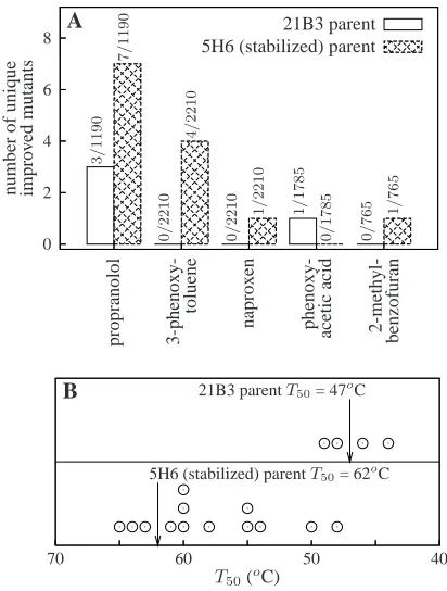
To experimentally test the effect of stability on the evolvability of real proteins, we ran-domly mutated two variants of a cytochrome P450 BM3 (also known as CYP102A1) heme domain peroxygenase [59] and screened for mutants with new or improved activity on five substrates. The cytochrome P450 superfamily contains members involved in important biochemical processes such as drug metabolism and steroid biosynthesis [60, 61]. P450 BM3 catalyzes sub-terminal hydroxylation of medium- and long-chain fatty acids [62]. The 21B3 variant is a laboratory-evolved version of the P450 BM3 heme domain that efficiently hydroxylates 12-p-nitrophenoxycarboxylic acid (12-pNCA, structure shown in Figure 3.3), utilizing hydrogen peroxide in place of the NADPH cofactor and oxygen [59]. The 5H6 variant was created by laboratory evolution of 21B3, selecting for mutants that were more thermostable while retaining activity on 12-pNCA [63]. We quantified the stabilities of the enzymes by the temperature (T50) at which half of the protein irreversibly denatured after
a 10 minute incubation. Because denaturation is irreversible, theseT50 values are not
equi-librium thermodynamic measurements, and so cannot be directly related to ∆Gf. However, theT50values were highly correlated with the stability to irreversible denaturation by urea,
supporting the notion that they reflect universal aspects of protein stability rather than unique characteristics of the process of irreversible thermal denaturation (Figure 3.12 and related discussion in the Methods section). As measured by the T50 values, the 21B3
en-zyme is only marginally stable (T50 = 47oC), while 5H6 is much more stable (T50 = 62oC)
(melting curves are shown in Figure 3.7). The 5H6 enzyme differs from 21B3 at eight residues (out of 464 total). Both variants displayed nearly the same activity (measured as total turnovers) on 12-pNCA and all other substrates examined in this work.
Base pairs sequenced 58,719
Total mutations 182
Mutation frequency (%) 0.31±0.02 Avg. mutations per gene 4.5±0.3 % synonymous mutations 28 % nonsynonymous mutations 63 % frameshift/nonsense mutations 9 Mutation types (%)
A →T, T→A 25
A →C, T→G 6
A →G, T →C 53
G →A, C →T 10
G →C, C→G 0
G →T, C→A 3
frameshift 3
Table 3.1: Mutation frequencies in error-prone PCR libraries. Statistics are for all 41 ran-domly chosen mutants. Standard errors are calculated assuming Poisson counting statistics.
number of mutations
n u m b er o f m u ta n ts 0 0 1 2 2 3 4 4 5
6 8 10 12
21B3 5H6
A
number of mutations
n u m b er o f m u ta n ts 0 0 2 2 4 4 6 6 8 8 10 12 B base pair cu m u la ti v e fr ac tio n 0.0 0.2 0.4 0.6 0.8 1.0
100 400 700 1000 1300
actual uniform
[image:36.612.204.444.116.327.2]C
Threshold Pr (F |21B3) Pr (F |5H6) Pr (A|F; 21B3) Pr (A|F; 5H6)
0.10 0.44 0.69 0.98 0.96
0.25 0.39 0.66 0.98 0.96
0.50 0.33 0.61 0.96 0.94
0.75 0.29 0.57 0.92 0.93
0.90 0.24 0.51 0.93 0.90
Table 3.2: Retention of folding and function. The probabilities that 21B3 and 5H6 mutants fold (F), and that those folded mutants are active (A) on 12-pNCA at different thresholds for folding and activity. The probabilities are determined from the six plates of each parent shown in Figure 3.8. The folding and activity status of each mutant is determined relative to the four parents on the plate. If the CO binding difference spectrum A447 - A490 is greater
than the threshold times the parental median, the mutant is considered folded. Folded mutants are classified as active if the 12-pNCA A398 value is greater than the threshold
times the parental median.
NO2
O
O OH
O CH3
O OH
NH CH3
CH3
12-pNCA 3-phenoxytoluene propranolol
O OH
O H3CO
H3C H
OH
O
O CH3
phenoxyacetic acid naproxen 2-methylbenzofuran Figure 3.3: The substrates on which the P450 mutants were screened for activity.
protein to fold (61% of 5H6 mutants contained at least half the folded protein of the parent versus 33% for 21B3, raw data are shown in Figure 3.8 and Table 3.2). Most of these folded mutants retained at least half the parental activity on 12-pNCA (94% and 96% for 5H6 and 21B3), indicating that mutations that disrupted parental function generally did so by preventing the formation of properly folded protein. This confirms the experimental findings of [7] that more stable proteins are more robust to mutations.
hydrox-0 2 4 6 8 n u m b er o f u n iq u e im p ro v ed m u ta n ts p ro p ra n o lo l 3 -p h en o x y -to lu en e p h en o x y -ac et ic ac id 2 -m et h y l-b en zo fu ra n n ap ro x en 21B3 parent 5H6 (stabilized) parent
A 3 / 1 1 9 0 7 / 1 1 9 0 0 / 2 2 1 0 0 / 2 2 1 0 4 / 2 2 1 0 1 / 2 2 1 0 1 / 1 7 8 5 0 / 1 7 8 5 1 / 7 6 5 0 / 7 6 5
70 60 50 40
T50(oC)
5H6 (stabilized) parentT50= 62oC
21B3 parentT50= 47oC
[image:38.612.225.431.90.362.2]B
Figure 3.4: Increased stability enhances evolvability of the P450 BM3 heme domain. (A) The stable 5H6 protein yielded more mutants with new or improved activity than the marginally stable 21B3 protein. The counts above the bars give the number of improved mutants out of the total number of mutants screened. (B) Some of the improved 5H6 mutants were greatly destabilized relative to the parent protein, while the stabilities of the improved 21B3 mutants clustered around those of the parent protein (circles show T50
values for improved mutants).
ylation activity using the 4-aminoantipyrene (4-AAP) assay to measure the total amount of product after completion of the reaction [67], and determined that neither 21B3 nor 5H6 had detectable activity on the first three substrates, both had equal weak activity on propranolol, and 21B3 had trace activity on 2-methylbenzofuran(Table 3.3). We used consistent quantitative criteria to identify mutants that had either acquired new activity or improved by more than 50% over the parental level in the 4-AAP assay. We screened 8,160 mutant-substrate pairs for each parent. From these, we identified 13 improved mutants of 5H6 and 4 improved mutants of 21B3 (Figure 3.4A, Table 3.3). All the improved mutants had unique protein sequences (given in Table 3.5). Thus, we found over three times more improved mutants in the 5H6 library than in the 21B3 library.
pro-tein, we measured the stabilities of all improved mutants (melting curves are in Figure 3.7). Figure 3.4B shows that none of the improved 21B3 mutants was destabilized by more than 3oC, but that the thermostable 5H6 parent produced improved mutants that were destabi-lized by as much as 14oC. We identified specific beneficial but destabilizing substitutions that could be made only in the stabilized parent. For example, neither 21B3 nor 5H6 exhib-ited activity on naproxen, presumably because the negatively charged naproxen molecule does not enter the hydrophobic P450 BM3 substrate binding pocket. Mutating leucine 75 in the substrate binding pocket to arginine allowed 5H6 to hydroxylate naproxen by pro-viding a compensating positive charge for the naproxen molecule (Figure 3.5). However, burial of this arginine residue in the hydrophobic binding pocket substantially destabilized the 5H6 mutant (∆T50 =−8oC). When we made the same substitution to 21B3, we could
only recover inactive and improperly folded protein (as indicated by a carbon monoxide difference spectrum peak at 420 nm [68] as shown in Figure 3.9). The F275S substitution (located 12˚A from the substrate [69]) is another example of a beneficial substitution which could be made only in the stabilized parent. This substitution conferred 3-phenoxytoluene activity on the 5H6 parent, but decreased theT50by 7oC. When we made this substitution
in 21B3 we again could not recover any folded protein (Figure 3.10). In contrast, the F205L substitution (located near the substrate binding pocket [69]) found in a 21B3 mutant with improved activity on propranolol did not have a substantial effect on stability, and slightly improved the activity of both 21B3 and 5H6 when introduced into those sequences (Figure 3.11).
3.4
Discussion
A
B
H3CO
H3C H
OH
[image:40.612.224.420.184.521.2]O
level of individual mutations. This apparent tradeoff is at least partly due to the simple fact that most randomly chosen mutations (functionally beneficial or not) are destabilizing [77, 78, 79, 27]. In addition, residues in a protein’s active site often must satisfy functional constraints (such as maintaining buried charges or cavities in a protein’s interior) that make them poorly optimized for stability [80, 81, 82]. Therefore, mutating active-site residues often enhances stability at the cost of function [80, 81, 82], and likewise acquiring new functions can require destabilizing mutations (as is the case for our L75R mutation in P450, which confers activity on naproxen by burying a positive charge). However it remains unclear whether active-site constraints intensify the tradeoff between stability and functional evolution, since a seemingly opposite argument can be made that mutations to an active site that is already poorly optimized for stability should be less destabilizing than typical mutations (they could even enhance stability if, for example, they confer function on smaller substrates by reducing the size of a cavity in a protein’s interior). If functionally innovative mutations tend to be more destabilizing than random mutations, then extra protein stability should enhance the rate of functional innovation more than it enhances the mutational robustness of the native function. In our lattice protein simulations, extra stability increased the rate of functional innovation by nearly 400% while it increased mutational robustness by only 50%; however, we feel our lattice model is too crude to confidently extrapolate conclusions involving residue-level properties to real proteins. In our P450 experiments, extra stability also increased the number of functionally improved mutants (from 4 to 13) more than it increased mutational robustness (by a factor of 1.8); however, here the statistics are too poor to conclude that functional innovation is improved significantly more than mutational robustness. Therefore, in our minds it remains unclear whether extra protein stability promotes evolvability simply by improving the tolerance to all mutations (some of which happen to be functionally beneficial), or whether the effect is further amplified by a tendency for functionally innovative mutations to be especially destabilizing.
mutations, causing stability to drift towards the minimum evolutionary requirement [41, 83]. Naturally evolving proteins must therefore wait for functionally neutral mutations to stabi-lize the structure in order to counterbalance the effects of other destabilizing but functionally beneficial mutations [36]. In this sense, a protein’s stability represents a hidden dimension in evolution: extra stability is neutral with respect to selection for protein function, but it can be crucial in allowing a protein to tolerate mutations that confer beneficial phenotypes. We have shown that protein engineering by directed evolution is more effective if direct selection for extra stability is used to increase a protein’s evolvability. The extent to which natural evolution might also select for evolvability has been the subject of much recent theoretical speculation [51, 55, 84]. We suggest that one possible mechanism by which nat-ural evolution could increase evolvability would be to stabilize proteins undergoing adaptive evolution or provide systems to buffer the effects of destabilizing protein mutations.
3.5
Methods
3.5.1 Model lattice protein simulations
A model lattice protein was represented as a chain ofN = 20 monomers of 20 types corre-sponding to the natural amino acids. A model protein could occupy any of the 41,889,578 conformations (representing 910,972 unique contact sets) corresponding to all length 20 self-avoiding walks on a two-dimensional lattice. A conformationC had an energy of
E(C) = N X i=1
i−2
X j=1
Cij(C)×ǫ(Ai,Aj),
where Cij(C) is one if residues i and j are nearest neighbors in conformation C and zero otherwise, and ǫ(Ai,Aj) is the interaction energy between residue types Ai and Aj, given by Table 5 of reference [46]. Energies are in units of kBT, and T = 1.0 for all simulations.
A model protein folds to a target conformation Ct with stability
whereQ(T) is the partition sum
Q(T) = X
{Ci}
exp [−E(Ci)/T].
Protein were considered stably folded to Ct if and only if ∆Gf(Ct)≤0.0.
A model protein’s activity was represented by binding to a small rigid ligand, much as in [45]. If a model protein did not stably fold to a unique conformation, it was considered inactive. If it did stably fold to a unique conformation, then we computed the binding energy of the ligand as the sum of the pairwise interactions between the ligand and protein residues, searching over all possible rotational and translational positions of the ligand. The binding affinity of the ligand to the folded conformation is given by the association constant
Ka, calculated as the exponential of the negative binding energy.
To create the model protein shown in Figure 3.1A, we first chose the target conformation of the protein, and then performed an adaptive walk from a random starting sequence until we found a sequence that folded to the conformation. We evolved this sequence for 10,000 generations with a population size of 10 and a mutation rate of 5×10⁻⁴ per
3.5.2 P450 mutant libraries.
We used error-prone PCR to create mutant libraries of the marginally stable 21B3 [59] and the thermostable 5H6 [63] variants of the cytochrome P450 BM3 heme domain. The tem-plate DNA was the appropriate gene cloned into the pCWori [86] plasmid as described in references [59] and [63]. We confirmed the sequences of the 21B3 and 5H6 genes by sequenc-ing them with the primers pCWori for ( 5’-GAAACAGGATCCATCGATGCTTAGGAGGTCAT-3’,pCWori rev (5’-GCGTATCACGAGGCCCTTTCGTCTTCAAGC-3’), and pCWori mid rev ( 5’-CCAGCTTGTGGCCAACCCGAC-3’). The sequences matched those that were reported [59, 63],
with 21B3 containing ten amino acid substitutions relative to the wildtype P450 BM3 heme domain (I58V, F87A, H100R, F107L, A135S, M145V, N239H, S274T, K434E, and V446I in the numbering scheme where residue one is the threonine after the cleaved N-terminal me-thionine) and 5H6 containing eight amino acid substitutions relative to 21B3 (L52I, S106R, V145M, A184V, L324I, V340M, I366V, and E442K) as well as the removal of one histidine from the C-terminal His tag.
The error-prone PCR reactions for the two parents were carried out in parallel using iden-tical conditions to ensure the same mutation rate for both. The reactions were 100 µl and contained 13 ng of template plasmid (corresponding to 3 ng of gene), 0.5µM of the oligonu-cleotide primers (pCWori for andpCWori rev clone, 5’-GCTCATGTTTGACAGCTTATCATCG-3’), 200µM dATP and dGTP, 500µM dTTP and dCTP, 7 mM MgCl2, 200µM MnCl2, 1X
Applied Biosystems PCR Buffer, and 5 units ofTaq. PCR conditions were 95oC for 5 min. followed by 16 cycles of 30 s at 95oC, 30 s at 51oC, and 60 s at 72oC. Gel electrophoresis versus a known standard indicated that this yielded PCR product at a concentration of≈12 ng/µl, for a PCR efficiency ofλ= 0.45. The PCR products were cloned into pCWori with
BamHI and EcoRI, electroporated into a catalase-free strain of E. coli [86], and plated on LB plates containing 100 µg/ml ampicillin. Transformation of a control ligation with no insert indicated that the background rate of plasmid self ligation was less than 1%.
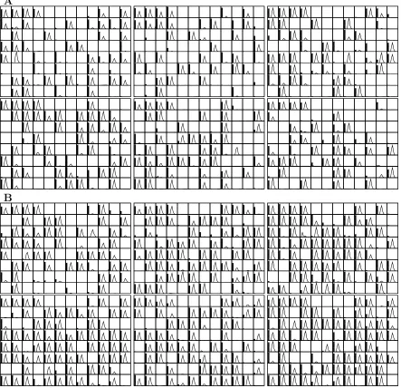
non-sense mutations). The 5H6 clones contained a total of 87 mutations in the 29,799 sequenced base pairs, with 23 synonymous mutations, 55 nonsynonymous mutations, and 9 mutations leading to premature truncation of the gene. The distributions of mutations in the two libraries were statistically indistinguishable (Figure 3.2A). After confirming that the muta-tion rates in the two libraries were indistinguishable, we combined the sequencing results for further analysis (Table 3.1). Figure 3.2B shows that the distribution of mutations is consistent with the theoretical distribution for error-prone PCR [48, 64], which gives the probability that a mutant in a library with an average ofhmntimutations per gene hasmnt
nucleotide mutations as
Pr (mnt) = (1 +λ)−n n
X
k=0
n k
λk(kx)
mnte−kx
mnt!
, (3.1)
wherenis the number of PCR cycles,λis the PCR efficiency, andx= hmntnλi(1+λ). We also confirmed that the mutations were distributed uniformly along the gene sequence (Figure 3.2C). If each position in the gene is equally likely to be mutated, then among 41 sequenced clones, 156.3 positions should be mutated once, 9.7 positions should be mutated twice, and 0.4 positions should be mutated three times. This in good agreement with the observed values of 148, 13, and 1.
3.5.3 Screening for improved mutants
Single mutant colonies were picked from transformation plates with sterile toothpicks and used to inoculate sterile 1 ml deep-well plates (Falcon) with each well containing 400 µl of liquid LB with 100 µg/ml ampicillin. As controls, wells A1, A2, A3, and A4 were always inoculated with the parent (21B3 or 5H6); wells A5, A6, A7, and A8 were always inoculated with a negative control (the pCWori plasmid lacking a P450 gene); and well H12 was not inoculated. Each of the remaining 87 wells contained a different mutant. These deep-well plates were grown for 20-24 hours in a humidified shaker (Kuhner ISF-1-W) at 215 or 225 rpm, 30oC, and 80% relative humidity. To express the proteins, a pipetting robot (Beckman Multimek 96) was used to transfer 100µl per well from these LB deep well plates to 2 ml deep-well plates (Falcon) containing 400 µl of terrific broth (TB) supplemented with 100
plates were also grown for 20-24 hours in the humidified shaker at 215-225 rpm, 30oC, and 80% relative humidity. After this growth, the cells were harvested by centrifugation at 6,100 g for 10 min at 4oC, and stored at -20oC. The LB deep-well plates were stored at 4oC so that improved mutants could be streaked from the plates.
To perform assays, the pelleted cells were resuspended in 600µl of lysis buffer (100 mM EPPS pH 8.2 with 0.5 mg/ml lysozyme and 2 U/ml deoxyribonuclease) per well with the pipetting robot. The plates were incubated for one hour at 37oC, and then centrifuged at 6,100 g for 10 min at 4oC to pellet cell debris. The pipetting robot was used to add 80µl per well of this clarified lysate to 96-well microtiter plates. To assay for folded protein, high– throughput carbon monoxide (CO) binding difference spectra were measured as described in [66] with the modifications that 80µl of clarified lysate in the EPPS buffer was used, and that the heme was reduced by the addition of 20µl of 0.1 M sodium hydrosulfite in 1.3 M potassium phosphate, pH 8.0. Blank spectra were read prior to binding CO with a Spectra Max Plus 384 plate reader (Molecular Devices) at every 10 nm from 400 to 500 nm, as well as at the points 447 nm (the absorbance peak for both 21B3 and 5H6) and 490 nm. After 5-10 minutes of incubation with CO, the absorbance readings were read at these points.
Mutants were screened for the retention of activity on 12-p-nitrophenoxycarboxcylic acid (12-pNCA, Figure 3.3) using a slightly modified version of the method described in [59]. A 6X stock of 12-pNCA was prepared by combining 3.6 ml of 4.17 mM 12-pNCA in DMSO with 6.4 ml of 100 mM EPPS (pH 8.2) immediately before use. Twenty µl of this 6X 12-pNCA stock was added to the 80 µl of clarified lysate in each well of the microtiter plate, and the plate reader was used to mix the plate and blanked at 398 nm. To initiate the reaction, 20 µl of a 6X stock of hydrogen peroxide (6X stock was 24 mM H2O2 in 100
mM EPPS pH 8.2, made immediately before use) was added to each well and mixed with the plate reader. The amount of final product was quantified by reading the absorbance at 398 nm after 20-30 minutes.


![Figure 3.2: Distribution of mutations in the two P450 error-prone PCR libraries. (A) Thedistribution of mutations among 20 randomly chosen 21B3 mutants and 21 randomly chosenKolmogorov-Smirnov tests [65], and represent the probability that the samples or t](https://thumb-us.123doks.com/thumbv2/123dok_us/9416117.443511/36.612.204.444.116.327/distribution-mutations-libraries-thedistribution-mutations-chosenkolmogorov-represent-probability.webp)