Clifford R. Jack, Jr.1.2 Roushdy S. Boulos' Bharat A. Mehta' Daksha Bhansali' James I. Ausman3 Fernando G. Diaz3 Suresh C. Patel'
Received March 31, 1986; accepted after r evi-sion September 22,1986.
Presented at the annual meeting of the American Society of Neuroradiology, San Diego, January
1986.
, Department of Diagnostic Radiology, Division of Neuroradiology, Henry Ford Hospital, Detroit. MI 48202.
2 Present address: Department of Radiology, Mayo Clinic, Rochester, MN 55905. Address reprint requests to C. R. Jack, Jr.
3 Department of Neurosurgery, Henry Ford Ho s-pital, DetroIt. MI 48202.
AJNR 8:211-219, March/April 1987 0195-6108/87/0802-0211
© American Society of Neuroradiology
211
Cerebral Angiography in
Brainstem Revascularization
Surgical management of patients with vertebrobasilar insufficiency has been devel-oped within the past decade. Cerebral angiography plays a crucial role in identifying potential surgical candidates and in directing the surgical approach. Fifty-two patients underwent brainstem revascularization procedures at Henry Ford Hospital between November 1979 and August 1985. Twelve occipital artery to anterior inferior cerebellar artery bypasses, five occipital artery to posterior inferior cerebellar artery bypasses, four intracranial vertebral endarterectomies, 29 superficial temporal to superior cere-bellar artery bypasses, and two superficial temporal to posterior cerebral artery by-passes were performed. The preoperative angiograms in these patients were analyzed to illustrate how angiographic localization of vascular disease directs the surgical approach. We report the results of postoperative angiograms. Technical features of the various surgical procedures, the role of the neuroradiologist, and several features of the angiographic technique used with these patients are described.
Within the past decade, surgical treatment has become available to patients with vertebrobasilar insufficiency. The first extracranial to intracranial bypass procedure for posterior fossa revascularization was performed by Ausman et al. [1] in 1975. Since then, other innovative surgical procedures directed at posterior fossa revas-cularization have been devised [2, 3]. Cerebral angiography plays a pivotal role in the management of these patients by first identifying that subgroup of patients with vertebrobasilar insufficiency who are surgical candidates, and second in directing the surgical approach. Despite this, the role of cerebral angiography in posterior fossa revascularization remains unreported in the radiologic literature.
Materials and Methods
Patient Population
All 52 patients in this series were clinically diagnosed as having vertebrobasilar insufficiency on the basis of having two or more of six signs and symptoms r 4J: (1) motor and/or sensory symptoms in the same attack, (2) ataxia, (3) diplopia, (4) dysphagia, (5) homonomous hemianopsia, and (6) perioral numbness and dizziness. Most patients had four or five signs and/or symptoms consistent with the clinical diagnosis. The frequency of vertebrobasilar transient ischemic attacks (TIAs) in this group ranged from several per day to two per year for a period of 1 day to 6 years. All had tried anti platelet agents, and 70% were on anticoagulants without relief of symptoms.
Between November 1979 and August 1985, these 52 patients underwent brainstem revascularization procedures at the Henry Ford Hospital neurosurgery department. An occip-ital artery to anterior inferior cerebellar artery (OA-AICA) bypass was performed in 12 patients, nine on the right and three on the left. An occipital artery to posterior inferior cerebellar artery (OA-PICA) bypass was performed in five patients, three on the left and two on the right. Four patients underwent an endarterectomy of the vertebral artery at the craniocervical junction,
212 JACK ET AL. AJNR:8, March/April 1987
artery (PCA) bypass in two. The average age was 59 years (range 42-70). There were 42 men and 10 women.
Preoperative Angiographic Technique
A four-vessel cerebral angiogram was obtained in all patients before surgery. The complete vertebrobasilar circulation from verte-bral origin to intracranial branches was imaged bilaterally in each case. The carotid circulation was evaluated bilaterally as was the interdependence of anterior and posterior circulation. Arch aortogra-phy was performed only if needed to confirm suspected occlusion of a vessel that could not be catheterized selectively. All arch studies after 1982 were performed with the intraarterial digital technique.
In the last 2 years, we have routinely used a biplane projection with the head turned 300 to the contralateral side to image the entire length of the vertebral artery with one injection. Vertebral origins were seen with selective subclavian injections. Magnification and subtraction techniques were used in all studies [5].
Postoperative Angiographic Technique
Postoperative angiograms were obtained in 51 patients, 48 by the femoral route and three axillary, at an average of 12 days after surgery (range 1-42 days). Postoperative studies were limited to injection of the ipsilateral external carotid circulation in bypass pa-tients and of the operated vertebral circulation in endarterectomy cases [5J.
Preoperative Angiographic Evaluation
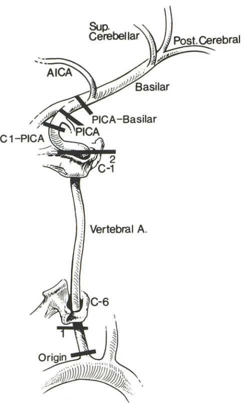
For purposes of this study, each vertebral artery was divided into six segments (origin, anatomic first segment, anatomic second seg-ment, C1 to PICA, PICA origin, and PICA to basilar) (Fig. 1) [6]. Each segment was evaluated for percentage stenosis of the vessel diam-eter. The diameters of the stenotic segment(s) and the nearest uninvolved portion of the same vessel were measured directly on the films. Percentage stenosis was calculated as % stenosis = 100 x stenotic segment/uninvolved segment. If a particular lesion involved two adjacent arterial segments, it was included in both. The percent-age of the entire group with a lesion was calculated for each segment. Patients in whom the artery was occluded proximal to a given segment were not included in the statistics of that particular segment. Therefore, the number of patients included in the statistics for each segment varies, with the lower numbers in the more distal vertebral segments.
The average percentage stenosis in those patients with lesions in a given segment was calculated for each segment. Patients in whom the vertebral artery ended in PICA were averaged into the PICA to basilar segment as 100% stenosis. In only one patient was a vertebral artery occluded at the origin and reconstituted distally. This patient was included as 100% stenosis at the origin, not included in the first-or second-segment statistics and counted as 0% stenosis beyond C1.
For the purposes of this study, the basilar artery was divided into four segments: (1) proximal (to AICA), (2) AICA origin, (3) middle basilar (distal to AICA origin), and (4) basilar tip (including origin of SCAs and PCAs). The frequency and severity of lesions in each segment were calculated in a manner identical to those for the vertebral arteries.
Two of the 31 upper brainstem revascularization cases had pre-operative angiograms done elsewhere and are not included in this evaluation.
Vertebral A,
Fig. 1.-Division of vertebral artery into six parts: (1) origin, (2) anatomic first segment-from origin to entry of vertebral into foramen transversarium of C6; (3) anatomic second segment-foramen of C6-C1; (4) C1 to PICA origin; (5) PICA origin; (6) PICA origin to vertebrobasilar junction.
Postoperative Angiographic Evaluation
A postoperative angiogram was obtained in all bypass patients. Seven items were assessed in each angiogram: (1) Bypass patency (defined as any filling of the recipient vessel by the bypass) [7]. The presence of antegrade (2) and retrograde (3) filling of the recipient vessel. (4) Filling of the basilar artery via the bypass. (5) Hypertrophy of the donor vessel. (This was graded by visual inspection as none, moderate, or marked; moderate enlargement roughly corresponds to less than 50% and marked greater than 50% increase in diameter of the donor vessel after bypass. Changes in magnification between pre- and postoperative studies were considered when grading.) (6) Evidence of arterial narrowing at the anastomotic site. (7) The number of vertebrobasilar branches (including the recipient vessel) filled via bypass flow was recorded. Patients in whom the bypass was oc-cluded are not inoc-cluded in the results of the arterial narrowing or donor hypertrophy categories.
[image:2.613.317.559.82.485.2]postopera-AJNR:8, March/April 1987 ANGIOGRAPHY IN BRAINSTEM REVASCULARIZATION 213
tive angiography. These studies were evaluated for patency and
morphology of the endarterectomized segment.
Clinical Evaluation
Each patient's postoperative clinical status (asymptomatic,
im-proved, no change, worse, died) was recorded at the time of hospital
discharge. Peri operative complications were recorded after surgery.
These data were retrieved from a computerized file and correlated
with angiographic results for purposes of this study.
Results
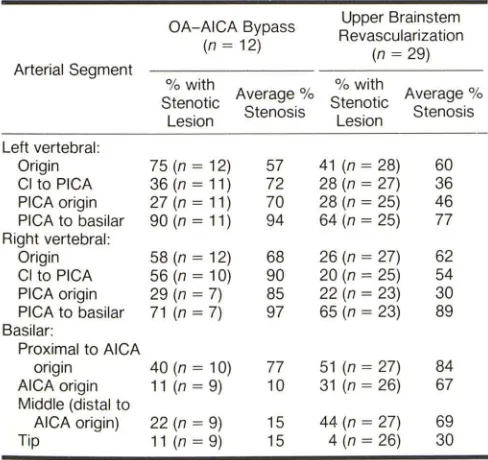
Preoperative angiographic results are in tabular form only for the 12 OA-AICA patients and for 29 of the upper brainstem revascularization patients (Table 1). Preoperative angiograms in the 12 OA-AICA patients demonstrated lesions occurring with the greatest frequency and severity in the PICA to basilar segment. Lesions were slightly less common at the vertebral origin, in the C1 to PICA segment, PICA origin, and proximal basilar. Lesions were uncommon elsewhere. Three right and three left vertebral arteries ended in PICA.
Preoperative angiograms in the five OA-PICA patients dem-onstrated stenotic lesions occurring with greatest frequency and severity in the C1 to PICA segment and at the vertebral origin. Lesions were rare elsewhere, particularly (and contrary to the OA-AICA patients) in the proximal basilar. The vertebral artery opposite the bypass ended in PICA in two patients. Each patient had multiple focal stenoses or a single fairly long segment of high-grade stenosis of the C1 to PICA segment on the side of the bypass.
TABLE 1: Preoperative Angiographic Findings in OA-AICA
Bypass and in Upper Brainstem Revascularization
OA-AICA Bypass Upper Brainstem
Revascularization
(n = 12)
(n = 29)
Arterial Segment
% with % with
Stenotic Average % Stenotic Average %
Lesion Stenosis Lesion Stenosis
Left vertebral:
Origin 75(n=12) 57 41 (n = 28) 60
CI to PICA 36 (n = 11) 72 28 (n = 27) 36
PICA origin 27 (n = 11) 70 28 (n = 25) 46
PICA to basilar 90 (n = 11) 94 64 (n = 25) 77
Right vertebral:
Origin 58 (n = 12) 68 26 (n = 27) 62
CI to PICA 56 (n = 10) 90 20 (n = 25) 54
PICA origin 29 (n = 7) 85 22 (n = 23) 30
PICA to basilar 71 (n = 7) 97 65 (n = 23) 89
Basilar:
Proximal to AICA
origin 40 (n = 10) 77 51 (n = 27) 84
AICA origin 11 (n = 9) 10 31 (n = 26) 67
Middle (distal to
AICA origin) 22 (n = 9) 15 44 (n = 27) 69
Tip 11 (n = 9) 15 4 (n = 26) 30
Note.-OA = occipital artery: AICA = anterior inferior cerebellar artery; PICA
= posterior inferior cerebellar artery.
All four endarterectomy patients had a short unifocal seg-ment of high-grade stenosis of the C1 to PICA segment, with the opposite vertebral being occluded (two patients) or ending in PICA (two patients).
Preoperative angiograms analyzed in 29 upper brainstem revascularization patients demonstrated stenotic lesions in the vertebral arteries occurring with greatest frequency and severity at the vessel origin and in the PICA to basilar segment (Table 1). Stenotic lesions occur with significant frequency and severity in the proximal and middle basilar artery. Be-cause of distal basilar filling via patent posterior communicat-ing arteries in certain cases of proximal basilar occlusion, the
number of patients in each basilar segment does not
system-atically decline distally in Table 1. Two left and 10 right vertebral arteries end in PICA.
Results of the postoperative studies in the 48 bypass patients are summarized in Table 2. In general (and particu-larly in the OA-AICA group) the bypass procedures were highly successful from a technical standpoint (high patency and low rate of angiographically identifiable complications,
such as arterial narrowing). A high percentage of patients also had good bypass performance (basilar filling and donor hy-pertrophy). Selective external carotid injections produced no apparent compromise of bypass integrity in any patient.
Postoperative angiograms in two of the three endarterec-tomy patients studied demonstrated successful removal of the stenotic plaque and excellent basilar filling. The vertebral artery was occluded at the endarterectomy site in the third patient.
Overall, 69% (36/52) of patients were asymptomatic or improved after surgery. The percentage of improved or asymptomatic patients by procedure was PICA, 80%; AICA,
75%; SCA, 71 %; and vertebral endarterectomy, 25%. Excellent correlation was seen between clinical outcome and the presence of arterial narrowing at a bypass anasto-mosis. This complication was seen in only three (9%) of 33 adequately angiographed, improved patients vs five (36%) of 14 unimproved patients with a patent bypass/endarterec-tomy. Bypass or endarterectomy occlusion was present in
TABLE 2: Postoperative Angiographic Results in
OA-AICAf>A-PICA, and Upper Brainstem Bypass
OA-AICA OA-PICA Upper
Brainstem
Finding
(n
=
12) (n = 5)(n
=
31)Bypass patent (%) 100 80 94
Antegrade filling of recipient
vessel (%) 100 80 94
Retrograde filling of recipient
vessel (%) 100 80 68
Basilar artery filling (%) 82 60 52
Donor hypertrophy (%) 64 75 57
Arterial narrowing at
anasto-motic site (%) 9 50 17
No. of basilar branches filling 3.6 2.6 2.2
[image:3.612.53.297.475.705.2] [image:3.612.318.557.573.703.2]214 JACK ET AL. AJNR:8, March/April 1987
only two (6%) of 35 improved patients vs two (13%) of 16
unimproved patients.
Overall mortality was four (8%) of 52. All four of these patients were in the SCA group. The bypass was occluded in one and in spasm in two of these four.
A major perioperative complication (infarct) was
encoun-tered in seven patients. Four of these seven eventually died.
Relatively minor perioperative complications that were all
successfully treated without apparent long-term sequelae
were present in 15 patients (meningitis/CSF leak, seven;
seizure, four; subdural hematoma, two; and cerebral or
cer-ebellar edema, two). Postoperative seizures were all well
controlled medically. No perioperative morbidity was
encoun-tered in 30 of 52 patients.
All patients were on anti platelet agents postoperatively. All asymptomatic and improved patients were taken off antico-agulants.
Discussion
In 1946, Kubic and Adams [8] published the first widely
accepted description of the clinical syndrome of
vertebroba-silar insufficiency. This clinical syndrome was further defined by Millikan et al. [9] in 1955 and by Cartlidge et al. [4] in 1977.
Proposed etiologies for the symptoms of vertebrobasilar
insufficiency fall into three categories: (1) thromboembolic, (2)
hemodynamic, and (3) local perforating vessel disease
[10-12]. Medical therapy for vertebrobasilar insufficiency is based
on the premise that symptoms are primarily thromboembolic in nature. The first specific form of therapy for treatment of
symptoms was systemic anticoagulation recommended by
Millikan et al. [9] in 1955. Both a decreased mortality and decreased incidence of stroke have been reported with this
form of therapy [13]. However, these study groups lacked
angiographic correlation of suspected vascular disease. Also,
other possible causes of symptoms suggestive of
vertebro-basilar insufficiency were not rigorously excluded in these
patients. Anticoagulant and antiplatelet therapy have not
proved to be of value in any clinical study that contains
randomized patients or patients who have undergone
angiog-raphy [10]. In fact, no therapeutic technique, medical or
surgical, has been proved to be unequivocably beneficial in the treatment of vertebrobasilar insufficiency (10). Recently
published results from the extracranial-intracranial bypass
study group find no reduction in stroke rate after ST A-middle
cerebral artery (MCA) bypass for patients with symptomatic
atherosclerotic disease of the internal carotid or middle
cere-bral artery (14). Patients with vertebrobasilar insufficiency
were not included in this study, which does not purport to
evaluate the efficacy of posterior fossa bypass. The clinical results in these 52 patients may justify undertaking a random-ized trial comparing surgical with medical therapy for verte-brobasilar insufficiency. Sixty-nine percent of these patients
were either less symptomatic or asymptomatic after surgery.
Most important, these surgical results were achieved in a
patient population that had already failed on medical therapy.
Several investigators have noted a qualitative difference
between TIAs in the anterior and posterior circulation: TIAs
are more likely to be of hemodynamic origin in the posterior circulation and of thromboembolic origin in the anterior circu-lation [11, 12, 15]. Surgical therapy for vertebrobasilar insuf-ficiency is based on the premise that symptoms are usually of hemodynamic origin [15]. If the flow deficit distal to a
stenotic lesion is corrected, then symptoms will be also.
Hemodynamic changes are caused by a deficiency of blood flow distal to a stenotic lesion or lesions that may be located at any level(s) in the vertebrobasilar system. Ischemia induced by dysautoregulation distal to a stenotic lesion is also thought to playa key role in patients whose symptomatology is highly sensitive to postural changes [11]. Surgical correction of stenoses is also directed at prevention of permanent deficit
resulting from stroke. Thirty-five percent of patients with
vertebrobasilar TIAs will progress to stroke if untreated [4].
This percentage was derived from a nonangiographed popu-lation with vertebrobasilar insufficiency defined by clinical
symptoms only, and may be higher if patients without
angio-graphically proven lesions are excluded. Thompson et al. [16]
found a 44% mortality within 3 months in 22 patients with
angiographically proven basilar occlusion. TIAs preceded
brainstem infarcts in 50% of these patients.
A variety of surgical approaches to'stenotic lesions of the
extracranial vertebral artery has been described. These
in-clude bypass with both arterial and vein graft donor vessels,
vertebral transposition, endarterectomy, a variety of
angio-plastic procedures, and decompressive procedures [6].
The surgical options for correction of intracranial vertebral
and basilar lesions are much more limited, however.
Extra-cranial to intraExtra-cranial bypass procedures or intraExtra-cranial ver-tebral endarterectomy are the only surgical treatments
avail-able for lesions of the intracranial vertebrobasilar system [10,
17, 18]. Bailoon angioplasty of basilar stenoses has been
reported, but results have been unsatisfactory [19].
Until 1975, systemic anticoagulation was the only treatment
available to patients with vertebrobasilar insufficiency caused by lesions of the posterior intracranial circulation. In 1975, Ausman et al. [1] reported the first case of posterior fossa revascularization. In this patient with bilateral distal vertebral
artery occlusion an OA-PICA bypass was performed. The
principle in posterior fossa revascularization is to bypass distal to a stenotic or occlusive lesion [20-22]. To this end, the ST A-SCA bypass was introduced in 1979 for lesions proximal to the SCA origin [2).The OA-AICA was introduced in 1981 for lesions proximal to the AICA origin [3].
The best site for anastomosis is that segment of the recipient vessel that has the largest diameter and fewest
brainstem branches. Anatomic studies show this site to be
the peri mesencephalic segment of the SCA, the segment of
the AICA just beyond the seventh and eighth nerves in the
cerebeilopontine angle cistern, and the caudal loop of the
PICA [22]. Recipient vessel diameters in these locations vary
from 1.0 to 1.8 mm [22].
Ail 31 upper brainstem revascularization procedures were
performed via a right temporal craniotomy to avoid the left
temporal speech area. Use of the PCA as the recipient vessel
AJNR:8, March/April 1987 ANGIOGRAPHY IN BRAINSTEM REVASCULARIZATION 215
vessel occlusion [12, 21). Should the SCA become occluded distal to its brainstem branches, the potential for production
of a serious deficit is minimized by a rich collateral network
over the cerebellar hemisphere [12). Two patients in this series underwent an ST A-PCA rather than an ST A-SCA
bypass. In these cases the latter procedure was planned
preoperatively. However, upon surgical exposure, the right
SCA was found to be too small or the peri mesencephalic loop too low-lying for a technically successful anastomosis. The PCA was then chosen as the alternative recipient vessel.
Before the late 1970s, detailed angiography of the posterior
fossa was avoided because of fear of complications and the
knowledge that the angiographic findings would probably not
change treatment, as medical therapy was the only option
available. However, with the relatively recent availability of
these surgical procedures and with technical advances in
cerebral angiography that can detail posterior fossa, angiog-raphy in patients with vertebrobasilar insufficiency is now justified [2, 10, 23].
Caplan and Rosenbaum [23] studied a group of patients
with vertebrobasilar insufficiency who had angiographic and
autopsy correlation and found that the clinical presentation
did not allow accurate determination of the site or the nature
of vascular disease. Lesions producing symptoms of
verte-brobasilar insufficiency may be located at any level from the
extracranial vertebral arteries to the median and paramedian
basilar artery branches [23]. A patient with symptoms on a
vascular basis may have gross large-vessel occlusive disease,
or may have an entirely normal angiogram. In the latter case,
A
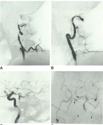
Fig. 2.-0A-AICA bypass for bilateral distal vertebral occlusion.
A, lateral right carotid injection. Preoperativ, angiogram shows basilar filling (arrows) via pat-ent right posterior communicating artery.
B, Anteroposterior late arterial phase of same
run shows large right AICA (arrows). En face projection "elongates" basilar artery.
C and 0, Anteroposterior (C) and lateral (D) right external carotid injection. Postoperative an-giogram shows patent end-to-side anastomosis between OA and AICA (small arrows) just be-yond porus acusticus. Antegrade (large arrows) and retrograde (arrowheads) recipient vessel and basilar filling are seen.
B
symptoms are presumably caused by plaque formation at the
origin of, or arteriolar hyalinization of, one or more small basilar
perforating arteries [23]. Furthermore, cerebral angiography
is the only reliable method by which the nature and location
of the vascular disease causing symptoms can be accurately
determined [10,23].
Results of the preoperative studies in this group of patients
demonstrate the important role angiography plays in
identi-fying that subgroup of patients with vertebrobasilar
insuffi-ciency who are surgical candidates. Furthermore, the final
decision as to which surgical procedure will be used is
pri-marily dictated by the angiographic findings. It is incumbent
on the neuroradiologist performing the preoperative
angio-gram to demonstrate all areas of stenosis in the vertebroba
-silar system and the relationship of the lesions to potential
recipient vessels. Lesions of the anterior circulation and the
interdependence of anterior and posterior circulation must be
demonstrated. Also, suitable donor vessels must be identified
and characterized as to size, length, and mobility.
Preoperative angiograms in 40% of patients who
under-went OA-AICA bypass demonstrated high-grade stenosis of
the basilar artery proximal to the AICA origin. The other 60%
had more proximal disease generally involving the PICA to
basilar segments. These findings narrow the surgical options
to one choice: the OA-AICA bypass (Fig. 2). This distribution
of atherosclerotic lesions is fairly common, resulting in the
greater frequency of OA-AICA procedures than OA-PICA
bypass or vertebral endarterectomy, for lower brainstem
vas-cular disease.
[image:5.612.53.559.429.729.2]216 JACK ET AL. AJNR:8, March/April1987
Patients who were candidates for both vertebral endarter-ectomy and OA-PICA bypass had one vertebral artery
oc-cluded or ending in PICA and the other stenotic at the C1 to
PICA segment. If the lesion is of short length and unifocal at
angiography the patient is an endarterectomy candidate (Fig. 3). A long lesion or more than one lesion makes the patient
an OA-PICA candidate (Fig. 4). Because of a larger-caliber
conduit and greater potential flow, endarterectomy is
theo-retically preferable to OA-PICA bypass when technically pos-sible.
Stenotic lesions of the mid basilar artery were present in
almost one-half of the upper-brainstem revascularization
pa-tients (14 of 29) (Fig. 5). The SCA is the only viable recipient
vessel available for purposes of bypassing lesions in this
location. Most of these patients also had additional more
proximal lesions.
Nearly one-half (15 of 29) of the upper-brainstem revascu-larization patients, however, did not have middle basilar
ste-nosis. Instead, multiple, highly stenotic or occlusive lesions of
the proximal basilar or vertebral artery (or a vertebral artery
ending in PICA) were found. This prevented adequate
pre-operative visualization of the entire vertebral basilar system,
particularly potential AICA or PICA recipient vessels. Because of proximal obstructing lesions in this group, only the basilar tip was imaged preoperatively (via posterior communicating collateral flow). Therefore, the only potential recipient vessel that could definitely be identified preoperatively was the SCA (Fig. 6).
This illustrates one of the major limitations of preoperative
A
Fig. 3.-Vertebral endarterectomy. A, Anteroposterior left vertebral injection. Pre-operative angiogram shows short unifocal plaque (arrow) narrowing C1 to PICA segment.
8 and C, Anteroposterior (8) and lateral (C) left vertebral injection with head turned to right. Postoperative angiogram shows patent endar-terectomized C1-PICA segment.
angiography in patients with vertebrobasilar insufficiency. In
order to direct the surgical approach logically, the entire
vertebrobasilar system and the relationship between all
le-sions and potential recipient vessels should be imaged.
Pa-tients who will benefit the most from surgery are generally those with the most severe stenotic/occlusive disease.
How-ever, it is precisely this group in whom visualization of the
entire vertebrobasilar circulation is most difficult, and the status of the vessels beyond a lesion or lesions may remain
unknown. In cases of extracranial vertebral occlusion, delayed
filming can be invaluable in imaging reconstituted circulation distal to the site of occlusion. In cases of intracranial verte-brobasilar occlusion, the only information about the status of the basilar artery may come from collateral posterior com-municating flow, hence the importance of thorough evaluation of the anterior circulation in these patients.
All arteriographic studies of posterior fossa circulation in this series of patients were performed with magnification and subtraction film technique. The role of intraarterial digital subtraction angiography (DSA) in the evaluation of patients with severe obstructive/occlusive disease of the vertebroba-silar circulation has not been investigated thoroughly as yet. It is possible that intraarterial DSA might give information regarding potential recipient vessels located distal to severe obstructive or occlusive lesions that is not available with conventional film technique. However, our preliminary expe-rience with intraarterial DSA would indicate that this technique may not adequately resolve stenoses of the small potential recipient vessels in the posterior circulation. This possible
[image:6.612.55.557.438.733.2]AJNR:8, March/April 1987 ANGIOGRAPHY IN BRAINSTEM REVASCULARIZATION 217
Fig. 4.-0A-PICA bypass.
A, Anteroposterior right vertebral injection with head turned to left. Preoperative angiogram shows two areas of discrete narrowing (arrows) proximal to PICA origin (arrowhead).
8 and C, Anteroposterior (8) and lateral (C) selective right occipital artery injection. Postop-erative study shows patent OA-PICA anasto-mosis (solid arrows). Faint but definite filling of basilar artery (open arrows) is typical of early postoperative OA-PICA bypass angiogram.
D, Anteroposterior right external carotid injec-tion in different patient. Postoperative OA-PICA study shows more intense basilar opacification (seen in only one OA-PICA patient).
c
limitation must be either disproved or overcome before intra-arterial DSA alone is considered an acceptable radiologic
approach in posterior fossa revascularization.
Twenty-two of the total 52 patients had one vertebral artery
ending in PICA. The high incidence of this "normal variant" in
a group with symptoms of vertebrobasilar insufficiency
sug-gests that this is not an entirely benign anomaly. Vertebral artery ending in PICA imposes a significant liability on patients who develop atherosclerotic disease in the vertebrobasilar system.
Good correlation is seen between an unsatisfactory surgical result at postoperative angiography and an unsatisfactory clinical outcome. The bypass/endarterectomy was occluded
or in spasm in seven (44%) of 16 unimproved patients vs
only five (14%) of 35 improved patients. The degree of arterial
narrowing at the anastomotic site in the three improved
patients with spasm presumably allowed sufficient bypass
flow to improve symptoms. It is difficult to rationalize how
two patients improved with an occluded bypass.
B
D
Bypass/endarterectomy patency was documented in 33 of
35 improved patients. However, basilar filling via the bypass
was demonstrated in only 21 of this group. The discrepancy
between filling of the target vasculature and clinical outcome
was addressed by Anderson et al. [24], who found the entire
sylvian group filled in only seven of 11 postoperatively
asymp-tomatic ST A-MCA bypass patients. They concluded that
"these patients had a lesser or only intermittent need for
additional flow. The new collateral channel may have supplied
this need by establishing a continuous and unobstructed
source of blood ... " Seven of the 16 unimproved patients
suffered a perioperative stroke. The bypass was in spasm in
one of the other nine unimproved patients. In eight of these
nine unimproved patients (without perioperative stroke) a
well-functioning bypass did not produce clinical improvement.
Bypass flow may have not met demand in these patients, or
TIAs in this small percentage of patients may have resulted from embolic episodes. Alternatively, some or all of these
[image:7.614.225.559.81.518.2]A
B
Fig. 5.-STA-SCA bypass for midbasilar stenosis.
A, Anteroposterior preoperative vertebral angiogram shows
circumfer-ential midbasilar lesion. Right AICA is filled but is unacceptable as potential recipient vessel as its origin is proximal to bulk of plaque (arrowhead).
SCA is only acceptable recipient vessel seen (arrow).
B, Lateral right external carotid injection. Postoperative angiogram
A
B
c
D
-I
r
c
shows anastomosis of STA to SCA (solid arrow). STA is of large caliber
(open arrow) after bypass.
C, Anteroposterior right external carotid injection. Postoperative angio-gram shows antegrade (solid arrow) and retrograde (open arrow) filling of
recipient vessel. Filling of four basilar branches (both PCAs and SCAs) via bypass is seen.
Fig. 6.-STA-SCA bypass for bilateral distal vertebral artery occlusion.
A and B, Right (A) and left (B) vertebral artery occlusion in C1 to basilar segment at
preopera-tive angiography.
C, Lateral carotid injection. Preoperative an-giogram shows filling of upper basilar artery via posterior communicating artery. SCA is only vi-able recipient vessel seen preoperatively.
[image:8.614.59.558.44.268.2] [image:8.614.55.389.337.742.2]AJNR:8, March/April 1987 ANGIOGRAPHY IN BRAINSTEM REVASCULARIZATION 219
or more small brainstem perforating branches. In such a patient an increase in gross basilar flow via a bypass would not eradicate symptoms caused by local disease. The findings at preoperative cerebral angiography, therefore, may not be an entirely accurate indicator of the demand for posterior fossa blood flow augmentation in patients with vertebrobasilar insufficiency. For this reason, a preoperative diagnostic method that accurately measures cerebral perfusion may be needed in addition to conventional angiography to aid in the appropriate selection of candidates for these procedures.
Attempts to measure regional blood flow noninvasively have been reported using both 133Xe inhalation with external colli-mation and stable xenon-enhanced CT scanning [25-27]. To date, the value of these techniques as preoperative screening tests in potential posterior fossa bypass candidates is unclear. Positron emission tomography (PET) has been shown to
demonstrate regional changes in cerebral metabolism after STA-MCA bypass [28]. However, a group of metabolic find-ings on PET scanning that reliably predicts a patient's re-sponse to bypass surgery has not been identified [24J.
It should be emphasized that this study represents only short-term angiographic follow-up of these patients. Prelimi-nary results indicate that intracranial filling and donor hyper-trophy may increase with time in posterior fossa revasculari-zation, as is the case with supratentorial bypass procedures
[7,29].
In summary, no therapeutic method, medical or surgical, has yet been proved to be unequivocably beneficial in
treat-ment of vertebrobasilar insufficiency. Newly developed sur-gical approaches to the vertebrobasilar circulation show promise as alternatives to medical treatment. In this series of patients a favorable postoperative angiographic result gen-erally correlates well with a favorable postoperative clinical result and vice versa. To our knowledge this is the first report
in the radiologic literature on the subject of angiography in posterior fossa revascularization. Radiologists playa vital role in the preoperative evaluation of patients with vertebrobasilar insufficiency. The type of surgical procedure for which the
patient is best suited is determined by angiography. Postop-erative angiography provides a safe and reliable method of evaluating anatomically the performance of the procedure.
ACKNOWLEDGMENTS
We thank Dorrine Dalley and Brenda Maxwell for manuscript preparation.
REFERENCES
1. Ausman JI, Lee MC, Klassen AC, et at. Stroke: what's new, cerebral revascularization. Minn Med 1976;58:223-227
2. Ausman JI, Lee MC, Chater N, et at. Superficial temporary artery to superior
cerebellar artery anastomosis for distal basilar artery stenosiS. Surg Neurol
1979;12:277-282
3. Ausman JI, Diaz FG, de los Reyes RA, et at. Anastomosis of OCCipital
artery to anterior inferior cerebellar artery for vertebrobasilar junction stenosis. Surg Neuro/1981;16(2):99-102
4. Cartlidge N, Whisnant JP, Elveback LR. Carotid and vertebral-basilar
transient cerebral ischemic attacks. Mayo Clin Proc 1977;52: 117-120 5. Boulos R, Patel S, Mehta B, et at. Cerebral angiography in posterior fossa
revascularization. Henry Ford Hosp Med J 1985;33:74-81
6. Diaz F, Ausman J. Surgical reconstruction of vascular lesions of the vertebral basilar circulation. Stroke 1984;19(4):19-24
7. Latchaw R, Ausman J, Lee M. Superficial temporal-middle cerebral artery
bypass. J Neurosurg 1979;51 :455-465
8. Kubic CS, Adams RD. Occlusion of the basilar artery-a clinical and
pathological study. Brain 1946;69:73-121
9. Millikan CH, Siekert RG, Shick RM. Studies in cerebrovascular disease: lit. The use of anticoagulant drugs in the treatment of insufficiency or throm-bosis within the basilar arterial system. Mayo Clin Proc 1955;30: 116-126 10. Ausman JI, Schrontz CE, Pearce JE, et at. Vertebrobasilar insufficiency: a
review. Arch Neuro/1985;42:803-808
11. Naritomi H, Sakai F, Meyer J. Pathogenesis of transient ischemic attacks within the vertebral arterial system. Arch Neuro/1979;36:121-128
12. Sundt T, Piepgras DG, Houser 0, et at. Interposition saphenous vein grafts
for advanced occlusive disease and large aneurysms in the posterior circulation. J Neurosurg 1982;56: 205-215
13. Whisnant J, Cartlidge N, Elveback L. Carotid and vertebral-basilar transient
ischemic attacks: effect of anticoagulants, hypertension, and cardiac dis-orders on survival and stroke occurrence-a population study. Ann Neurol
1978;3:107-115
14. The EC/IC Bypass Study Group. Failure of extracranial-intracranial arterial bypass to reduce the risk of ischemic stroke. Results of an international
randomized triat. N Engl J Med 1985;313(19): 1191-1200
15. Hadley M, Masferrer R, Zabramski J, et at. Management of vertebrobasilar insufficiency in cerebral revascularization for stroke. In: Spetlzer R, ed.
New York: Thieme-Stratton, 1985:475-487
16. Thompson JR, Simmons CR, Hasso AN, et at. Occlusin of the intradural vertebrobasilar artery. Neuroradiology 1978;14: 219-229
17. Allen GS, Cohen R, Preziosi RC. Microsurgical endarterectomy of the intracranial vertebral artery for vertebrobasilar transient ischemic attacks. Neurosurgery 1981 ;8(1): 56-59
18. Ausman J, Diaz F, Pearce J, et at. Endarterectomy of the vertebral artery
from C2 to posterior inferior cerebellar artery intracranially. Surg Neurol 1982;18:400-404
19. Piepgras DG, Sundt TM, Forbes GS, et at. Balloon catheter transluminal
angioplasty for vertebrobasilar ischemia in vertebrobasilar arterial occlusive
disease. In: Berger, Bauer, eds. New York: Raven, 1984:215-224 20. Ausman JI, Diaz F, de los Reyes R, et at. Extracranial-intracranial
anasto-moses in the posterior circulation in vertebrobasilar occlusive disease. In:
Berger, Bauer, eds. New York: Raven, 1984:313-319
21. Ausman JI, Diaz F. Microsurgical reconstruction of the posterior circulation.
Head Neck Surg 1984;92: 102-108
22. Shrontz C, Dujovny M, Ausman JI, et at. Vertebrobasilar reconstruction:
microanatomical considerations. Surg Forum 1985;36:506-507
23. Caplan L, Rosenbaum A. Role of cerebral angiography in vertebrobasilar occlusive disease. J Neurol Neurosurg Psychiatry 1975;38:601-612
24. Anderson RE, Reichman OH, Davis 00. Radiological evaluation of
tem-poral artery-middle cerebral artery anastomosis. Radiology 1974;113:
73-79
25. Drayer B, Gur 0, Wolfson S, et at. Regional blood flow in the posterior
fossa. Xenon enhanced CT scanning. Acta Neurol Scand {Suppl]
1979;60[2]:218-219
26. Meyer J. Application of cerebral blood flow: ischemic cerebrovascular
disease. In Moossy J, Reinmuth 0, eds. Cerebrovascular diseases. New York: Raven, 1981: 125-141
27. Yonas H, Good W, Gur 0, et at. Mapping cerebral blood flow by xen
o-nenhanced computed tomography: Clinical experience. Radiology
1984;152:435-442
28. Powers W, Martin W, Herscovitch P, et at. Extracranial-intracranial bypass
surgery: hemodynamic and metabolic effects. Neurology 1984;34:
1168-1174
29. Shrontz C, Diaz F, Patel S, et at. Long-term angiographic follow-up after extracranial-intracranial anastomosis for vertebrobasilar ischemia. Pr