J. Exp. Biol. (197a), 57. 701-707
^t't/i 5 text-figures
Wrxnted in Great Britain
THE EYE AND COLOUR CHANGE IN THE MINNOW
(PHOXINUS PHOXINUS L)
BY MICHAEL J. GENTLE*
Zoology Department, Bedford College, Regent's Park, London
{Received 17 March 1972)
INTRODUCTION
The eye is the primary sense organ responsible for background adaptation, and a number of workers in this field have stated that the colour of the animal is a reaction to the ratio of direct/reflected light entering the eye. Attempts have been made to define this ratio and the retinal regions involved. Sumner & Keys (1929), using
Hypsopsetta guttulats and varying the light, and Sumner (1933), using false celloidin
corneas in Fundulus parvipinms, concluded that the shade the fish assume on a given background is determined by the relative luminosity of the upper and lower portions of the visual fields, the latter acting in a positive sense and the former in a negative sense. In a long series of experiments Butcher & Adelmann (1937) and Butcher (1937a,
b, 1938, 1939) using Fundulus heteroclitus concluded that stimulation of the upper
retina resulted in lightening, of the lower darkening, and they formed distinct regions in the retina. He also stated that the shade the fish assumed depended on the quantita-tive ratio of the direct/reflected light entering the eye. Butcher's experiments were very extensive but unfortunately no quantitative measure of the colour of the fish was made. Butcher also found that single and double cones were present in the dorsal retina but only double cones were present in the ventral retina and thus demonstrated anatomical and physiologicaljdifferentiation in the retina.
Hogden & Landgrebe (1940) considered that the ratio must not be construed in any quantitative sense. They also located the retinal regions involved in chromatic adaptation in Gasterosteus aculeatus, using the refractive index of the lens, general optics of the eye and superior and inferior illumination on various backgrounds. They concluded that the photoreceptors concerned with black background responses were located in the floor of the retina below the optic nerve and the white background receptors were located in a restricted region in the central retina above and below the optic nerve. With respect to colour change the dorsal region was neutral.
No work has been done on the minnow and the work to be described was designed to see whether the minnow had anatomically distinct areas in the retina and to examine the effects of retinal removal on the ability of the fish to perform background adaptation.
702
Dorsal
Ventral
M. J. GENTLE
1-5 mm
Fig. i. Drawing of a section of the eye showing the dorso-ventral axis marked and the parts of the eye cut when the knife is orientated at an angle to this axis.
MATERIALS AND METHODS
The fish used were 6 cm long adults of both sexes collected from the river Chess near Rickmansworth. They were kept throughout the year in the laboratory in large sinks where the temperature ranged from 15 to 20 °C.
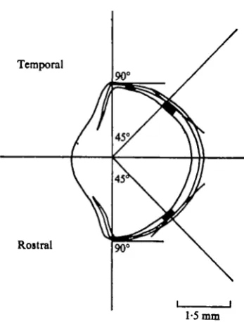
Counts were made of the retinal elements using tangential sections of the retina. The eye was dissected out of the fish and fixed in aqueous Bouin. It was embedded in paraffin wax, sectioned at 4 /im and stained with haematoxylin and eosin. In previous work (Lyall, 1956, 1957^) on the minnow pieces of the retina were removed before sectioning but this method makes orientation difficult when such a small eye is used. In the present work the whole eye was sectioned. A small tie was placed in the extreme dorsal part of the eye before it was removed from the animal. The eye was then blocked out so that it was clearly visible from the outside of the wax block. On the outside of the wax it was possible to mark the dorso-ventral axis (Fig. 1) and, provided the rostral-temporal axis was parallel to the front of the block, the cutting angle could be measured and scored directly on the wax block. The block was then mounted on the microtome chuck so that the knife cut parallel to the marked cutting angle. By mounting the block at various angles all parts of the retina were cut (Fig. 1, 2). Counts were made of the retinal elements from an area of o-oi mm2, and from each region at least 3-4 different counts were made.
Eye and colour change in the minnow
7°3
Temporal
Rostral
[image:3.451.141.315.65.295.2]1-5 mm
Fig. a. Drawing of a section of the eye at A-B (Fig. i) showing the angles used to obtain tangential sections of the rostral and temporal fields of the retina.
away from the surrounding skin and an incision made at the base of the iris into the vitreous humor. The retina was removed and the conjunctiva re-attached to the sur-rounding skin by fine nylon stitches. At the end of the experiment the eyes were removed and the remaining retina was examined histologically for any degenerative changes and to determine the extent of the retina removed.
The methods used to record the colour of the fish and the methods for testing the colour change were described in a previous paper (Gentle, 1971).
RESULTS
Retinal receptors
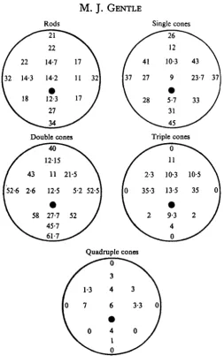
The results from the retinal counts are shown in Fig. 3. The rods were very difficult to see and the values presented for them are therefore only approximate. The general distribution of the rods follows a pattern characteristic of many other vertebrates in that they occur in large numbers at the periphery of the retina and decrease in number centrally.
The double and single cones showed a tendency to become reduced in number in the central regions of the retina. This reduction in number centrally was associated with the increase in number of triple and quadruple cones which were found in the peripheral parts but form the dominant cone types in the central regions. The quad-ruple cones were never very numerous in the retina.
32 143 14-2 11 32 37 27 9 23-7 37
Fig. 3. A series of drawings of the retina looked at from the front of the eye showing the approximate positions where the receptors were counted and the mean numbers present.
Retinal removal
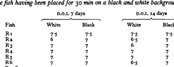
The left optic tract of 24 minnows was sectioned and the fish were allowed to recover for at least 24 h. These fish were then tested to confirm that they could perform normal colour change. In three fish a cut was made over the dorsal part of the right eye (R7, 8, 9) and in another six fish (Ri, 2, 3, 4, 5, 6) the dorsal part of the retina was removed. The extent of the retina removed is shown in Fig. 4 and the colour of the fish after 30 min on a white and black background 7 and 14 days after operation in Table 1. The removal of the ventral retina was performed in seven fish, the parts removed being shown in Fig. 5 and their colour on black and white backgrounds in Table 2.
Eye
and c o b
change
in
the
n t k o w705
&able I . The colour of thefuh with dorsal retinal r-ed m 7 and 14 days after operation,
the f i h having been placed for 30 min on a black and white background
D.O.I. 7 day8 D.O.I. I 4 d a y
r L
\ P
Fish White Black White Black
Table 2. The colour of thefih with ventraIretM removed at 7 ad 14 days
afLtr
operation,thefih hating beenplacedfor 30 min on a black or white background
D.O.I. 7 days D.O.I. 14 days
r \ P
Fish White Black White Black
ventral retina. The results from the control iish showed that the operation had no effect on the ability of the
fish
to change colour.In the histological sections of the eye 14 days after surgery there was no evidence of haemorrhage in the vitreous humor and the e gretina appeared to be normal. The results from the experimental fish showed that fairly large cuts in the ventral retina ( R I ~ , 15, 16) had very little effect on colour change, whereas larger ventral removals showed a D.O.I. of 4-6 on a black background and 2-4 on the white. The
ability of the fish to adapt did not exceed 1-2 degrees on the D.O.I. scale and, with
increasing removal of the ventral retina dorsalwards, there was a reduction in the ability to pale. The ability to darken remained the same regardless of increasing removal of the ventral retina. None of the lish showed any ability to pale by more than
I degree (R2, 3) following the removal of the dorsal retina. This inability to pale was
also seen in the fish in which only a very small part of the retina was removed (R7,8, 9). Four fish showed a slight recovery by the 14th day after operation. The changes in the tint of the skin were responses to the background colour and could not be produced by varying the overall light intensity.
D I S C U S S I O N
[image:5.474.88.377.93.204.2]706 M. J. GENTLE
RI
RIO R l l
0 8 mm 0-8 mm
Fig. 4 Fig. S
Fig. 4. A series of drawings showing the extent of the dorsal retina removed (stippled area) in fish R i, a, 3,4, s, 6 and 7 (R8 and 9 being the same as 7) as viewed from the front of the eye. Fig. 5. A series of drawings showing the extent of the ventral retina removed (stippled area) in finVi R io, i i , ia, 13, 14 (R15 and 16 are the same as 14) as viewed from the front of the eye.
numbers of cones are calculated in terms of a single cone unit (i.e. double = 2, triple = 3, quadruple = 4) then the number of cones in any given area of the retina did not differ significantly throughout the retina. This finding supports the hypothesis that these multiple cone types are formed by the fusion of double and single cones (Lyall, 1957ft) which is in turn based on the assumption that the retina of the minnow grows from its edge as in the trout (Lyall, 1957a).
The extreme dorsal part of the retina appeared to have a marked effect on the ability of the fish to change colour, and here the minnow appears to differ from Gasterosteus
aculeatus (Hogben& Landgrebe, 1940), where it appears to be neutral in colour change.
Eye and colour change in the minnow 707
SUMMARY
1. Counts were made of the retinal receptors and observations were made of the colour of the minnow, Phoxinus phoxinus L., following the surgical removal of parts of the dorsal and ventral retina.
2. It was found that there were greater numbers of retinal receptors in the temporal field than in the rostral field of the eye.
3. There were very few triple and quadruple cones but a large number of double and single cones in the ventral retina compared to the dorsal.
4. Surgical removal of the dorsal retina or only part of it resulted in the fish being fully dark-adapted on a black or white background.
5. Surgical removal of the ventral retina resulted in the fish assuming an inter-mediate colour on a white background and a darker tint on a black background.
The author wishes to express his thanks to Professor N. Millott, who kindly provided laboratory facilities, and to Dr E. G. Healey, who supervised the work and offered much encouragement and advice. Finally, he is indebted to the S.R.C. for financial support during the tenure of their studentship.
The work presented here was part of the work approved by London University for the degree of Doctor of Philosophy.
REFERENCES
BUTCHER, E. O. (1937a). The structure and distribution of the rods and cones in the eye of Fundulus
heteroclitus. Bull. Mt. Desert Isl. Biol. Lab. pp. 18-19.
BUTCHER, E. O. (19376). Rods and cones in the retina of Fundulus heteroclitus, and the regions of the retina related to different chromatophoric responses. Anat. Rec. 70, 56.
BUTCHER, E. O. (1938). Structure of the retina of Funduhu heteroclitus and regions of the retina associ-ated with different chromatic responses.^, exp. Zool. 79, 275-97.
BUTCHER, E. O. (1939). The illumination of the eye necessary for different melanophoric responses of
Funduhu heteroclitus. Biol. Bull. mar. biol. Lab., Woods Hole 77, 258-67.
BUTCHER, E. O. & ADELMANN, H. B. (1937). The effect of covering and rotating the eye on the melano-phore responses in Funduhu heteroclitus. Bull. Mt. Desert 1st. Biol. Lab. pp. 16-18.
ENOSTROM, K. (1963). Cone types and cone arrangements in teleost retinae. Acta Zool. 44, 179—343. GENTLE, M. J. (1971). The central nervous control of colour change in the minnow (Phoxinus phoxinus
L). I. Blinding and the effects of tectal removal on normal and blind fish. J. exp. Biol. 54, 83-91. HOOBBN, L. & LANDGREBE, F. (1940). The pigmentary effector system. IX. Receptor fields of the
teleo-stean visual response. Proc. Roy. Soc. B 138, 317-43.
LYALL, A. H. (1956). Occurrence of triple and quadruple cones in the retina of the minnow (Phoxinus
laevis). Nature, Lond. 177, 1086-^7.
LYALL, A. H. (1957a). The growth of the trout retina. Q.J. microsc. Sri. 98, 101-10. LYALL, A. H. (19576). Cone arrangement in teleost retinae. Q.J. microsc. Sci. 98, 189-201. SUMNER, F. B. (1933). The differing effects of different parts of the visual field upon the chromatophore
responses in fishes. Biol. Bull. mar. biol. Lab., Woods Hole 65, 266-82.