Original Article
Over-expression of lysine-specific
demethylase 1 predicts tumor progression
and poor prognosis in human esophageal cancer
Lujun Chen*, Yun Xu*, Bin Xu, Haifeng Deng, Xiao Zheng, Changping Wu, Jingting Jiang
Department of Tumor Biological Treatment, The Third Affiliated Hospital, Soochow University, Changzhou 213003, Jiangsu, China. *Equal contributors.
Received September 24, 2014; Accepted November 26, 2014; Epub December 1, 2014; Published December 15, 2014
Abstract: Lysine specific demethylase 1 (LSD1), the first characterized histone demethylase, roles importantly in
epigenetic regulation of carcinogenesis and cancer progression. In the present study, we examined LSD1 expres-sion in 103 cases of esophageal cancer tissues, and further investigated its relationship to patient’s clinical param-eters and post-operative prognosis. We found that the positive LSD1 immunochemical staining was predominantly observed on the nuclei and cytoplasm of esophageal cancer cells, while negative or very weak in adjacent normal
tissues. The intensity of LSD1 immunostaining was significantly correlated to the tumor size (P = 0.013), nodal me-tastasis (P = 0.002), distant metastasis (P = 0.025), and TNM stage (P = 0.010), whereas it was not correlated to patient’s gender, age and tumor invasion depth. The overall survival rate of patients with low LSD1 expression was better than those with high LSD1 expression (P = 0.014). We also showed that the tumor size (P = 0.003) as well as the TNM stage (P = 0.007) was a useful prognostic predictor for esophageal cancer. However, when the gender, age,
tumor size, TNM stage and LSD1 expression level were involved in the multivariate proportional hazards regression analysis in a Cox’s model, we showed that the tumor size (P = 0.013) and the TNM stage (P = 0.032) could be used as independent risk factors to predict patient’s postoperative prognosis, but LSD1 expression level as well as other factors could not independently predict patient’s outcome. Thus, our results indicated that LSD1 was involved in cancer progression and metastasis in human esophageal cancer, and could be a potential prognostic predictor for this malignancy.
Keywords: LSD1, immunohistochemistry, prognosis, esophageal cancer
Introduction
Esophageal cancer is one of the most common types of human malignancies, and it ranks as the sixth cause of cancer deaths in the past year [1]. Esophageal cancer could be divided into two major types, adenocarcinomas and squamous cell carcinomas according to the his-tological classification [2-4]. And the esopha -geal squamous cell carcinoma constitutes more than 90% of all esophageal cancer cases worldwide [2, 5]. As of now, numerous types and combinations of surgery, chemotherapy and radiotherapy, as well as other therapeutic strategies, have been used over recent decades to the treatment of esophageal cancer, but those patients usually undergo local recur-rence or distant metastasis after curative
oper-ation, due to the aggressive nature of this can-cer type [2-4, 6]. Therefore, it is of great importance for us to investigate the molecular mechanism of the initiation and progression, and further find out the novel therapeutic tar -gets for this cancer.
pro-approved by the ethics committee of the hospital.
Immunohistochemistry procedures
Immunohistochemical staining was performed by using the Dako EnVisionTM technique accord-ing to the manufacturer’s instructions (Dako, Glostrup, Denmark). In brief, formalin-fixed, paraffin-embedded tissues were cut into 3 μm-thick sections, and were then dewaxed in xylene, rehydrated and graded ethanol solu-tions. Antigen was retrieved by heating the tis-sue sections at 100°C for 30 min under EDTA solution. Sections were cooled and immersed in presence of 0.3% hydrogen peroxide for 20 min to block endogenous peroxidase activity, and then rinsed in PBS for 5 min, blocked with 3% BSA at room temperature for 20 min, and then incubated with primary polyclonal anti-body against LSD1 (diluted in 1:300, Novus Biologicals, Littleton, CO, USA) at 4°C overnight. liferation, migration, and invasion in various
cancer cells, and the abnormal expression of LSD1 in human cancer tissues significantly associated with cancer recurrence and poor prognosis [10, 11]. Moreover, Ding et al. [12] also demonstrated that the positive expression of LSD1 and negative expression of E-cadherin correlated with metastasis and poor prognosis of colon cancer patients, suggesting that the over-expression of LSD1 might contribute to poor outcomes of cancer patients via regulat-ing the epithelial mesenchymal transition, which plays an essential role in the initial step of cancer metastasis.
In the present study, we examined LSD1 expres-sion in 103 cases of esophageal cancer tissues by using immunohistochemistry, and further investigated its relationship to patient’s clinical parameters and post-operative prognosis, in order to explore its value of clinical application in diagnosis and therapeutics against this malignancy.
Patients and methods
Patients and tissue samples
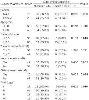
[image:2.612.91.383.98.425.2]Esophageal cancer samples were collected from 103 patients who underwent surgical resection between January 2001 and March 2005 in the hospital (75 men and 28 women, median age at diagnosis was 58 years). No patients received pre-operative chemotherapy or radiotherapy. All cases were confirmed as the esophageal squamous cell carcinoma (ESCC), and the tumor-node-metastasis (TNM) stages were assigned according to the American Joint Com- mittee on Cancer Criteria [13]. Patients’ clinical param-eters are shown in the Table 1, and patients’ survival intervals were dated to the end of Nov 2013. In addition, 5 cases of normal esopha-gus tissues from the non-malignant portion were col-lected and used as controls. The present study was Table 1. Correlation between clinical parameters and LSD1
expres-sion level in esophageal cancer tissues
Clinical parameters Cases LSD1 immunostaining P-value
H-score ≤ 235 H-score > 235 χ2
Gender
Male 75 65 (86.7%) 10 (13.3%) 0.016 0.900 Female 28 24 (85.7%) 4 (14.3%)
Age (years)
< 60 63 55 (87.3%) 8 (12.7%) 0.110 0.740
≥ 60 40 34 (85.0%) 6 (15.0%)
Tumor size (cm)
< 3.5 38 37 (97.4%) 1 (0.6%) 6.159 0.013
≥ 3.5 65 52 (83.9%) 13 (16.1%) Tumor invasion depth (T)
T1 + T2 59 53 (89.8%) 6 (10.2%) 1.378 0.241 T3 + T4 44 36 (81.8%) 8 (18.2%)
Nodal metastasis (N)
Yes 49 37 (75.5%) 12 (24.5%) 9.450 0.002
No 54 52 (96.3%) 2 (3.7%) Distant metastasis (M)
Yes 16 11 (68.8%) 5 (31.2%) 5.029 0.025
No 87 78 (89.7%) 9 (10.3%) TNM stage*
I 13 13 (100.0%) 0 (0.0%) 6.621 0.010
II 56 50 (89.3%) 6 (10.7%) III 18 15 (83.3%) 3 (16.7%) IV 16 11 (68.8%) 5 (31.2%)
A negative control was performed by omitting the primary antibodies. The sections were then incubated with HRP-labeled goat anti mouse/rabbit secondary antibody (Ready to use, Dako, Glostrup, Denmark) for 30 min at 37°C. Diaminobenzene was used as the chro -mogen and hematoxylin as the nuclear counter-stain. The sections were dehydrated, cleared and mounted.
Evaluation of LSD1 expression in esophageal cancer tissues
All slides were examined independently by two pathologists who were not informed patients’ clinical data. The LSD1 immunostaining intensi-ties were assessed according to the H-score
method described by our previous reports [3, 4, 14]: H-score = (% tumor cells unstained × 0) + (% tumor cells stained weak × 1) + (% tumor cells stained moderate × 2) + (% tumor cells stained strong × 3). The H-scores ranged from 0 (100% negative tumor cells) to 300 (100% strong staining tumor cells). Results from the two pathologists were averaged and used in the statistical analysis. In the present study we ranks intensity of the immunochemical staining
as, low intensity (H-score ≤ 235) and high inten -sity (H-score > 235), the cut-off value = 235 was selected by using the minimum P-value seek in the log-rank survival analysis, which was conducted according to the method from the literatures and our previous studies [3, 4, 14-16].
Statistical analyses
Statistical analysis was performed using the GraphPad Prism 5.0 software package (GraphPad Software, Inc., San Diego, USA). Paired Student’s t-test, the Wilcoxon signed rank test or the survival analysis were used where appropriate. The Cox model was ana-lyzed by the SPSS13.0 software package. A
p-value of < 0.05 was deemed significant.
Results
Immunochemical staining of LSD1 in esopha-geal tissues
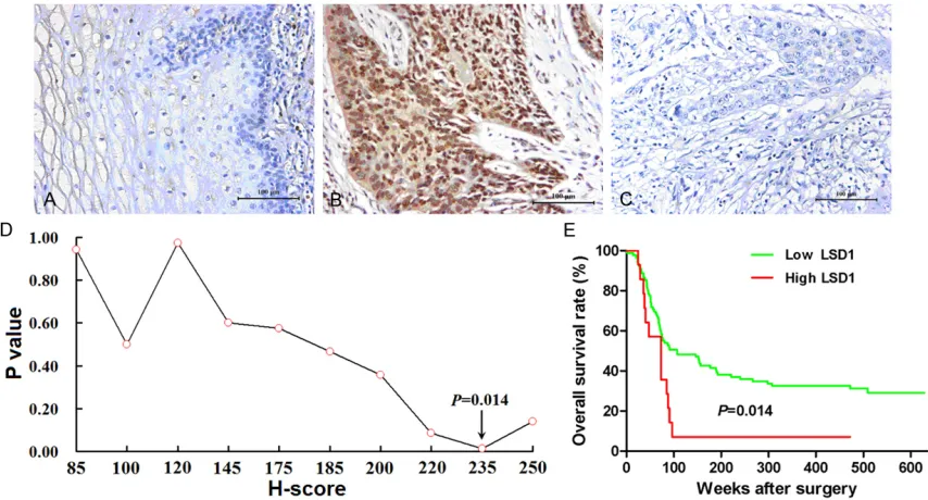
[image:3.612.93.520.73.303.2]As shown in Figure 1, positive LSD1 immuno-chemical staining was predominantly observed on the nuclei and cytoplasm of esophageal
Figure 1. LSD1 expression in human esophageal cancer and its prognostic value. A. LSD1 was negatively or weakly expressed on normal esophagus tissues. B. LSD1 was positively expressed on the nuclei and the cytoplasm in esophageal cancer cells. C. Negative control. D. The minimum P-value seek was performed by using the log-rank survival analysis, and when the cut-off value of H-score = 235 was selected, the overall survival rate of patients with lower LSD1 expression (H-score ≤ 235) was better than those with higher LSD1 expression (H-score > 235) (P = 0.014, Hazard Ratio = 2.12, 95% CI: 1.15-3.89. E. The survival curve was shown when the cut-off value H-score
cancer cells (Figure 1B), while none or very weak staining was found in normal esophageal tissues (Figure 1A). We could found LSD1 immunostaining could be found in all the speci-mens of esophageal cancer, and according to the evaluation of LSD1 immunostaining inten-sity by H-score method, we found and charac-terized that, 89 out of 103 (86.4%) cases of esophageal cancer patients with low intensity (H-score ≤ 235), and 14 out of 103 (13.6%) cases of esophageal cancer patients with high intensity (H-score > 235).The cut-off value of
H-score = 235 was selected according to the minimum P-value seek in the log-rank survival analysis (Figure 1D and 1E), which has been described in the literatures and our previous studies [3, 4, 14-16].
LSD1 immunochemical staining in relation to patients’ clinical data and prognoses
As shown in Table 1, the intensity of LSD1 immunostaining was significantly correlated to the tumor size (P = 0.013), nodal metastasis (P
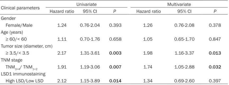
= 0.002), distant metastasis (P = 0.025), and TNM stage (P = 0.010), whereas it was not cor-related to patient’s gender, age and tumor inva-sion depth. When the cutoff value of H-score = 235 was selected, the overall survival rate of patients with low LSD1 expression was better than those with high LSD1 expression (P = 0.014, Hazard Ratio = 2.12, 95% CI: 1.15-3.89, Figure 1D and 1E). We also showed that the tumor size (P = 0.003) as well as the TNM stage (P = 0.007) was also a useful prognostic predic-tor for esophageal cancer (Table 2). However, when the gender, age, tumor size, TNM stage and LSD1 expression level were involved in the
multivariate proportional hazards regression analysis in a Cox’s model, we showed that the tumor size (P = 0.013) and the TNM stage (P = 0.032) could be used as independent risk fac-tors to predict patient’s postoperative progno-sis, while LSD1 expression level as well as other factors could not independently predict esoph-ageal cancer patient’s outcome (Table 2). Discussion
Epigenetic alteration is a current hallmark in cancer biology. The epigenetic mechanisms role importantly in carcinogenesis and cancer progression, which include DNA methylation, histone modifying enzymes and their histone modifications [17]. As of now, many attractive targets from the epigenetic research of cancer biology have been developed as novel prognos-tic biomarkers, and further explored for drug development [18]. It has been demonstrated that some histone-modifying enzymes, such as lysine-specific demethylase 1 (LSD1) and his -tone deacetylase 2 (HDAC2), are involved in the initiation and progression of many human can-cers [19]. In the present study, we examined LSD1 expression in esophageal cancer tissues by using immunohistochemistry, and further investigated its relationship to patient’s clinical parameters and post-operative prognosis. Our immunohistochemistry study results showed that positive LSD1 immunochemical staining could be found in all the specimens of esophageal cancer, while it was negative or very weak in adjacent normal esophagus tis-sues. Based on the classification of low/high expression levels of LSD1 in esophageal can-Table 2. Prognostic factors in Cox’s proportional hazards model
Clinical parameters Univariate Multivariate
Hazard ratio 95% CI P Hazard ratio 95% CI P
Gender
Female/Male 1.24 0.76-2.04 0.393 1.26 0.76-2.08 0.378 Age (years)
≥ 60/< 60 1.11 0.70-1.76 0.658 1.05 0.65-1.70 0.847
Tumor size (diameter, cm)
≥ 3.5/< 3.5 2.17 1.31-3.61 0.003 1.98 1.16-3.37 0.013
TNM stage
TNM3+4/ TNM1+2 1.91 1.19-3.06 0.007 1.74 1.05-2.88 0.032
LSD1 immunostaining
High LSD/Low LSD 2.12 1.15-3.89 0.014 1.34 0.69-2.60 0.397
[image:4.612.91.524.84.245.2]cer tissues, we found that the LSD1 expression in esophageal cancer was significantly corre -lated to the tumor size, nodal metastasis, dis -tant metastasis, and TNM stage, whereas it was not correlated to patient’s gender, age and tumor invasion depth. Shinya et al. [20] sug-gested that the over-expression of LSD1 con-tributes to human carcinogenesis through chro-matin regulation in various cancers. Yu et al.
[21] also showed that LSD1 expression in human esophageal cancer significantly associ -ated with nodal metastasis status. In human hepatocellular carcinoma, over-expression of LSD1 significantly associated with tumor stage and tumor grade [11]. And in human colon can-cer, the LSD1 expression level significantly associated with tumor stage and distant metas-tasis [12]. Thus, in combination with the results from other groups, our results suggested that LSD1 expression in esophageal cancer was involved in the initiation and progression of this malignancy, particularly contributed to the nodal metastasis and the distant metastasis. The prognostic value of LSD1 has been a well-concerning focus in human cancers. Yuan et al.
[22] reported that the high expression of LSD1 associates with cancer cell proliferation and unfavorable prognosis in tongue cancer. Over-expression of LSD1 in hepatocellular carcino-ma tissues could also predict poorer outcome of the patients [11]. Ding et al. [12] showed that the colon cancer patients with lower LSD1 expression favor better post-operative progno-ses than the patients with higher LSD1 expres-sion. In our present study, we found that the LSD1 expression level as well as tumor size, tumor stage could predict patients’ survival by using the log-rank survival analysis, and the esophageal cancer patients with lower LSD1 expression represented better prognoses than the patients with higher LSD1 expression. However, in the COX model analysis, we could only found that tumor size and TNM stage could be used as the independent risk factors for pre-dicting esophageal cancer patients’ survival, while the LSD1 expression level couldn’t. In future, an expanded sample of esophageal cancer was needed to further investigate its application in predicting prognosis as an inde-pendent risk factor in this malignancy.
It is needed to be concerned that LSD1 roles importantly in the alteration of cancer cell phe-notype, especially in the regulation of epitheli-al-to-mesenchymal transition (EMT) [23]. In
human colon cancer, the LSD1 expression was negatively associated with E-cadherin expres-sion, suggesting an essential role of LSD1 in regulating the EMT of cancer cells [12]. Lin et al. [24] demonstrated that the transcription factor Snai1 could interact with and recruit LSD1 to epithelial gene promoters, and in the absence of LSD1, Snai1 failed to repress E-cadherin, indicating that the LSD1 could be a potential therapeutic target for prevention of EMT-associated tumor invasion. We have previ-ously demonstrated that the EMT status in human esophageal cancer significantly associ -ated with invasion, metastasis and prognosis (data not shown). In future, the detailed mecha-nism of LSD1 in regulating the EMT of esopha-geal cancer cells might help us to further reveal the aggressive nature of this malignancy. Acknowledgements
This work was supported by grants from the National Natural Science Foundation of China (NSFC) (81171653; 81301960), Natu- ral Science Foundation of Jiangsu Provin- ce (BK2011246 and BK2011247), Jiangsu Provincial Innovation Award BC2012093 by the Bureau of Science and Technology of Jiangsu Province, Sanitation Bureau Guidance Project of Changzhou (WZ201303) and Jiangsu Pro-vincial Health Bureau of medical research (H201350).
Disclosure of conflict of interest
None.
Address correspondence to: Dr. Changping Wu or Dr. Jingting Jiang, Department of Tumor Biological
Treatment and Oncology, The Third Affiliated Hospital of Soochow University, Changzhou 213003,
Jiangsu, China. E-mail: [email protected] (CPW); [email protected] (JTJ)
References
[1] Siegel R, Naishadham D and Jemal A. Cancer statistics, 2013. CA Cancer J Clin 2013; 63: 11-30.
[2] Chen L, Luo G, Tan Y, Wei J, Wu C, Zheng L, Zhang X and Xu N. Immunolocalisation of tis-sue factor in esophageal cancer is correlated with intratumoral angiogenesis and prognosis of the patient. Acta Histochem 2010; 112: 233-239.
MT2-MMP correlates positively to angiogenesis of human esophageal cancer. Anticancer Res 2010; 30: 4363-4368.
[4] Chen L, Sun J, Wu H, Zhou S, Tan Y, Tan M, Shan B, Lu B and Zhang X. B7-H4 expression associates with cancer progression and pre-dicts patient’s survival in human esophageal squamous cell carcinoma. Cancer Immunol Immunother 2011; 60: 1047-1055.
[5] Stoner GD and Gupta A. Etiology and chemo-prevention of esophageal squamous cell carci-noma. Carcinogenesis 2001; 22: 1737-1746. [6] Berger AC, Farma J, Scott WJ, Freedman G,
Weiner L, Cheng JD, Wang H and Goldberg M. Complete response to neoadjuvant chemora-diotherapy in esophageal carcinoma is
associ-ated with significantly improved survival. J Clin
Oncol 2005; 23: 4330-4337.
[7] Shi Y, Lan F, Matson C, Mulligan P, Whetstine JR, Cole PA, Casero RA and Shi Y. Histone de-methylation mediated by the nuclear amine oxidase homolog LSD1. Cell 2004; 119: 941-953.
[8] Chen Y, Yang Y, Wang F, Wan K, Yamane K,
Zhang Y and Lei M. Crystal structure of human
histone lysine-specific demethylase 1 (LSD1).
Proc Natl Acad Sci U S A 2006; 103: 13956-13961.
[9] Amente S, Lania L and Majello B. The histone LSD1 demethylase in stemness and cancer transcription programs. Biochim Biophys Acta 2013; 1829: 981-986.
[10] Kahl P, Gullotti L, Heukamp LC, Wolf S, Fried -richs N, Vorreuther R, Solleder G, Bastian PJ,
Ellinger J, Metzger E, Schule R and Buettner R. Androgen receptor coactivators lysine-specific
histone demethylase 1 and four and a half LIM domain protein 2 predict risk of prostate can-cer recurrence. Cancan-cer Res 2006; 66: 11341-11347.
[11] Zhao ZK, Yu HF, Wang DR, Dong P, Chen L, Wu
WG, Ding WJ and Liu YB. Overexpression of
ly-sine specific demethylase 1 predicts worse
prognosis in primary hepatocellular carcinoma patients. World J Gastroenterol 2012; 18: 6651-6656.
[12] Jie D, Zhongmin Z, Guoqing L, Sheng L, Yi Z, Jing W and Liang Z. Positive expression of LSD1 and negative expression of E-cadherin correlate with metastasis and poor prognosis of colon cancer. Dig Dis Sci 2013; 58: 1581-1589.
[13] Greene FL, Page DL and Fleming ID, et al. AJCC cancer staging manual. 6th edition. New York: Springer-Verlag; 2002.
[14] Chen L, Zhou Q, Xu B, Liu J, Shi L, Zhu D, Wu C and Jiang J. MT2-MMP expression associates with tumor progression and angiogenesis in human lung cancer. Int J Clin Exp Pathol 2014; 7: 3469-3477.
[15] Zhu X, Zhang J, Zhuang P, Zhu H, Zhang W, Xiong Y, Wu W, Wang L, Tang Z and Sun H. High expression of macrophage colony-stimulating factor in peritumoral liver tissue is associated with poor survival after curative resection of hepatocellular carcinoma. J Clin Oncol 2008; 26: 2707-2716.
[16] Galon J, Costes A, Sanchez-Cabo F, Kirilovsky
A, Mlecnik B, Lagorce-Pages C, Tosolini M,
Ca-mus M, Berger A, Wind P, Zinzindohoue F, Bru -neval P, Cugnenc PH, Trajanoski Z, Fridman WH and Pages F. Type, density, and location of immune cells within human colorectal tumors predict clinical outcome. Science 2006; 313: 1960-1964.
[17] Lund AH and van Lohuizen M. Epigenetics and
cancer. Genes Dev 2004; 18: 2315-2335. [18] Klose RJ and Zhang Y. Regulation of histone
methylation by demethylimination and de-methylation. Nat Rev Mol Cell Biol 2007; 8: 307-318.
[19] Derr RS, van Hoesel AQ, Benard A,
Goossens-Beumer IJ, Sajet A, Dekker-Ensink NG, de Kruijf
EM, Bastiaannet E, Smit VT, van de Velde CJ
and Kuppen PJ. High nuclear expression levels of histone-modifying enzymes LSD1, HDAC2
and SIRT1 in tumor cells correlate with de-creased survival and inde-creased relapse in breast cancer patients. BMC Cancer 2014; 14: 604.
[20] Hayami S, Kelly JD, Cho HS, Yoshimatsu M,
Unoki M, Tsunoda T, Field HI, Neal DE, Yamaue H, Ponder BA, Nakamura Y and Hamamoto R. Overexpression of LSD1 contributes to human carcinogenesis through chromatin regulation in various cancers. Int J Cancer 2011; 128: 574-586.
[21] Yu Y, Wang B, Zhang K, Lei Z, Guo Y, Xiao H,
Wang J, Fan L, Lan C, Wei Y, Ma Q, Lin L, Mao C, Yang X, Chen X, Li Y, Bai Y and Chen D. High
expression of lysine-specific demethylase 1
correlates with poor prognosis of patients with esophageal squamous cell carcinoma. Bio-chem Biophys Res Commun 2013; 437: 192-198.
[22] Yuan C, Li Z, Qi B, Zhang W, Cheng J and Wang Y. High expression of the histone demethylase LSD1 associates with cancer cell proliferation and unfavorable prognosis in tongue cancer. J Oral Pathol Med 2014; Jul 18. doi: 10.1111/ jop.12220. [Epub ahead of print]. PMID: 25040359.
[23] Lin Y, Dong C and Zhou BP. Epigenetic regula-tion of EMT: the Snail story. Curr Pharm Des 2014; 20: 1698-1705.