Case Report
Epithelioid angiosarcoma of the ilium: a case report
Mingxia Chen1,2, Wei Zhang2, Jianli Qu2, Lei Li2, Qing Sun1
1Department of Pathology, Qian-Fo-Shan Hospital Affiliated Shandong University, 16766 Jingshi Road, Jinan
250014, China; 2Department of Pathology, Yatai Yaitaishan Hospital, 91 Jiefang Road, Yatai 264001, China Received October 16, 2014; Accepted December 1, 2014; Epub December 1, 2014; Published December 15, 2014
Abstract: Bone epithelioid angiosarcoma (EA) is rare and characterized by large, mildly to moderately pleomorphic epithelioid cells, with abundant eosinophilic cytoplasm, vesicular nuclei, and prominent nucleoli. The tumors may arise in various locations in bone and the patients may present with unifocal or multifocal osseous disease. We present a unifocal lesion case of EA of the ilium in a 62-year-old woman. A needle biopsy of the ilium was performed and first diagnosed poorly differentiated adenocarcinoma based on CKpan and CK18 immunopositivity. The tumor was treated initially with curettage followed by chemotherapy. The final diagnosis on the surgical specimen was epithelioid angiosarcoma.
Keywords: Epithelioid angiosarcoma, bone
Introduction
Bone epithelioid angiosarcoma (EA) is one of the intraosseous epithelioid vascular tumors. It is a rare high-grade sarcoma of intraosseous vascular endothelial origin and is a rare variant of angiosarcoma [1]. It can affect any portion of the skeleton. The long tubular bones of the lower extremities are the most commonly in- volved [2, 3]. Patients may present with unifo-cal or multifounifo-cal osseous disease. The tumor cells frequently express epithelial markers as well as endothelial cell markers, which may lead to a misdiagnosis of metastatic carcino-ma, especially when the poorly differentiated tumor cells were happened to be encountered with the needle biopsy specimen.
Case report
A 62 years old female presented persistent pain in the low back and buttocks last month after sprain, exacerbation of sitting and walking. In addition, the right buttock pain was more serious than the left. An X-ray revealed an osteolytic lesion (Figure 1A), which showed a high-low mixed signal in both T1WI and T2WI, as well as cystic long T1, long T2 signal on MRI images in the right ilium and adjacent sacroiliac joint. On fat suppressed image, the lesion was
hyperintense (Figure 1B). Computed tomogra-phy scan (CT) display, lesions were osteolytic, border is not clear, the size of 10.2 cm × 6.2 cm × 5.3 cm, extending into the adjacent soft tissue, erosion of sacroiliac joint surface (Figure 1C). B ultrasound examination has nothing to do with the internal organs, chest computer tomography in normal.
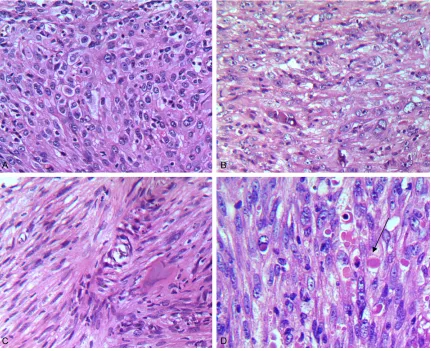
intra- and extra- cellular eosinophilic hyaline droplets or globules were observed (Figure 2D). The stroma varied from thin fibrovascular con-nective tissue to densely collagenous areas
[image:2.612.93.522.72.189.2]associated with multifocal chronic inflammato-ry cell infiltrates. Extensive hemorrhage and hemosiderin deposits, focal necrosis, and cys-tic changes were also distinct.
Figure 1. A. Radiographs showing the osteolytic lesion in the right ilium. C. Computed tomography scan revealing an osteolytic lesion with ill-defined margins and extending into the proximal soft tissue. B. Magnetic resonance image revealing a cystic, destructive soft tissue lesion of the right ilium.
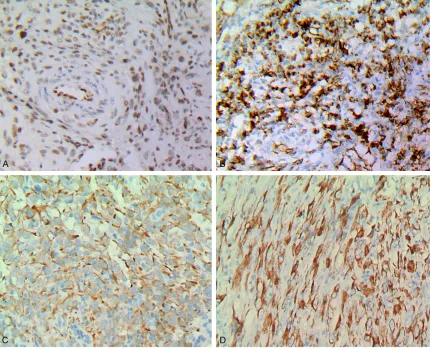
[image:2.612.92.523.251.602.2]The results of immunohistochemistry excised tumors displayed, vimentin and the friend leu-kemia integration (FLI-1) diffuse staining (Fi- gure 3A). Some tumor cells expressed CD31 (Figure 3B). Keratin AE1/AE3 and CK18 were focal positive (Figure 3C, 3D). However, CD34, epithelial membrane antigen (EMA), smooth muscle actin (SMA), desmin, S-100 protein and HMB45 were negative. Mitosis is high, Ki-67 is about 50%.
According to the clinical features, image data, especially the histological and immunohisto-chemical results, the diagnosis of intraosseous epithelioid angiosarcoma was confirmed. Discussion
Bone epithelioid angiosarcoma (EA) is a rare primary malignant bone tumor of vascular ori-gin, the incidence rate of less than 1% [4]. More common in young and old, sixty and eighty are two small peaks [2, 5]. Incidence of males is
slightly higher than that of females. In addition, tumors tend to involve lower extremity long bone, such as the femur and tibia [2, 6]. Pain is the most common complaint. X-ray characteris-tics of bone angiosarcoma are not specific, ill-defined osteolytic lesions are the prominent appearance, commonly involving the adjacent soft tissue.
[image:3.612.92.523.73.422.2]Epithelioid angiosarcoma is a highly malignant tumor under the microscope often has flakes or wide distribution of cords, cells were large and round, pleomorphic epithelioid, with abundant eosinophilic cytoplasm, vesicular nuclei and sig- nificant nucleolus, and exhibit multiple and hi- gh mitotic proliferation activity. Epithelioid angi- osarcoma of bone matrix, in addition to seeing the reaction of the bone, but also can have a variety of inflammatory cells, including chronic inflammatory cells, neutrophils and eosinophils [7-9]. The classic case, you can often see the blood-filled cavities and vacuole-like structures
located within the cytoplasm. However, these appearances may not be easily identified if the tumor cells are poorly differentiated. In particu-lar, when a completely epithelioid focus is en- countered with scant biopsy specimen, it is easily misdiagnosed.
Most of angiosarcoma is positive immunohisto-chemical CD31 expression [2, 9, 10], so that CD31 is considered the most sensitive and specific for all types of angiosarcoma of con-ventional markers. CD34 is highly sensitive, is expressed in approximately 90% of the heman-gioma. However, CD34 is rather non-specific [11]. FLI-1 is both a sensitive and specific mark-er of blood vessels, which is supmark-erior to CD31, CD34, and von Willebrand factor [12] as a mar- ker of tumor blood vessels as the core is still in its coloring with respect to the membrane (CD- 31 and CD34) or cytoplasm (von Willebrand fa- ctor) coloring, FLI-1 is easier to recognize [12]. To avoid misdiagnosis, requires the use of mul-tiple markers for diagnosis of vascular endothe-lial sarcoma, because there are a significant proportion of metastatic cancer can be CD34-positive (15%) and CD31-CD34-positive (38%) [13].
In the present case, there was no irregularly anastomosing vessel formation and the tumor cells were poorly differentiated. In some areas the tumor cells were positive for CD31 and in the other areas negative. When the poorly dif-ferentiated tumor cells were negative for CD31, Fli-1 was positive. When CKpan and CK18 posi-tive happened to be encountered with the nee-dle biopsy specimen, it is easily to make an erroneous diagnosis. Therefore, under this cir-cumstance, the utility of the other endothelial markers, such as vwf and Factor VIII, especially Fli-1 is very helpful for the differential diagno-sis. Although the significance was not clear, the presence of intra- and extra-cellular eosinophil-ic hyaline droplets or globules was another fea-ture of this case. Maybe they can provide an evident for diagnosis of bone angiosarcoma. The differential diagnosis includes other prima-ry vascular tumors, especially Epithelioid hema- ngioendothelioma (EHE) and pseudomyogenic hemangioendothelioma (PMH) as well as meta-static carcinoma. EHE may show focal high-gr- ade areas, with cellular atypia and a sheeted architecture, but most cells reside in small ne- sts and trabeculae. The mitotic count is rather low (2 mitoses per 10 high-power fields), the
epithelioid angiosarcoma has a greater degree of cytologic atypia and mitoses, focal areas of vessel formation, and a sheeted growth pattern comprising most of the malignancy.
Like epithelioid angiosarcoma, PMH may be composed of epithelioid cells with abundant eosinophilic cytoplasm, vesicular nuclei and pr- ominent nucleolus although more spindle cells
showing generally mild nuclear atypia and little mitotic activity was also noted. It is commonly composed of plump spindle cells. EA and PMH should be based within the cytoplasm of tumor cells without cavities, no vascular channels li- ned with epithelium and low proliferation index (Ki-67) to distinguish [14, 15].
Treatment of angiosarcoma of bone usually includes wide resection, radiotherapy and ch- emotherapy.
In conclusion, bone epithelioid angiosarcoma is a rare vascular tumor. When poorly differenti-ated tumor cells were met with biopsy speci-mens, it is easy to be misdiagnosed. Therefore, H & E stained sections initial evaluation and joint use of multiple antibodies is a necessary condition for the diagnosis of epithelioid ang- iosarcoma.
Acknowledgements
This study was supported by the National Na- tural Science Foundation of China (No. 81272420), the Scientific and Technological Development Projects in Shandong Province of China (No. 2011GSF11838), Shandong Pro- vince Natural Science Foundation (No. ZR2012HM085), and the Scientific and Technological Development Projects of Jinan City (No. 201202039).
Disclosure of conflict of interest
None.
Address correspondence to: Dr. Qing Sun, Depart- ment of Pathology, Qian-Fo-Shan Hospital Affiliated Shandong University, 16766 Jingshi Road, Jinan 250014, China. Tel: 531-89268155; Fax: +86-531-89263647; E-mail: [email protected]
References
[2] Verbeke SL, Bertoni F, Bacchini P, Sciot R, Fle- tcher CD, Kroon HM, Hogendoorn PC and Bovee JV. Distinct histological features charac-terize primary angiosarcoma of bone. Histo-pathology 2011; 58: 254-264.
[3] Palmerini E, Maki RG, Staals EL, Alberghini M, Antonescu CR, Ferrari C, Ruggieri P, Mavrogenis A, Bertoni F, Cesari M, Paioli A, Marchesi E, Picci P and Ferrari S. Primary Angiosarcoma of Bone: A Retrospective Analysis of 60 Patients From 2 Institutions. Am J Clin Oncol 2013; [Epub ahead of print].
[4] Deshpande V, Rosenberg AE, O’Connell JX and Nielsen GP. Epithelioid angiosarcoma of the bone: a series of 10 cases. Am J Surg Pathol 2003; 27: 709-716.
[5] Saglik Y, Yildiz Y, Atalar H and Basarir K. Pri-mary angiosarcoma of the fibula: a case re -port. Acta Orthop Belg 2007; 73: 799-803. [6] Mortazavi SM, Wenger D, Asadollahi S, Shariat
Torbaghan S, Unni KK and Saberi S. Periosteal osteoblastoma: report of a case with a rare histopathologic presentation and review of the literature. Skeletal Radiol 2007; 36: 259-264. [7] Baliaka A, Balis G, Michalopoulou-Manolout-siou E, Papanikolaou A and Nikolaidou A. Pri- mary angiosarcoma of bone. A case report. Hippokratia 2013; 17: 180-182.
[8] Hasegawa T, Fujii Y, Seki K, Yang P, Hirose T, Matsuzaki K and Sano T. Epithelioid angiosar -coma of bone. Hum Pathol 1997; 28: 985-989.
[9] Hart J and Mandavilli S. Epithelioid angiosar -coma: a brief diagnostic review and differential diagnosis. Arch Pathol Lab Med 2011; 135: 268-272.
[10] Chen Y, Shen D, Sun K, Bao D, Song Q, Wang G, Chen D, Yan T and Guo W. Epithelioid angio-sarcoma of bone and soft tissue: a report of seven cases with emphasis on morphologic diversity, immunohistochemical features and clinical outcome. Tumori 2011; 97: 585-589. [11] Yang Z, Tao H, Ye Z and Yang D. Multicentric
epithelioid angiosarcoma of bone. Orthopedics 2012; 35: e1293-1296.
[12] Folpe AL, Chand EM, Goldblum JR and Weiss SW. Expression of Fli-1, a nuclear transcription factor, distinguishes vascular neoplasms from potential mimics. Am J Surg Pathol 2001; 25: 1061-1066.
[13] Gill R, O’Donnell RJ and Horvai A. Utility of im -munohistochemistry for endothelial markers in distinguishing epithelioid hemangioendo-thelioma from carcinoma metastatic to bone. Arch Pathol Lab Med 2009; 133: 967-972. [14] Hornick JL and Fletcher CD. Pseudomyogenic
hemangioendothelioma: a distinctive, often multicentric tumor with indolent behavior. Am J Surg Pathol 2011; 35: 190-201.