Evolutionary Relationship of Wheat Protein Disulphide
Isomerase (PDI) Gene Promoter Sequence Based on
Phylogenetic Analysis
Arun Prabhu Dhanapal1,2
1Plant Genetic Resources, Scuola Superiore Sant’ Anna, Rome, Italy; 2Division of Plant Sciences, University of Missouri, Columbia, USA.
Email: [email protected], [email protected]
Received December 12th,2011; revised January 10th, 2012; accepted January 24th, 2012
ABSTRACT
Protein disulphide isomerase (PDI) is an oxidoreductase enzyme abundant in the endoplasmic reticulum (ER). In plants, PDIs have been shown to assist the folding and deposition of seed storage proteins during the biogenesis of protein bodies in the endosperm. Cloning and characterization of the complete set of genes encoding PDI and PDI like proteins in bread wheat (Triticum aestivum cv. Chinese Spring) and the comparison of their sequence, structure and expression with homologous genes from other plant species were reported in our previous publications. Promoter sequences of three homoeologous genes encoding typical PDI, located on chromosome group 4 of bread wheat, and PDI promoter sequence analysis of Triticum urartu, Aegilops speltoides and Aegilops tauschii had also been reported previously. In this study, we report the isolation and sequencing of a ~700 bp region, comprising ~600 bp of the putative promoter region and 88 bp of the first exon of the typical PDI gene, in five accessions each from Triticum urartu (AA), Aegilops speltoides (BB) and Aegilops tauschii (DD). Sequence analysis indicated large variation among sequences belonging to the different genomes, while close similarity was found within each species and with the corresponding homoeologous PDI sequences of Triticum aestivum cv. CS (AABBDD) resulting in an overall high conservation of the sequence con- ferring endosperm-specific expression.
Keywords: Protein Disulfide Isomerase (PDI) Promoter; Wheat; Phylogenetic Analysis
1. Introduction
Wheat is adapted to temperate regions of the world and was one of the first crops to be domesticated. Spread over all continents, it is today one of the most important food source for human beings. Bread wheat belongs to the tribe Triticeae, and has three genomes A, B and D, each organised in seven homoeologous chromosome groups. The diploid progenitors of the three genomes A, B, and D have been identified in Triticum urartu, Ae- gilops speltoides and Aegilops tauschii, although the progenitor of B genome is still matter of debate [1]. Throughout their evolutionary history, multiple poly- ploidization events occurred between species of the
Triticum and Aegilops genera and human manipulation of wild species led to the domestication of different culti- vated lineages [2-5]. The first hybridisation event com- bined the genome of the wild species Triticum urartu
(AA genome) and that of Aegilops speltoides (BB ge- nome) to give rise to the tetraploid (AABB) Triticum turgidum ssp. dicoccoides. Subsequent molecular studies
identified Triticum urartu as the donor of the A genome of emmer wheat (AABB), durum wheat (AABB), and common wheat (AABBDD) [6-10]. The second poly- ploidisation event occurred between the new tetraploid species (AABB) and diploid Aegilops tauschii (DD ge- nome) to give rise to hexaploid wheat (AABBDD) Triti- cum aestivum [8,11]. A recent study revealed lower lev- els of polymorphism in the D genome than in the A and B genome, indicating that possibly the gene flow from tetraploid to hexaploid species was more frequent than that from the diploid Aegilops tauschii [12].
for >90% of the total protein content in the wheat grain. Different quality of grain storage proteins produced by the same genotype under different environmental condi- tion has stimulated research on protein folding and as- sembling.
Secretory protein folding and disulphide bond forma- tion takes place within the Endoplasmic reticulum (ER lumen), but the precise mechanisms involved, and the role of other proteins such as molecular chaperones, are not fully understood [13]. PDI plays an important role in assisting protein folding and assembly, catalyzing thiol- disulfide oxidation, reduction and isomeration, this latter occurring directly by intramolecular disulfide rearrange- ment or through cycles of reduction and oxidation [14]. During the maturation of the secretory proteins, disulfide bonds cross-linking specific cysteines are added to stabi- lize a protein or to join covalently different polypeptides. These bonds are crucial for the stability of the final pro- tein structure, thus mispairing of cysteine residues can prevent proteins from attaining their native conformation and lead to misfolding [15].
Typical PDI is the most prominent member of a family of related proteins (PDI-like) characterised by one, two or three thioredoxin-like active domains [16]. It has been cloned and sequenced in many plant species, such as alfalfa [17], barley [18], maize [19], castor bean [20], soyabean [21-24], common and durum wheat [25,26]. A detailed knowledge of the complexity and diversity of the genes encoding PDI and PDI-like proteins in Arabi- dopsis thaliana, wheat and other plant species was de- scribed by [27,28].
Wheat genes coding for typical PDI in bread wheat have been located in homoeologous chromosome group four [29]. Analyses performed on 23 species of Triticum
and Aegilops [30], indicated that PDI restriction frag- ments were highly conserved within each species and confirmed that plant PDI is encoded either by one or few copy sequences, respectively in diploid and polyploid species. The nucleotide sequences of the three genes lo- cated respectively on genomes A, B, and D (designed as GPDI-4A, GPDI-4B and GPDI-4D) were 3561 bp, 3527 bp and 3466 bp long. The comparison of typical PDI gene sequences of wheat, rice and Arabidopsis showed a significant conservation of the exon/intron structure across the three species [31]. More detailed study on the complexity and diversity of the genes encoding PDI and PDI-like proteins in wheat and other plant species also showed a significant conservation of the exon/intron structure [28].
The expression analysis of the typical PDI homoeolo- gous genes located on chromosomes 4A, 4B and 4D of bread wheat cv. Chinese Spring (CS) (Ciaffi et al.,2006) showed that the PDI transcripts, although constitutively
present at a low level in all the analyzed tissues, are equally abundant in the developing caryopses but are differentially expressed in spikelets, roots and leaves [25, 31]. The PDI-4A transcription was higher in spikelets, that of PDI-4B were higher in roots while the PDI-4 D transcripts were more abundant in leaves. The transcript- tion levels of the three genes were higher in the early stage of seed development (6 - 14 DAA) and decreased during middle to late stage of (18 - 34 DAA) grain filling [31]. Within the upstream putative promoter region of the three homoeologous genes cloned from bread wheat cv CS, respectively 1352 bp for PromPDI-4A, 1370 bp for PromPDI-4B and 1292 bp for PromPDI-4D long, several cis-acting elements involved in endosperm specific ex- pression were detected, consistently with the higher PDI expression detected in the kernels. PDI promoter se-quence analysis of Triticum urartu, Aegilops speltoides
and Aegilops tauschii was given by [32].This paper re-ports variability in a ~700 bp region, comprising ~600 bp of the 5’ upstream putative promoter region and 88 bp of the first exon of the typical PDI gene from the diploid species Triticum urartu (AA) Aegilops speltoides (BB) and Aegilops tauschii (DD). For simplicity we will fur- ther refer to the amplified ~700 bp region shortly as the ~700 bp target region.
2. Materials and Methods
2.1. Plant Material and DNA Extraction
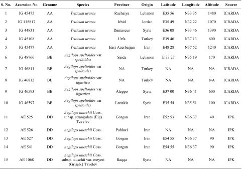
A total of 15 accessions, 5 each from Triticum urartu
(AA), Aegilops speltoides (BB) and Aegilops tauschii
(DD) were used in this study (Table 1). Eight plants per accessions were grown and analysed for a total of 120 plants. Flag leaves were collected at heading stage from plants grown in green house (January-June 2008), im- mediately frozen in liquid nitrogen and kept at −80˚C until use. About 200 mg of leaf tissue was ground in liq- uid nitrogen and genomic DNA was extracted using Sigma Gen Elute Plant Genomic DNA Kit (G2N-350, Sigma Aldrich, St. Louis, Mo.).
2.2. Primers Design and PCR Amplification
Table 1. Accession numbers and geographical origin of the wheat samples used in this study.
S. No. Accession No. Genome Species Province Origin Latitude Longitude Altitude Source
1 IG 45475 AA Triticum urartu Rachaiya Lebanon E35 56 N33 35 1480 ICARDA
2 IG 115817 AA Triticum urartu Irbid Jordan E35 49 N32 22 1070 ICRADA
3 IG 44831 AA Triticum urartu Damascus Syria E36 08 N33 46 1390 ICARDA
4 IG 45108 AA Triticum urartu Urfa Turkey E39 46 N37 13 600 ICARDA
5 IG 45477 AA Triticum urartu East Azerbaijan Iran E48 28 N37 52 1240 ICARDA
6 IG 48766 BB Aegilops speltoidesspeltoides var Saida Lebanon E 33 27 N35 19 170 ICARDA
7 IG 46811 BB Aegilops speltoidesspeltoides var NA Turkey NA NA NA ICRADA
8 IG 46812 BB Aegilops speltoidesligustica var NA Turkey NA NA NA ICARDA
9 IG 46593 BB Aegilops speltoidesligustica var Aleppo Syria E37 00 N36 41 600 ICARDA
10 IG 46597 BB Aegilops speltoidesspeltoides var Lattakia Syria E35 54 N35 51 100 ICARDA
11 AE 525 DD subsp. strangulata (Eig) Aegilops tauschii Coss. Tzvelev
Gorgan Iran E52 53 N36 37 40 IPK
12 AE 526 DD Aegilops tauschii Coss. Pahlavi Iran NA NA NA IPK
13 AE 527 DD Aegilops tauschii Coss. Gorgan Iran E54 55 N36 37 90 IPK
14 AE 541 DD Aegilops tauschii Coss. Gorgan Iran E54 55 N36 37 90 IPK
15 AE 1068 DD
Aegilops tauschii Coss. subsp. tauschii var. meyeri
(Griseb.) Tzvelev
Raqqa Syria NA NA NA IPK
species. Further amplification reactions were carried out in the 120 plants using the primer pair PDIAPDF2- PDIAPDR1 and directly sequenced using the PDIAPDF2 primer (Table 2).
PCR reaction mixture included 10 ng/μl genomic DNA, 0.20 mM dNTPs 5 ul, 0.05 units/µl of Taq 0.50 µl (go-taq, Promega), 0.40 μM primer 2.0 µl (0.20 µM per each), 5× buffer 10 µl and 25.5 µl of ddH2O used per reaction (final volume of 50 μl). PCR condition included initial denaturation step at 95˚C for 3 min, then 32 cycles of (95˚C for 1min, 59˚C for 35 sec, 72˚C for 1 min) and final elongation at 72˚C for amplification of the se- quences from the A, B and D Genome.
2.3. Cloning and Sequencing
One conserved primer pair (PDIAPDF2-PDIAPDR1) and one genome specific primer pair (Table 2) were used to amplify, clone and sequence the DNA extracted from one plant each of the 3 wild relatives. PCR products of ex- pected size were excised from the gel, purified using the High Pure Purification kit (Roche) according to manu- facturer’s instructions, and cloned into the pGEM-T easy
plasmid vector (Promega). Plasmids were transformed by heat shock into Escherichia coli strain DH5α. Bacteria
were plated onto LB medium containing ampicillin, X- Gal and IPTG, and recombinant plasmids were identified by blue/white screening. For each primer combination two independent PCR reactions were performed and a total of 6 clones were sequenced. Plasmid DNA for se- quencing reaction was prepared from 3 ml overnight cultures using a plasmid miniprep kit (Qiagen). Sequenc- ing was performed on both strands by the ABI PRISM 377 capillary sequencer (PE Applied Biosystem) using an ABI Prism Dye Terminator sequencing kit (PE Ap- plied Biosystem) and sequenced both with vector and sequence specific primers. The target region was further analysed in all 120 plants using the conserved primer pair PDIAPDF2-PDIAPDR1 and directly sequenced.
2.4. Data Analysis
Table 2. List of primers used for the isolation of PDI partial promoter analysis.
PCR Target for Direct Sequencing Primer Forward Reverse
PDIAPDF2 5’-ACTCCAAATTTGGAACGGG-3’
PDIAPDR1 5’-GGTGAGCACTGCCTCGGG-3’
PCR Target for Cloning Primer Forward Reverse
PDIAPDF2 5’-ACTCCAAATTTGGAACGGG-3’
PDIAPDF3 5’-CAAACATAAATATTTCCGCCA-3’
PDIAPDF5 5’-TCCTAGTCCTGAACCGGCAAC-3’
PDIAPDR1 5’-GGTGAGCACTGCCTCGGG-3’
PDIAPDR5 5’-GAAGTTGTCGGCGTGCA-3’
PDIAPDR6 5’-ATGAAGGGGTGCTTGGCGAT-3’
sequences revealed that plants within accessions of all three wild species were identical. So among 120 se- quences analyzed, a total of 15 sequences (one for each accession) from the three wild species (5 accessions each × 3 wild species = 15 sequences) were further used analyses along with the 3 homoeologous sequences from CS obtained from NCBI database (GPDI-4A, AJ868102; GPDIA-4B, AJ868103; GPDI-4D, AJ868104; [31]). So totally 18 sequences were used for multiple alignment and other analysis. The 18 sequences were multiple aligned with Clustal X software version 2.011 [33], IUB as DNA weight matrix with default parameters. A phy- logenetic tree was constructed on obtained data by using the neighbour-joining (NJ) method [34], using MEGA version 4 with neighbour joining option and 1000 boot- strap [35]. Two measures of nucleotide polymorphism were calculated [36]: nucleotide diversity (π) based on the average number of pairwise comparisons in a sample [37] and Watterson’s estimator (θ) based on the number of segregating sites [38] using DnaSP v5 soft-ware [39]. Sequences obtained from this work were submitted in EMBL Nucleotide Sequence Database with sequence ID starting from FN677020 to FN677037.
3. Results
3.1. Phylogenetic Analysis
The evolutionary relationship between 18 sequences, comprising part of the putative promoter as well as part of the first exons was studied by phylogeny reconstruct- tion. These included 5 sequences each from Triticum urartu, Aegilops speltoides, Aegilops tauschii for a total of 15 new sequences and the three homoeologous se- quences from CS previously reported by [31]. The phy- logenetic tree was constructed using three different me- thods namely the neighbour-joining (NJ) method, Mini-
mum Evolution (ME) and Maximum Parsimony (MP) methods. As the results of the three methods were similar, only NJ tree is presented here (Figure 1).
All sequences were grouped into two major clusters, one containing the sequences derived from the A genome, and one containing two subcluster each formed respec- tively by the B and D genome sequences. The sequences from Aegilops speltoides (BB) closely clustered together with the sequence located on the chromosome 4B of CS.
Aegilops speltoides is native to the eastern Mediterranean and Middle East region and consists of two sub species, sub species speltoides (awnless lemma except for apical spikelet) and sub species ligustica (awned lemma) [40, 41]. Both sub species were present in this study. Se- quences from accessions IG48766 (Aegilops speltoides
var speltoides, Lebanon) IG46812 (Aegilops speltoides
var ligustica, Turkey), IG46811 (Aegilops speltoides var
speltoides, Turkey), IG46593 (Aegilops speltoides var
ligustica, Syria) and IG46597 (Aegilops speltoides var
speltoides, Syria) were all similar but not identical and clustered independently. The homoeologous B genome of hexaploid wheat is closely related to the genome of
Aegilops speltoides that is proposed to be its wild pro- genitor [1,11,42].
The sequences from the Aegilops tauschii (DD) accessions closely clustered with that located on the 4D chromosome of CS and in particular three accessions from Iran, AE525 (Aegilops tauschii Coss. subsp.
B B C S + EX
IG 4 8 7 6 6 - 1 L e b a n o n
IG 4 6 8 1 2 -3 Tu r k e y
IG 4 6 8 1 1 - 2 Tu r k e y
IG 4 6 5 9 7 -5 S y r i a
IG 4 6 5 9 3 - 4 S y r i a
D D C S + EX
A E5 2 5 - 1 Ir a n
A E5 2 6 - 2 Ir a n
A E5 2 7 - 3 Ir a n
A E5 4 1 - 4 Ir a n
A E1 0 6 8 -5 S y r i a
A A C S +EX
IG 4 5 4 7 5 - 1 L e b a n o n
IG 1 1 5 8 1 7 - 2 J o r d a n
IG 4 4 8 3 1 - 3 S y r i a
IG 4 5 1 0 8 - 4 Tu r k e y
IG 4 5 4 7 7 -5 Ir a n
51
97 80
89
69
100
98
71
100
98
Aegilops speltoides(BB)
Aegilops taushcii(DD)
[image:5.595.91.505.94.363.2]Triticum uratu(AA)
Figure 1. Phylogenetic tree of PDI gene sequences (partial promoter and partial part of first exon) of Triticum urartu (AA), Aegilops speltoides (BB) and Aegilpos taushcii (DD).
sions of emmer wheat to produce bread wheat [40,43, 44].
As expected also all the accessions of Triticum urartu
(AA) closely clustered with the sequence from CS lo- cated on chromosome 4A. The accessions from Lebanon
(IG45475), Turkey (IG45108), Jordan (IG115817) and Syria (IG44831) formed a single group with 100% se- quence identity, the accession from Iran (IG45477) stayed separately.
3.2. Test for Neutrality
For test of neutrality, level of nucleotide diversity and population mutation, as estimated by π [37] and θ [38], were evaluated for deviation from an equilibrium neutral model. To test for the operation of natural selection in the region under study, we applied following neutrality tests [45,46] potentially able to detect deviations of the ob- served data from the expectations under the neutral model. Neutrality test conducted on the 5 sequences from each Triticum urartu, Aegilops speltoides and Aegilops tauschii showed high level of nucleotide diversity and Watterson estimator value (π = 0.00954, θ = 0.01110) for
Aegilops speltoides when compared to Triticum urartu (π
= 0.00112, θ = 0.00135) and Aegilops taushcii (π = 0.00168, θ = 0.00135) (Table 3). Neutrality tests such as Tajima’s D Fu, Li’s D and Fu and Li’s D gave negative values for accessions from Triticum urartu and Aegilops
speltoides and positive values for Aegilops taushcii but none of the values resulted significant (Table 3). Hap- lotype diversity observed in Triticum urartu, Aegilops speltoides and Aegilops tauschii sequences was 0.400, 1.000 and 0.600 respectively.
4. Discussion
It is well known that cultivated wheat has formed from throughout several polyploidization processes [43]. Phy- logenetic analysis of 18 sequences showed a genome specific clustering of the analysed sequences.
Studies of haplotype diversity in plants are limited and so far restricted to a few species, such as Arabidopsis, soybean, barley, and maize [47-51]. Haplotype diversity observed in Triticum urartu, and Aegilops tauschii
Table 3. Estimates of nucleotide diversity and test of statistics for selection in AA Genome (Triticum urartu), BB Genome (Aegilops speltiodes) and DD Genome (Aegilops tauschii).
Region Length (bp)# π (per site) θ (per site) D FD FF
AA Genome (Triticum urartu) 713 0.00112 0.00135 −0.97256$ −0.97256$ −0.95440$
BB Genome (Aegilops speltoides) 696 0.00954 0.01110 −1.03096$ −1.03096$ −1.09993$
DD Genome (Aegilops tauschii) 714 0.00168 0.00135 1.45884** 1.45884** 1.43161*
Note: π: nucleotide diversity, θ: Watterson’s estimator, D: Tajima’s D (Significance: Not significant, P > 0.10); FD-Fu and Li’s D test (Significance: Not sig-nifi- cant, P > 0.10); FF-Fu and Li’s F test (Significance :Not significant, P > 0.10); *Significant (P < 0.05); **Significant (P < 0.02); $Non significant; #Partial
promoter and partial part of first exon.
diversity were interpreted as results of recent selective sweeps [52].
Neutrality tests [Tajima’s D, [46]; Fu and Li’s D,[45]; Fu and Li F, [45] conducted on Triticum urartu, Aegilops speltoides and Aegilops tauschii showed negative values for accessions from Triticum urartu and Aegilops spel- toides and positive values for Aegilops taushcii acce- ssions, but none of them were significant. Similar results were obtained in Mustard Genus (Leavenworthia) for the (cytosolic phosphoglucose isomerase) PgiC Locus [54].
5. Conclusion
Former studies in wheat had been restricted to the char- acterization of the genes encoding the typical PDI and their promoter, and the cloning and characterization of the complete set of genes encoding PDI and PDI like proteins in bread wheat (Triticum aestivum cv. Chinese Spring) and the comparison of their sequence, structure and expression with homologous genes from other plant species, which is of special interest for its involvement in determining the bread making qualities and technological properties of flour. The interest of extending the study to promoters of wild species is due to high conservation of gene, due to their relevant metabolic functions, as well as to the interesting expression pattern found in previous work. This study did not detect any variation within ac- cession in all the three genomes studied. This might be due to the genetic variation in gene bank accessions does not reflect the genetic diversity of their original popu- lation. It would be more useful if further studies involve large number of accessions and less number of plants per accessions. Future studies should involve characterisa- tion of proximal and distal end of the Typical PDI pro- moter in diploid and tetraploid progenitor from diverse geographic origin of world to determine the diversity among larger number of accessions.
6. Acknowledgements
I thank Professor Enrico Porceddu and Dr. Elisa Laura di Aloisio for reviewing this manuscript. I thank Shalini Narasimhan for helping me in editing and finalizing the
manuscript.
REFERENCES
[1] B. S. Gill, R. Appels, A. M. Botha-Oberholster, C. R. Buell, J. L. Bennetzen, B. Chalhoub, F. Chumley, J. Dvorák, M. Iwanaga, B. Keller, W. Li, W. R. McCombie, Y. Ogihara, F. Quetier and T. Sasaki, “A Workshop Report on Wheat Genome Sequencing: International Genome Research on Wheat Consortium,” Genetics, Vol. 168, No. 2, 2004, pp. 1087-1096. doi:10.1534/genetics.104.034769
[2] H. Kihara, “Uber Cytolosgische Studien bei Einigen Ge- treidearten. I. Spezies-Bastarde des Weizens und Weizen- mggen-Bastarde,” Botanical Magazine (Tokyo), Vol. 33, 1919, pp. 17-38.
[3] H. Kihara, “Cytologische und Genetische Studien bei Wichtigen Getreidearten mit Besonderer Rucksicht auf das Verhalten der Chromosomen und die Sterilität in den Bastarden,” Memoirs of the College of Science, Kyoto Imperial University, Vol. 8, 1924, pp. 1-200.
[4] K. Sax, “Sterility in Wheat Hybrids. II. Chromosome Be- havior in Partially Sterile Hybrids,” Genetics, Vol. 7, No. 6, 1922, pp. 49-68.
[5] R. Morris and E. R. Sears, “The Cytogenetics of Wheat and Its Relatives,” In: K. S. Quisenberry and L. P. Reitz, Eds., Wheat and Wheat Improvement, American Society of Agronomy, Madison, 1967, pp. 19-87.
[6] B. R. Baum and L. G. Bailey, “The Origin of the A Ge- nome Donor of Wheats (Triticum: Poaceae): A Perspec- tive Based on the Sequence Variation of the 5S DNA Gene Units,” Genetic Resources and Crop Evolution, Vol. 51, No. 2, 2004, pp. 183-196.
doi:10.1023/B:GRES.0000020861.96294.f4
[7] J. Dvorak, P. Tetizi, H. B. Zhang and P. Resta, “The Evo- lution of Polyploid Wheats: Identification of the A Ge- nome Donor Species,” Genome, Vol. 36, No. 1, 1993, pp. 21-31.doi:10.1139/g93-004
[8] M. Feldman, “Origin of Cultivated Wheat,” In: A. P. Bonjean and W. J. Angus, Eds., The World Wheat Book: A History of Wheat Breeding, Intercept Ltd., London, 2001, pp. 3-56.
[9] J. Jiang and B. S. Gill, “Different Species-Specific Chro- mosome Translocations in Triticum timopheevi and
pp. 59-64. doi:10.1007/BF01539455
[10] H. Kihara, “Discovery of the DD-Analyser, One of the Ancestors of Triticum vulgare,” Agricuture and Horticul- ture, Vol. 19, 1944, pp. 889-890.
[11] J. Dvorak, M. C. Luo and Z. L. Yang, “Genetic Evidence on the Origin of T. aestivum L.,” In: A. Damania, Ed., The Origins of Agriculture and the Domestication of Crop Plants in the Near East, ICARDA, Aleppo, 1998, pp. 235-251.
[12] S. Chao, W. Zhang, E. Akhunov, J. Sherman, Y. Ma, M. C. Luo and J. Dubcovsky, “Analysis of Gene-Derived SNP Marker Polymorphism in US Wheat (Triticum aes-tivum L.) Cultivars,” Molecular Breeding, Vol. 23, No. 1, 2009, pp. 23-33. doi:10.1007/s11032-008-9210-6
[13] P. R. Shewry, J. A. Napier and A. S. Tatham, “Seed Stor-age Proteins: Structure and Biosynthesis,” Plant Cell, Vol. 7, No. 7, 1995, pp. 945-956.
[14] M. Schwaller, B. Wilkinson and H. F. Gilbert, “Reduc- tion-Reoxidation Cycles Contribute to Catalysis of Disul- fide Isomerisation by Protein-Disulfide Isomerase,” Jour- nal of Biological Chemistry, Vol. 278, No. 9, 2003, pp. 7154-7159.doi:10.1074/jbc.M211036200
[15] B. P. Tu and J. S. Weissman, “Oxidative Protein Folding in Eukaryotes: Mechanisms and Consequences,” Journal of Cell Biology, Vol. 164, No. 3, 2004, pp. 341-346.
doi:10.1083/jcb.200311055
[16] D. M. Ferrari and H. D. Soling, “The Protein Disulphide- Isomerase Family: Unravelling a String of Folds,” Bio- chemistry Journal, Vol. 339, No. 1, 1999, pp. 1-10.
doi:10.1042/0264-6021:3390001
[17] B. S. Shorrosh and R. A. Dixon, “Molecular Cloning of a Putative Plant Endomembrane Protein Resembling Ver- tebrate Protein Disulfide-Isomerase and a Phosphatidy- linositol-Specific Phospholinase,” Proceedings of Natio- nal Academy of the Sciences, Vol. 88, No. 23, 1991, pp. 10941-10945.
[18] F. Chen and P. M. Hayes, “Nucleotide Sequence and Developmental Expression of Duplicated Genes Encod- ing Protein Disulfide Isomerase in Barley (Hordeum vul- gare L.),” Plant Physiology, Vol. 106, No. 4, 1994, pp. 1705-1706. doi:10.1104/pp.106.4.1705
[19] C. P. Li and B. A. Larkins, “Expression of Protein Disul- fide Isomerase Is Elevated in the Endosperm of the Maize Floury-2 Mutant,” Plant Molecular Biology, Vol. 30, No. 5, 1996, pp. 873-882. doi:10.1007/BF00020800
[20] S. J. Coughlan, C. Hastings and R. J. Winfrey, “Molecu- lar Characterization of Plant Endoplasmic Reticulum: Identification of Protein Disulfide-Isomerase as the Major Reticuloplasmin,” European Journal of Biochemistry, Vol. 235, No. 1-2, 1996, pp. 215-224.
doi:10.1111/j.1432-1033.1996.00215.x
[21] K. Iwasaki, S. Kamauchi, H. Wadahama, M. Ishimoto, T. Kawada and R. Urade, “Molecular Cloning and Charac- terization of Soybean Protein Disulfide Isomerase Family Proteins with Non Classic Active Center Motifs,” FEBS Journal, Vol. 276, No. 15, 2009, pp. 4130-4141.
doi:10.1111/j.1742-4658.2009.07123.x
[22] S. Kamauchi, H. Wadahama, K. Iwasaki, Y. Nakamoto, K. Nishizawa, M. Ishimoto, T. Kawada and R. Urade, “Mo- lecular Cloning and Characterization of Two Soybean Protein Disulfide Isomerases as Molecular Chaperones for Seed Storage Proteins,” FEBS Journal, Vol. 275, No. 10, 2008, pp. 2644-2658.
doi:10.1111/j.1742-4658.2008.06412.x
[23] H. Wadahama, S. Kamauchi, M. Ishimoto, T. Kawada and R. Urade, “Protein Disulfide Isomerase Family Pro- teins Involved in Soybean Protein Biogenesis,” FEBS Journal, Vol. 274, No. 3, 2007, pp. 687-703.
doi:10.1111/j.1742-4658.2006.05613.x
[24] H. Wadahama, S. Kamauchi, Y. Nakamoto, K. Nishizawa, M. Ishimoto, T. Kawada and R. Urade, “A Novel Plant Protein Disulfide Isomerase Family Homologous to Ani- mal P5: Molecular Cloning and Characterization as a Functional Protein for Folding of Soybean Seed Storage Proteins,” FEBS Journal, Vol. 275, No. 3, 2008, pp. 399-410. doi:10.1111/j.1742-4658.2007.06199.x
[25] M. Ciaffi, A. R. Paolacci, L. Dominici, O. A. Tanzarella and E. Porceddu, “Molecular Characterization of Gene Sequences Coding for Protein Disulfide Isomerase (PDI) in Durum Wheat (Triticum turgidum ssp. durum),” Gene, Vol. 265, No. 1-2, 2001, pp. 147-156.
doi:10.1016/S0378-1119(01)00348-1
[26] Y. Shimoni, G. Segal, X. Zhu and G. Galili, “Nucleotide Sequence of a Wheat cDNA Encoding Protein Disulfide Isomerase,” Plant Physiology, Vol. 107, No. 1, 1995, pp. 281. doi:10.1104/pp.107.1.281
[27] N. L. Houston, C. Fan, Q. Y. Xiang, J. M. Schulze, R. Jung and R. S. Boston, “Phylogenetic Analyses Identify 10 Classes of the Protein Disulfide Isomerase Family in Plants, Including Single-Domain Protein Disulfide Isom- erase-Related Proteins,” Plant Physiology, Vol. 137, No. 2, 2005, pp. 762-778. doi:10.1104/pp.104.056507
[28] E. d'Aloisio, A. R. Paolacci, A. P. Dhanapal, O. A. Tan-zarella, E. Porceddu and M. Ciaffi, “The Protein Disulfide Isomerase Gene Family in Bread Wheat (T. aestivum L.),”
BMC Plant Biology, Vol. 10, 2010, pp. 101.
doi:10.1186/1471-2229-10-101
[29] M. Ciaffi, L. Dominici, O. A. Tanzarella and E. Porceddu, “Chromosomal Assignment of Gene Sequences Coding for protein Disulphide Isomerase (PDI) in Wheat,” Theor- itical and Applied Genetics, Vol. 98, No. 3-4, 1999, pp. 405-410. doi:10.1007/s001220051086
[30] M. Ciaffi, L. Dominici, E. Umana, O. A. Tanzarella and E. Porceddu, “Restriction Fragment Length Polymorphism (RFLP) for Protein Disulfide Isomerase (PDI) Gene Se- quences in Triticum and Aegilops Species,” Theoritical and Applied Genetics, Vol. 98, No. 1-2, 2000, pp. 220- 226. doi:10.1007/s001220051472
[31] M. Ciaffi, A. R. Paolacci, E. d’Aloisio, O. A. Tanzarella and E. Porceddu, “Cloning and Characterization of Wheat PDI (Protein Disulfide Isomerase) Homoeologous Genes and Promoter Sequences,” Gene, Vol. 366, No. 2, 2006, pp. 209-218. doi:10.1016/j.gene.2005.07.032
tauschii,” Plant Genetic Resources, Vol. 9, No. 2, 2011, pp. 338-341. doi:10.1017/S147926211100013X
[33] M. A. Larkin, G. Blackshields, N. P. Brown, R. Chenna, P. A. McGettigan, H. McWilliam, F. Valentin, I. M. Wal- lace, A. Wilm, R. Lopez, J. D. Thompson, T. J. Gibson and D. G. Higgins, “Clustal W and Clustal X Version 2.0,”
Bioinformatics, Vol. 23, No. 21, 2007, pp. 2947-2948.
doi:10.1093/bioinformatics/btm404
[34] N. Saitou and M. Nei, “The Neighbor-Joining Method: A New Method for Reconstructing Phylogenetic Trees,”
Molecular Biology and Evolution, Vol. 4, No. 4, 1987, pp. 406-425.
[35] K. Tamura, J. Dudley, M. Nei and S. Kumar, “MEGA4: Molecular Evolutionary Genetics Analysis (MEGA) Soft- ware Version 4.0,” Molecular Biology and Evolution, Vol. 24, No. 8, 2007, pp. 1596-1599.
doi:10.1093/molbev/msm092
[36] F. Tajima, “Evolutionary Relationship of DNA Sequences in Finite Populations,” Genetics, Vol. 105, No. 2, 1983, pp. 437-460.
[37] M. Nei, “Molecular Evolutionary Genetics,” Columbia Uni- versity Press, New York, 1987.
[38] G. A. Watterson, “On the Number of Segregating Sites in Genetic Models without Recombination,” Theoretical Pop- ulation Biology, Vol. 7, No. 2, 1975, pp. 256-276.
doi:10.1016/0040-5809(75)90020-9
[39] P. Librado and J. Rozas, “DnaSP v5: A Software for Com- prehensive Analysis of DNA Polymorphism Data,” Bio- informatics, Vol. 25, No. 11, 2009, pp. 1451-1452.
doi:10.1093/bioinformatics/btp187
[40] G. Kimber and M. Feldman, “Wild Wheat, an Introduc- tion,” College of Agriculture University of Missouri Spe- cial Report, Vol. 353, Columbia, 1987, p. 146.
[41] M. van Slageren, “Wild Wheats: A Monograph of Aegi- lops L. and Amblyopyrum (Jaub. & Spach) Eig (Poa- ceae),” Wageningen Agriculture University Papers, Vol. 7, 1994, pp. 513.
[42] J. Dvorak, M. C. Luo, Z. L. Yang and H. B. Zhang, “The Structure of the Aegilops tauschii genepool and the Evo- lution of Hexaploid Wheat,” Theoritical and Applied Ge- netics, Vol. 97, No. 4, 1988b, pp. 657-670.
[43] P. Gitte, O. Seberg, M. Yde and K. Berthelsen, “Phyloge- netic Relationships of Triticum and Aegilops and Evi- dence for the Origin of the A, B, and D Genomes of Com- mon Wheat (Triticum aestivum),” Molecular Phylogenet- ics and Evolution, Vol. 39, No. 1, 2006, pp. 70-82.
doi:10.1016/j.ympev.2006.01.023
[44] T. Simonite, “Ancient Genetic Tricks Shape Up Wheat
Turning Back the Evolutionary Clock Offers Better Crops for Dry Regions,” Nature, 2006.
[45] Y. X. Fu and W. H. Li, “Statistical Tests of Neutrality of Mutations,” Genetics, Vol. 133, No. 3, 1993, pp. 693-709. [46] E. Tajima, “Statistical Test for Testing the Neutral Muta- tion Hypothesis by DNA Polymorphism,” Genetics, Vol. 123, No. 3, 1989, pp. 585-595.
[47] D. Charlesworth, C. Bartolome, M. H. Schierup and B. K. Mable, “Haplotype Structure of the Stigmatic Self-Incom- patibility Gene in Natural Populations of Arabidopsis Iy- rata,” Molecular Biology and Evolution, Vol. 20, No. 11, 2003, pp. 1741-1753. doi:10.1093/molbev/msg170
[48] N. C. Collins, T. Lahaye, C. Peterhansel, A. Freialdenho- ven, M. Corbitt and P. Schulze-Lefert, “Sequence Haplo- types Revealed by Sequence-Tagged Site Fine Mapping of the Ror1 Gene in the Centromeric Region of Barley Chromosome 1H,” Plant Physiology, Vol. 125, No. 3, 2001, pp. 1236-1247. doi:10.1104/pp.125.3.1236
[49] M. Nordborg, J. O. Borevitz, J. Bergelson, C. C. Berry, J. Chory, J. Hagenblad, M. Kreitman, J. N. Maloof, T. Noyes, P. J. Oefner, E. A. Stahl and D. Weigel, “The Ex-tent of Linkage Disequilibrium in Arabidopsis thaliana,”
Nature Genetics, Vol. 30, No. 2, 2002, pp. 190-193.
doi:10.1038/ng813
[50] A. Rafalski and M. Morgante, “Corn and Humans: Re-combination and Linkage Disequilibrium in Two Ge-nomes of Similar Size,” Trends in Genetics, Vol. 20, No. 2, 2004, pp. 103-111. doi:10.1016/j.tig.2003.12.002
[51] Y. L. Zhu, Q. J. Song, D. L. Hyten, C. P. Van Tassell, L. K. Matukumalli, D. R. Grimm, S. M. Hyatt, E. Fickus, N. D. Young and P. B. Cregan, “Single-Nucleotide Polymor- phisms in Soybean,” Genetics, Vol. 163, No. 3, 2003, pp. 1123-1134.
[52] R. M. Clark, G. Schweikert, C. Toomajian, S. Ossowski, G. Zeller, P. Shinn, N. Warthmann, T. Hu, G. Fu, D. A. Hinds, H. Chen, K. A. Frazer, D. H. Huson, B. Schölkopf, M. Nordborg, G. Rätsch, J. R. Ecker and D. Weigel, “Com- mon Sequence Polymorphisms Shaping Genetic Diversity in Arabidopsis thaliana,” Science, Vol. 317, No. 5836, 2007, pp. 338-342. doi:10.1126/science.1138632
[53] G. Zeller, R. M. Clark, K. Schneeberger, A. Bohlen, D. Weigel and G. Rätsch, “Detecting Polymorphic Regions in Arabidopsis thaliana with Resequencing Microarrays,”
Genome Research, Vol. 18, No. 6, 2008, pp. 918-929.
doi:10.1101/gr.070169.107