RESEARCH ARTICLE
COMPUTED TOMOGRAPHY AND ACOUSTIC RHINOMETRY TECHNIQUES FOR EVALUATION OF
THE NASAL VOLUME CHANGES FOLLOWING RAPID MAXILLARY EXPANSION
Mario Cappellette Jr,
*Lucia Hatsue Yamamoto Nagai, Fauze Ramez Badreddine, Raquel Mori
Gonçalves, Aparecida Keiko Akutsu Yuki, Reginaldo Raimundo Fujita
Department of Otolaryngology-head and Neck Surgery, Discipline of Pediatric Otolaryngology
UniversidadeFederaldeSãoPaulo-UNIFESPRuaCoronel Lisboa856, SãoPaulo-SP–Brasil CEP04020-041
ARTICLE INFO ABSTRACT
Background: Different exams can provide clinical information in mouth breathing children undergoing rapid maxillary expansion (RME) and to assess the effect of this procedure on nasal airway, however the correlation among these exams remains unclear.
Objective: Evaluate through two methods of exams, the volumetric changes of the anterior nasal cavity post-RME.
Methods: Nasal cavity changes in fifty mouth breathers, undergoing RME, were evaluated by acoustic rhinometry (AR); ten children were selected from the total sample and examined by acoustic rhinometry and computed tomography (CT). AR and CT were undertaken at pre-RME (T1) and 3 months post-RME (T2), and the correlation between AR and CT was estimated.
Results: Significant increase in nasal volume demonstrated by both methods in basal conditions revealed that RME has a great effect on the nasal valve area, which have a significant value for rhinology.
Conclusion: Correlation was observed between AR and CT in anterior nasal cavity.
Copyright©2017, Lucia Hatsue Yamamoto Nagai.This is an open access article distributed under the Creative Commons Attribution License, which permits
unrestricted use, distribution, and reproduction in any medium, provided the original work is properly cited.
INTRODUCTION
Nasal respiration contributes to the ideal development of the nasomaxillary complex.Many reports relate the restricted nasal function and its subsequent effects on dentofacial development (Doruk, 2007). However, other studies reported no correlation between mouth breathing and facial pattern or malocclusions (Coelho, 2010 and Frasson, 2006). Since the maxillary transverse deficiency is often found in children with abnormal breathing (Oliveira de Felippe, 2008), therapid maxillary expansion (RME)is an effective orthopedic procedure that has been widely used by orthodontists to increase the maxillary transverse dimensions and also increases nasal width and volume (Cross, 2000; Cross, 2002; Chung, 2004; Cappellette, 2008; Haralambidis, 2009; Iwasaki, 2012; Cappelette, 2017 and Cappellette, 2017) of young patients (Iwasaki, 2012). Because of that close relationship, some rhinologists referred patients for RME to treat facial skeletal characteristics such as a sharp nose or palatal hypoplasia based solely on the knowledge that maxillary development had some relationship
*Corresponding Author: Lucia Nagai,
Department of Otolaryngology-head and Neck Surgery, Discipline of Pediatric Otolaryngology Universidade Federalde São Paulo-UNIFESPRua Coronel Lisboa 856, SãoPaulo-SP–Brasil CEP04020-041.
on the development of the nasal cavity (Cappellette, 2008; Cappellette, 2017). The maxillary bones form part of the nasal
cavity’s anatomic structure; therefore, the RME would affect
the anatomy and the physiology of the nasal cavity (Oliveira de Felippe, 2008 and Basciftci, 2002), and it promotes the separation of the maxillary bones with a total increase in the
nasal cavity’s volume and could result in improvement in the patient’s ability to breathe through the nose. More controversial is the question of whether rapid maxillary expansion can achieve a shift from oral to nasal breathing modes. The examination of the upper airway plays an important role in the evaluation of the growth and general health of subjects with breathing disorders. Because of the great complexity of airway anatomy and function, several measurement methods have been proposed. These methods can complement each other in the assessment of changesin breathing function after RME (Eichenberger, 2014; Ghoneima, 2015). The anterior portion of the nasal cavity, the nasal valve, is an extremely important site of maximum resistance along the entire respiratory tract. Small changes in nasal valve size result in large changes in airflow resistance, which in turn affects nasal function (Miman, 2006; Lee, 2009). There is no gold standard for measuring the nasal airway (Magnusson, 2011). For decades, rhinologists have been trying to find an
ISSN: 0975-833X
International Journal of Current Research Vol. 9, Issue, 10, pp.58842-58849, October, 2017
INTERNATIONAL JOURNAL OF CURRENT RESEARCH
Article History: Received 22ndJuly, 2017
Received in revised form 08thAugust, 2017
Accepted 26thSeptember, 2017
Published online 17thOctober, 2017
Citation:Mario Cappellette Jr, Lucia Hatsue Yamamoto Nagai, Fauze Ramez Badreddine, Raquel Mori Gonçalves, Aparecida Keiko Akutsu Yuki, Reginaldo Raimundo Fujita, 2017.“Computed tomography and acoustic rhinometry techniques for evaluation of the nasal volume changes following rapid maxillary expansion”, International Journal of Current Research, 9, (10), 58842-58849.
Key words:
Rapid Maxillary Expansion. Rhinometry Acoustic. Computed Tomography. Nasal Cavity. Upper Airway. Nasal Volume. Nose. Nasal Geometry. Mouth Breathing. Orthodontics.
RESEARCH ARTICLE
COMPUTED TOMOGRAPHY AND ACOUSTIC RHINOMETRY TECHNIQUES FOR EVALUATION OF
THE NASAL VOLUME CHANGES FOLLOWING RAPID MAXILLARY EXPANSION
Mario Cappellette Jr,
*Lucia Hatsue Yamamoto Nagai, Fauze Ramez Badreddine, Raquel Mori
Gonçalves, Aparecida Keiko Akutsu Yuki, Reginaldo Raimundo Fujita
Department of Otolaryngology-head and Neck Surgery, Discipline of Pediatric Otolaryngology
UniversidadeFederaldeSãoPaulo-UNIFESPRuaCoronel Lisboa856, SãoPaulo-SP–Brasil CEP04020-041
ARTICLE INFO ABSTRACT
Background: Different exams can provide clinical information in mouth breathing children undergoing rapid maxillary expansion (RME) and to assess the effect of this procedure on nasal airway, however the correlation among these exams remains unclear.
Objective: Evaluate through two methods of exams, the volumetric changes of the anterior nasal cavity post-RME.
Methods: Nasal cavity changes in fifty mouth breathers, undergoing RME, were evaluated by acoustic rhinometry (AR); ten children were selected from the total sample and examined by acoustic rhinometry and computed tomography (CT). AR and CT were undertaken at pre-RME (T1) and 3 months post-RME (T2), and the correlation between AR and CT was estimated.
Results: Significant increase in nasal volume demonstrated by both methods in basal conditions revealed that RME has a great effect on the nasal valve area, which have a significant value for rhinology.
Conclusion: Correlation was observed between AR and CT in anterior nasal cavity.
Copyright©2017, Lucia Hatsue Yamamoto Nagai.This is an open access article distributed under the Creative Commons Attribution License, which permits
unrestricted use, distribution, and reproduction in any medium, provided the original work is properly cited.
INTRODUCTION
Nasal respiration contributes to the ideal development of the nasomaxillary complex.Many reports relate the restricted nasal function and its subsequent effects on dentofacial development (Doruk, 2007). However, other studies reported no correlation between mouth breathing and facial pattern or malocclusions (Coelho, 2010 and Frasson, 2006). Since the maxillary transverse deficiency is often found in children with abnormal breathing (Oliveira de Felippe, 2008), therapid maxillary expansion (RME)is an effective orthopedic procedure that has been widely used by orthodontists to increase the maxillary transverse dimensions and also increases nasal width and volume (Cross, 2000; Cross, 2002; Chung, 2004; Cappellette, 2008; Haralambidis, 2009; Iwasaki, 2012; Cappelette, 2017 and Cappellette, 2017) of young patients (Iwasaki, 2012). Because of that close relationship, some rhinologists referred patients for RME to treat facial skeletal characteristics such as a sharp nose or palatal hypoplasia based solely on the knowledge that maxillary development had some relationship
*Corresponding Author: Lucia Nagai,
Department of Otolaryngology-head and Neck Surgery, Discipline of Pediatric Otolaryngology Universidade Federalde São Paulo-UNIFESPRua Coronel Lisboa 856, SãoPaulo-SP–Brasil CEP04020-041.
on the development of the nasal cavity (Cappellette, 2008; Cappellette, 2017). The maxillary bones form part of the nasal
cavity’s anatomic structure; therefore, the RME would affect
the anatomy and the physiology of the nasal cavity (Oliveira de Felippe, 2008 and Basciftci, 2002), and it promotes the separation of the maxillary bones with a total increase in the
nasal cavity’s volume and could result in improvement in the patient’s ability to breathe through the nose. More controversial is the question of whether rapid maxillary expansion can achieve a shift from oral to nasal breathing modes. The examination of the upper airway plays an important role in the evaluation of the growth and general health of subjects with breathing disorders. Because of the great complexity of airway anatomy and function, several measurement methods have been proposed. These methods can complement each other in the assessment of changesin breathing function after RME (Eichenberger, 2014; Ghoneima, 2015). The anterior portion of the nasal cavity, the nasal valve, is an extremely important site of maximum resistance along the entire respiratory tract. Small changes in nasal valve size result in large changes in airflow resistance, which in turn affects nasal function (Miman, 2006; Lee, 2009). There is no gold standard for measuring the nasal airway (Magnusson, 2011). For decades, rhinologists have been trying to find an
ISSN: 0975-833X
International Journal of Current Research Vol. 9, Issue, 10, pp.58842-58849, October, 2017
INTERNATIONAL JOURNAL OF CURRENT RESEARCH
Article History: Received 22ndJuly, 2017
Received in revised form 08thAugust, 2017
Accepted 26thSeptember, 2017
Published online 17thOctober, 2017
Citation:Mario Cappellette Jr, Lucia Hatsue Yamamoto Nagai, Fauze Ramez Badreddine, Raquel Mori Gonçalves, Aparecida Keiko Akutsu Yuki, Reginaldo Raimundo Fujita, 2017.“Computed tomography and acoustic rhinometry techniques for evaluation of the nasal volume changes following rapid maxillary expansion”, International Journal of Current Research, 9, (10), 58842-58849.
Key words:
objective means of assessing the nasal airway that can be applied to a broad spectrum of patients. Airway changes induced by RME treatment have been studied by means of functional examinations such as rhinomanometry and acoustic rhinometry (AR). These diagnostic procedures indicate a significant decrease in nasal airway resistance with consequent improvement in nasal breathing (Compadretti, 2006). The anterior rhinoscopy risks distortion of the nasal vestibule and misinterpretation of the nasal valve structure and function. Nasal resistance as measured by computerized rhinomanometry is used to quantitate the effort in breathingthrough the nose (White, 1989). Although function of the valve can be evaluated by this technique, it does not provide a description of its geometry. Hilberg et al. (1989) introduced acoustic rhinometry (AR) as a useful tool for measuring the dimensions of the nasal cavity. This is a quick, painless, non-invasive, and reliable method that can be performed easily and requires minimal patient co-operation21. AR is suggested for characterizing the geometry of the nasal cavity, quantifying the dimensions of nasal obstructions and provides an objective measurement of the relationship between the cross-sectional area and volume of the nasal cavity. The method is based on the analysis of the sound reflection from the nasal cavity, taking into account the properties of the incident sound submitted to the nasal cavity along with associated reflected sound waves. The purposes of this study were to use 2 objective methodscomparing AR and computed tomography (CT) data to evaluate nasal volume changes and to propose an anatomical delimitation to evaluate the same anterior region of the nasal cavity using the images obtained through CT.
MATERIAL AND METHODS
The study sample comprised 50 patients (23female and 27male), regardless of malocclusion type or race with a mean age of8.6 years ranging from 5 to 12 yearsselected from the Pediatric Otorhinolaryngology Clinic. All patients were in primary, mixed or permanent dentition, with a diagnosis of mouth breathing and skeletal maxillary deficiencyby otorhinolaryngology and orthodontic evaluation. In order to check for the mouth breathing pattern, all patients were clinically examined and the presence of an adequate nasal cavity space was confirmed using anterior rhinoscopic examination by a single qualified otolaryngologist. Potential candidates foradenoidectomy or adenotonsillectomy, septum desviation, complete occlusion of the nasal cavity by nasal turbinates, anatomic alterations of the nasal septum, intranasal tumors or polyps, adenoid occupying more than 70% of the choanas, purulent secretions in the middle meatus or in the floor of the nose were excluded from the study. Syndromic patients or patients with craniofacial abnormalities such as Pierre Robin and Treacher Collins, among others and children who had been previously subjected to orthodontic treatment, and patients with dental or periodontal changes were not considered as part of the study. The orthodontic evaluation observed the narrowing of the upper arch and the incompatibility between maxilla and the WALA border of the mandible with or without posterior crossbite. Parents/legal guardians, for those who agreed to take part in the study, signed an informed consent form after proper explanation of the objectives, procedures, risks, discomforts and benefits of the research.The study protocol was approved by the local ethics committee and both the patient and parents were informed about the general aims of the study(at the São Paulo
Hospital/Universidade Federal de São Paulo NR 885/98) From the total of 50 patients of the sample whosewere evaluated by AR, 10 patients were referred to CT scans according to medical orientation. The subjects were divided into three groups:
G1
The sample comprised 50 patients (22 female and 28male), mean age of 8.6 years, maxillary hypoplasia clinically confirmed, was evaluated byARanalyses sound waves which are reflected within the nasal cavity. Acoustic pulses, which are generated by a spark, pass through the wave tube and enter the nasal passage through the nosepiece of the AR device. The sound, which is reflected as a wave, impacts against structures in its passage. These reflected waves are detected by a microphone and are then amplified, lowpass filtered, and digitized. The processed data are then converted into an area–
distance plot using a computer (Hilberg, 1989). The patient was instructed to hold your breath for 3 s to perform nasal measurement. The examination was performed following standards set by the International Committee of Rhinometry Standardization and Rhinometry (Hilberg, 2000 and Hilberg, 2002). The measurements were performed after a minimum of 20 minutes in an air-conditioned room at 20⁰ to 22⁰ Celsius
with relative humidity between 40% and 50% and background noise that does not exceed 60 dB, as indicated in the Standardization of Rhinometry Acoustics. The patients were allowed to rest for 30 minutes before the recordings commenced and the device was calibrated. Two area measures were evaluated from each nasal cavity: minimum cross-sectional area between 0 and 22 mm (MCA1) of the nostril and minimum cross-sectional area between 22 and 54 mm (MCA2) of the nostril. Similarly, two volume measures of each cavity were evaluated: the volume between 0 and 22 mm of the nostril corresponding to the nasal valve region (VOL1) and the volume between 22 and 54 mm of the nostril corresponding to the turbinate region (VOL2). The results were described in centimeters squared and centimeters cubed, respectively. All AR measurements were performed by the same otolaryngologist at the following time periods: T1 and T2 (Fig 1). The front portion of the nasal cavity is the narrowest and most resistant area to nasal airflow and comprises the inner valve, the anterior part of the turbinate, and the isthmus nasi (Nigro, 2005).
G2
The sample comprised 10 patients (6 female and 4 male), mean age of 9.1 years, evaluated by CT. All CT scans were performed in the Department of Diagnostic Imaging of the Institution, using a multislice device (Philips® Brilliance CT scanner 64 channels) in the supine position. The following scan parameters were used: 16 3 0.75-mm detector collimation (pitch, 0.6); 1mm slice thickness; 0.5mm increments; 0.75 second rotation time; 120 kV; 200 mAs. Both CT examination files (before and after treatment) were converted into DICOM (Digital Imaging Communication in Medicine) format, and Dolphin®3D software was used to read and evaluate patients’
upper airways. Volumetric measurements, comparisons between images of groups T1 and T2, and the nasal segmentation were carried out with the aid of Dolphin®
Imaging V. 11.7 using the "Airway Volume” tool. Within the
fill and calculate the volume. It works filling the structures in another color according to the structure density or Hounsfield Units (HU). The range inside the tool varies from 0 to 100 in which the operator must decide what kind of structures would be measured volumetrically. This is accessed through the tissue density in the image. The one operator selected the best threshold based on a visual analysis of the anatomical structures in the axial, sagittal, and coronal slices (Fig 2). All of the patients were analyzed with semiautomatic segmentations with fixed threshold protocols defined at the level 65 of 0 to 100 ranges, furthermore the level of 65 did not color any bone structures or soft tissue structures. After defining the tool at 65, the nasal cavity complex area was delimited in sagittal, coronal and axial slices. The anatomic boundaries and airway outlines used are identified in Table 1.
The next step to acquire the volumetric measurement was to insert points inside every slice of every structure inside every image where the measurement must be obtained. These points will fill all the structures in pink color in that CT slice. This operation is repeated in all sagittal, coronal and axial slices along the nasal cavity complex. After filling the entire nasal cavity complex with the all points, the operator must click the button "Calculate Volume" and then the computer shows the 3D image of the volume calculated in cubic millimeters and the operator can work with the new volume acquired. In order to measure the anterior portion of the nasal volume, the anatomic boundaries and airway outlines used are identified in Table 1. Their first CT scans and RA were performed before RME treatment. Pretreatment (T1) records, including CT scans and AR were taken for G2and AR for G1. Each patient
underwent a standardized protocol with RME in the form of the tooth-anchored device activated by means of a Hyrax expander with a soldered framework and orthodontic bands on first molarsand extended forward to the palatal surfaces of the deciduous canines only in the cases the first premolars were insufficiently erupted or supported by bilateral maxillary first premolars. After the expander was cemented, the 6 initial activations of the appliance were applied by the orthodontist. Subsequent activations were performed by the legal guardians, who were instructed to make two daily activations, with no interval between them. The degree of expansion was calculated for each patient, including a general bilateral overexpansion and buccal tipping of a half-cusp width and the reference was the WALA border on the mandible arch. This procedure went on until RME was achieved, within a period ranging from 15
to 20 days. After this period, the appliance was tied off with a ligature wire and it was kept in place as a passive retainer for at least 90 days (3 months), ranging from 91 to 106 days. This period of retention allowed for reorganization and reossification of the midpalatal suture after expansion. All patients did not receive brackets or wires on the maxillary arch until the T2 records were taken. Postexpansion (T2) CT scans and AR was taken 3 months the expander was inactivated and immediately after removal of the Hyrax appliance. The mean interval between T1 and T2 was 98 days (range, 91-106 days).
Statistical Analysis
[image:3.595.48.563.252.305.2]The AR device took a minimum of 10 successive mean rhinograms automatically for each measurement. All measurements were repeated three times for each patient by Table 1. Definition of airway boundaries
Anterior boundary Posterior boundary Superior boundary Inferior boundary Nasal cavity Line connecting the ANS to the tip of
the nasal bone
Line extending from Cli to the PNS
Line connecting Na to Cli Line extending from ANS to PNS
Anterior portion of the Nasal cavity
Line connecting the incisive canal to the tip of the nasal bone.
Line extending from Na to incisive canal
Line extending from Na to the tip of the nasal bone
[image:3.595.174.440.331.412.2]incisive canal
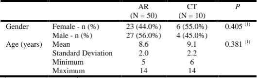
Table 2. Distribution of gender and age of children in the AR and CT groups
AR (N = 50)
CT (N = 10)
P
Gender Female - n (%) 23 (44.0%) 6 (55.0%) 0.405(1)
Male - n (%) 27 (56.0%) 4 (45.0%)
Age (years) Mean 8.6 9.1 0.381(1)
Standard Deviation 2.0 2.2
Minimum 5 6
Maximum 14 14
(1)
[image:3.595.116.499.457.592.2]Pearson’s Chi-square(2)Mann-Whitney Test. AR= acoustic rhinometry CT=computed tomography.
Table 3. Measurements and difference of nasal volumes (cm3) post–pre-RME time interval for AR
VOLUMES T1
(M±SD) T2 (M±SD)
Difference Mean (%)(1)
p(2) d of Cohen
Volume 1
Total nasal cavity (right+left) 2.59± 0.55 2.72±0.50 0.13 (+5.1%) 0.276 0.25 Right nasal cavity 1.25±0.28 1.31±0.20 0.05 (+4.1%) 0.494 0.21 Left nasal cavity 1.34±0.28 1.42±0.33 0.08 (+6.1%) 0.201 0.27 Volume 2
Total nasal cavity (right+left) 6.69±1.74 7.96±1.50 1.28 (+19.1%) 0.006 0.78 Right nasal cavity 3.05±0.98 3.91±1.07 0.857 (+28.1%) 0.018 0.84 Left nasal cavity 3.63±0.95 4.05±1.06 0.42 (+11.5%) 0.166 0.41 Volume 1 + Volume 2
Total nasal cavity (right+left) 9.28±2.16 10.68±1.90 1.41 (+15.2%) 0.010 0.69 Right nasal cavity 4.31±1.18 5.22±1.15 0.91 (+21.1%) 0.026 0.78 Left nasal cavity 4.97±1.14 5.47±1.37 0.50 (+10.1%) 0.140 0.40 M= mean; SD=standard deviation;
(1)
the same orthodontist and the mean value was used toreduce any possible errors. Volume changes due to expansion with the two methods were evaluated using a Wilcoxon matched signed ranks test. Correlation analysis was used to determine the correlation between the two methods. The statistical treatment of the data was performed with the Statistical Package for the Social Sciences (SPSS), version 22 for Windows®. Descriptive statistics including the mean (M), standard deviation (SD), and ranges were calculated for the measurements at T1 and T2. The data were tested for normality with the Shapiro-Wilk test. The Student paired t test was used to investigate the difference between the measurements before and after treatment. The evaluation of the dimension of the differences was made through analysis of the percentage variation of the means and the dimension measure of the Cohen's d effect size. For the classification of the effect size, the values proposed by Cohen (1992)25 were followed: d = 0.20 small effect; d = 0.50 medium effect; d = 0.80 large effect. The t-test for paired samples and the Intraclass Correlation Coefficient (ICC) were used to study the correspondence between tomography and rhinometry measurements.
A non-significant Student's t-test (p> 0.05) and a CCI greater than 0.75 (Fleiss, 1999) ensure matching between tomography and rhinometry measurements.
RESULTS
[image:4.595.100.502.326.390.2]The results showed that none of the variables had a normal distribution (p <0.05). For this reason, non-parametric tests were used: Mann-Whitney test for comparison between independent groups and Wilcoxon test for comparison between repeated measurements (pre-RME and post-RME comparison). The analysis of the significance of differences when qualitative variables was done with the Chi-square test (Table 2) Volumes measured by rhinometry (Table 3): significant differences were observed between T1 and T2 in volume 2 of the total nasal cavity (right + left) (p = 0.006, d = 0.78), volume 2 of the right nasal cavity (p = 0.018, d = 0.84), volume 1 + volume 2 of the right nasal cavity (P = 0.026, d = 0.78), and volume 1 + volume 2 of the total nasal cavity (P = 0.010, d = 0.69). In the remaining variables, differences between T1 and T2 were not significant (p> 0.05), with differences of small or medium size
Table 4. Measurements and difference of nasal volumes (cm3) post–pre-RME time interval for CT
VOLUMES T1
(M±SD) T2 (M±SD)
Difference Mean (%)(1) p
(2)
d of Cohen
Total nasal cavity 35.28±5.17 39.78±4.96 4.50 (+12.7%) < 0.001 0.89 Anterior nasal cavity (right and left) 2.05±0.65 2.64±0.76 0.59 (+28.7%) 0.006 0.84 Leftt anterior nasal cavity 1.09±0.41 1.33±0.35 0.24 (+22.1%) 0.003 0.63 Rightt anterior nasal cavity 0.94±0.27 1.36±0.41 0.42 (+44.8%) 0.005 1.20 M= mean; SD=standard deviation;
[image:4.595.95.511.449.774.2](1)p–difference between T1 and T2 means; (%)–perceptual variance of the means; (2)p–significance value of t test of Student for pared samples.
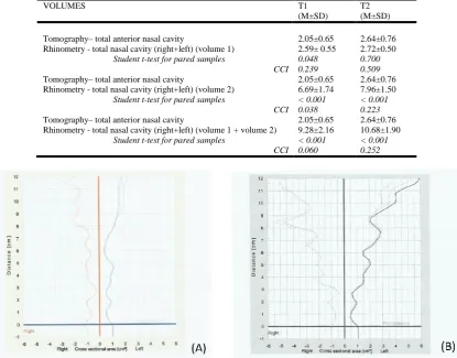
Table 5. Correspondence between measurements of rhinometry and tomography (cm3) in T1 and T2 (n=10)
VOLUMES T1
(M±SD)
T2 (M±SD)
Tomography–total anterior nasal cavity 2.05±0.65 2.64±0.76 Rhinometry - total nasal cavity (right+left) (volume 1) 2.59± 0.55 2.72±0.50
Student t-test for pared samples 0.048 0.700
CCI 0.239 0.509
Tomography–total anterior nasal cavity 2.05±0.65 2.64±0.76 Rhinometry - total nasal cavity (right+left) (volume 2) 6.69±1.74 7.96±1.50 Student t-test for pared samples < 0.001 < 0.001
CCI 0.038 0.223
Tomography–total anterior nasal cavity 2.05±0.65 2.64±0.76 Rhinometry - total nasal cavity (right+left) (volume 1 + volume 2) 9.28±2.16 10.68±1.90
Student t-test for pared samples < 0.001 < 0.001
CCI 0.060 0.252
(Cohen's d <0.40). Volumes measured by computed tomography (Table 4): The main increase, 44.8%, occurred in the right anterior nasal cavity: from 0.94 ± 0.27 in T1 to 1.36 ± 0.41 in T2 (p = 0.005; d = 1.20). The leftt anterior nasal cavity increased 22.1%, from 1.09 ± 0.41 to 1.33 ± 0.35 (p = 0.003; d = 0.63). The total anterior nasal cavity increased 28.7%, from 2.05 ± 0.65 to 2.64 ± 0.76 (p = 0.006; d = 0.84). The total nasal cavity increased 12.7%, from 35.28 ± 5.17 to 39.78 ± 4.96 (p <0.001; d = 0.89). The results of Table 5 show that there is no correspondence between the volumes evaluated by tomography and rhinometry. Only in the case of the total anterior nasal cavity (tomography) and volume 1 of the total nasal cavity: right + left (rhinometry) in T2, there were no significant differences (p = 0.700). In all other cases tested, the Student's t-test was significant (p <0.05), indicating significant differences between the volumes, and the CCI was very low (CCI <0.25).
Figure 2.
DISCUSSION
The relationship of nasal respiratory function with the development of dentofacial complex is controversial and genetic factors are likely to contribute to the presence of deficiency (Sakai, 2016).Studies show that patients with more severe deficiency showed lower airflow values and support the theory that mouth breathers have impaired nasal breathing due to the presence of transverse maxillary deficiency and narrower nasal base (Luzzi, 2013; Trevisan, 2015). These results suggest that RME for airway purpose alone is not justified; Warren et al (Warren, 1987). States that nasal respiration is subject to developmental considerations, both physical and behavioral. In recent years, RME has been added to the list of recommended procedures to improve nasal airway respiration (Doruk, 2007). In the literature, many authors (Compadretti, 2006; Sakai, 2016 and De Felippe, 2009), have emphasized the ability of RME to produce lateral expansion of the nasal cavity and to decrease nasal resistance.Small changes in nasal valve size result in large changes in airflow resistance,controversy in theliterature with regard to the existence of a relationshipbetween nasorespiratory function and dentofacialmorphology (Doruk, 2004). While some
authors supported RME as a means of reducing or eliminating a mouth-breathing posture, others remain sceptical of the influence of RME on the nasal airway. White et al. (White, 1989) found a statistically significant average reduction in nasal airway resistance of 48.7% and affirmed that such reduction was highly correlated to the nasal resistance level prior to RME. The maxillary bones form approximately 50%
of the nasal cavity’s anatomic structure (Badreddine, 2017). Therefore, treatment modalities that alter the morphology of the maxillary dental arch, such as RME whose effects have been noted in the midpalatal suture as well as in the neighboring structures such as the internasal, nasomaxillary, and frontomaxillary sutures (Cappelette, 2017), can affect the geometry and function of the nasal cavity (El H, 2014 and Babacan, 2016). The traditional explanation for the influence of RME on nasal volume is based on the separation of the lateral walls of the nasal cavity, which occurs concurrently during dental arch expansion (Doruk, 2007). The increase in the distance between the lateral walls of the nasal cavity increases nasal volume and enlarges the cross-sectional area of the nasal passage, facilitating breathing (Doruk, 2007). The RME indirectly causes a widening at the anterior nares, which contributesto reductions in nasal resistance (Warren, 1987 and Hartgerink, 1987). However, Wertz (Wertz, 1968), reported that no justification for airway enlargement existed for RME unless an obstruction was present in the anteroinferior aspect of the nose, the area mostfavorablyaffected by RME. Methods for evaluating nasal airway volume have included two-dimensional (2D) cephalometric radiographs (McNamara, 2015).
healthy tissues in a living patient. When evaluating a patient by AR, if it is asked to inspire nasally, often leading to a dynamic collapse of their nasal airway or internal nasal valve. On the other hand, the CT scans represent a moment in time that is captured while the patient is asked to hold his or her breath or breathe quietly. Therefore, this CT study considers the measurements performed in the bone nasal valve that could be more reliable since the sample was composed with children whose inspire control is difficulty due to a lack of cooperation.In this study, vasoconstrictor was not used in order to evaluatethe volume changes without reduction of nasal mucosa, moreover, it asked to hold your breath during AR procedure. According studies (Oliveira de Felippe, 2008; Parvez, 2000; Aras, 2010), the basal condition (no nasal decongestant) is more realistic when evaluating anatomic-functional variability. Several factors limit the accuracy of AR measurements (Cakmak, 2001). The most widelyproblem with acoustic-pulse analysis is the inability to accurately measure areas beyond narrow apertures and the sound loss to the paranasal sinuses may negatively affect the accuracy of AR measurements of more distal segments (Cakmak, 2003). Consequently, AR findings for the distal part of the nasal cavity may not be sufficiently accurate for clinical use.Djupesland and Rotnes (Djupesland, 2001), demonstrated that AR is not able to detect correctly constrictions and expansions shorter than 3-4 mm. The anterior part of the nasal cavity is the site of most interest for the rhinologist and orthodontist. The precision of AR in the anterior part of the nose, especially for the nasal valve area, makes this method valuable for rhinology (Cakmak, 2003). The limits proposed in this study to evaluate the anterior nasal cavity using images from CT, indicate that AR and CT findings for nasal valve area are significantly correlated.
In this study,both measurement methods demonstrated an increase of the anterior nasal cavityin the volume after RME, of AR 15.2% (volume 1 + volume 2); 19,1% (volume 2) and CT 28.7% (anterior nasal cavity).Therefore, the amount of increasing accomplished was greater anteriorly,of CT 12.7%in total nasal cavity. In the present study, significant increases of total volume by RA and anterior volume by CT between T1 and T2 revealed that RME has a great effect on the nasal valve area, which constitutes the greatest resistance while breathing. This is coherent with the aim of the treatment and these findings agree with others authors whose related significant increase in nasal valve volume after RME (Babacan, 2006). In their studies, Babacan et al (Babacan, 2006) reported a significant increase of 14.09% for total nasal volume after surgery RME and Wriedt et al (Wriedt, 2001) reported an average of 21.2% in total nasal volume and 29.1% statistically significant increases in nasal valve region. Nasal breathing is also influenced by the condition of the nasal mucosa (Sakai, 2012; Aras, 2010), reported greater percentages for AR measurements than this study, however, their study was carried out in patients with partial or near total nasal obstruction, showing that subjects who had greater nasal resistance, smaller increase in the nasal volume could lead dramatic changes in those parameters after RME. Several authors (Doruk, 2007; Sakai, 2016 and Terheyden, 2009), reported significant correlations between measurements by AR and CT in the anterior region of the nasal cavity. Contrarily, Baraldi et al (Baraldi, 2007), observed no statistically significant changes in volume when pre- and post-RME values were compared, nevertheless, their study was conducted on frontal cephalograms and by AR measurements and probably there
was a lack of datewhen evaluating the increase in the anterior region. Cankurtaran et al (Cankurtaran, 2007), undertook experimental studies to test the reliability of AR in determining nasal valve area and the results showed the technique to be reliable in quantifying changes in the anterior portion of the nasal cavity but not in relation to the cross-sectional area of the posterior nasal cavity.Christie et al (Christie, 2010), concluded, in their cone-beam computed tomography study, that nasal cavity width increases significantly (2.73 mm) after rapid maxillary expansion.According to Warren et al. (Warren, 1987), the dimensions of children’s airways increase
approximately 0.032 cm2/year and, in the current study, RME did not exceed 20 days, which is a relatively rapid treatment period and suggesting that the results would not be contaminated by significant growth during that period.The results show that volumes in the right side of the nasal cavity are significantly different than left side after RME (Table 3) This possibly could be caused bynasal anatomic variations in the selected sample. These studiesdemonstrate that CT may be a valuable tool in objectivelyassessing outcomes of functional nasal operations; however, neither study correlated the objective datato clinical findings. It is unclear whether the measurements of the changes in volume reflect the subjective sensations.
Conclusion
Acoustic rhinometry showed good correlation in relation to the proposed anterior delimitation of the nasal cavity on the CT images. The significant improvement of the anterior volume suggests that besides early orthopedic treatment with RME is beneficial in the treatment of maxillary constriction associated with mouth breathing in growing patients, the ERM treatment could contributed for development of facial skeletal avoiding both functional and structural imbalances.
REFERENCES
Aras, A., Akay, M.C., Çukurova, I., Günbay,T., Işıksal, E.and Aras, I. 2010. Dimensional changes of the nasal cavity after transpalatal distraction using bone-borne distractor: an acoustic rhinometry and computed tomography evaluation.
J Oral Maxillofac Surg 68(7):1487-1497.
Babacan, H., Doruk, C., Uysal, I.O. and Yuce, S. 2016. Effects of rapid maxillary expansion on nasal mucociliary clearance. Angle Orthodontist 86:250-254.
Babacan, H., Sokucu, O., Doruk, C., et al. 2006. Rapid maxillary expansion and surgically assisted rapid maxillary expansion effects on nasal volume. Angle Orthod., 76:66-71.
Badreddine, F.R., Fujita, R.R. and Cappellette, M. Jr. 2017. Short-term evaluation of tegumentary changes of the nose in oral breathers undergoing rapid maxillary expansion.
Braz J Otorhinolaryngol., 544:1-9.
Baraldi, C.E., Pretto, S.M. and Puricelli, E. 2007. Evaluation of surgically assisted rapid maxillary expansion using acoustic rhinometry and postero-anterior cephalometry. Int
J Oral Maxillofac Surg., 36:305-309.
Basciftci, F.A., Mutlu, N., Karaman, A.I., Malkoc, S. and Küçükkolbasi, H. 2002. Does the timing and method of rapid maxillary expansion have an effect on the changes in nasal dimensions? Angle Orthod 72:118-123.
Bicakci, A.A., Agar, U., Sökücü, O., Babacan, H. and Doruk, C. 2005. Nasal airway changes due torapid maxillary expansion timing. Angle Orthod, 75:1-6, 2005.
Bloom, J.D., Sridharan, S., Hagiwara, M., Babb, J.S., White, W.M. and Constantinides, M. 2012. Reformatted Computed Tomography to Assess the Internal Nasal Valve and Association With Physical Examination Arch Facial
Plast Surg 14:331-335.
Cakmak, O., Celik, H., Ergin, T. and Senaroğlu, L. 2001.
Accuracy of acoustic rhinometry measurements. Laryngoscope 111: 587–594.
Cakmak, O., Coşkun, M., Celik, H., Büyüklü, F.andOzlüoğlu,
L.N. 2003. Value of acoustic rhinometry for measuring nasal valve area. Laryngoscope 113:295-302.
Cankurtaran, M., Celik, H., Coşkun, M., Hizal, E. and Cakmak, O. 2007. Acoustic rhinometry in healthy humans: accuracy of area estimates and ability to quantify certain anatomic structures in the nasal cavity. Ann Otol Rhinol
Laryngol 116:906-916.
Cappelette, Jr M., Alves, F.E.M.M., Nagai, L.H.Y., Fujita, R.R. and Pignatari, S.S.N. 2017. Impact of rapid maxillary expansion on the volume of the nasomaxillary complex of mouth breathers. Dental Press J Orthod., 22:79-88.
Cappellette, Jr M., Nagai, L.H.Y., Gonçalves, R.M., Yuki, A.K., Pignatari, S.S.N. and Fujita, R.R. 2017. Skeletal effects of RME in the transverse and vertical dimensions of the nasal cavity in mouth breathing growing children.
Dental Press J Orthod., 22:61-69.
Cappellette, Jr M., Cruz, O.L., Carlini, D., Weckx, L.L. and Pignatari, S.S. 2008. Evaluation of nasal capacity before and after rapid maxillary expansion. Am J Rhinol., 22:74-77, 2008.
Christie, K.F., Boucher, N. and Chung, C.H. 2010. Effects of bonded rapid palatal expansion on the transverse dimensions of the maxilla: A cone-beam computed tomography study. Am J Orthod Dentofacial Orthop 137:S79-85.
Chung, C.H. and Font, B. 2004. Skeletal and dental changes in the sagittal, vertical, and transverse dimensions after rapid palatal expansion. Am J Orthod Dentofacial Orthop 126:569-575.
Coelho, A.R., Tanaka, O., Ribeiro, J.S., Machado, M.A. and Camargo, E.S. 2010. Transverse craniofacial dimensions in Angle Class II. Division 1 malocclusion according to breathing mode. Braz Oral Res., 24:70-75.
Cohen, J. 1992. "A Power Prime". Psycological Bulletin112:155-159.
Compadretti, G.C., Tasca, I., Alessandri-Bonetti, G., et al. 2006. Acoustic rhinometric measurements in children undergoing rapid maxillary expansion. Int J Pediatr
Otorhinolaryngol., 70:27-34.
Cross, D.L. and McDonald, J.P. 2000. Effect of rapid maxillary expansion on skeletal, dental, and nasal structures: a postero-anterior cephalometric study. Eur J
Orthod., 22:519-528.
De Felippe, N.L., Bhushan, N., Silveira, A.C., Viana, G. and Smith, B. 2009. Long-term effects of orthodontic therapy on the maxillary dental arch and nasal cavity. Am J Orthod
Dentofacial Orthop., 136:490.e1-490.e8.
Djupesland, P.G. and Røtnes, J.S. 2001. Accuracy of acoustic rhinometry.Rhinology 39:23-27.
Doruk, C., Sökücü, O., Biçakçi, A., Yilmaz, U. and Tas, F. 2007. Comparison of nasal volume changes during rapid maxillary expansion using acoustic rhinometry and computed tomography. Eur J Orthod., 29:251-255.
Doruk, C., Sökücü, O., Sezer, H. and Canbay, E. 2004. Evaluation of nasal airway resistance during rapid maxillary expansion using acoustic rhinometry. Eur J Orthod 26:397-401.
Eichenberger, M. and Baumgartner, S. 2014. The impact of rapid palatal expansion on children's general health: a literature review. Eur J Paediatr Dent., 15:67-71.
El, H. and Palomo, J.M. 2014. Three-dimensional evaluation of upper airway following rapid maxillary expansion: a CBCT study. Angle Orthod., 84:265-273.
Fleiss, J.L. 1999. "The Design and Analysis of Clinical Experiments". A Wiley-Interscience Publication. Wiley Classics Library Edition.
Frasson, J.M.D., Magnani, M.B.B.A., Nouer, D.F., Siqueira, V.C.V. and Lunardi, N. 2006. Comparative cephalometric study between nasal and predominantly mouth breathers.
Braz J Otorhinolaryngol 72:72-81.
Ghoneima, A., AlBarakati, S., Jiang, F., Kula, K. and Wasfy, T. 2015. Computational fluid dynamics analysis of the upper airway after rapid maxillary expansion: a case report.
Prog Orthod 16:10, 2015.
Görgülü, S., Gokce, S.M., Olmez, H., Sagdic, D. and Ors, F. 2011. Nasal cavity volume changes after rapid maxillary expansion in adolescents evaluated with 3-dimensional simulation and modeling programs. Am J Orthod Dentofacial Ortho., 140:633-640, 2011.
the nasal cavity induced by rapid maxillary expansion: a study on 3-dimensional computed tomography models. Am
J Orthod Dentofacial Orthop 136:815-821.
Hartgerink, D.V., Vig, P.S. and Abbott, D.W. 1987. The effect of rapid maxillary expansion on nasal airway resistence.
Am J Orthod. Dentofac Orthop., 92:381-389.
Hilberg, O., Jackson, A.C., Swift, D.L. and Pedersen, O.F. 1989. Acoustic rhinometry: evaluation of nasal cavity geometry by acoustic reflection. J Appl Physiol., 66: 295-303.
Hilberg, O. and Pedersen, O.F. 2000. Acoustic rhinometry: recommendations for technical specifications and standard operating procedures. Rhinol Suppl 16:3-17.
Hilberg, O. 2002. Objective measurement of nasal airway dimensions using acoustic rhinometry: methodological and clinical aspects. Allergy 57:5-39.
Iwasaki, T., Saitoh, I., Takemoto, Y., Inada, E., Kanomi, R., Hayasaki, H. and Yamasaki, Y. 2012. Improvement of nasal airway ventilation after rapid maxillary expansion evaluated with computational fluid dynamics. Am J Orthod
Dentofacial Orthop., 141:269-278, 2012.
Kjaergaard, T., Cvancarova, M. and Steinsvåg, S.K. 2008. Does nasal obstruction mean that the nose is obstructed ? Laryngoscope118:1476-1481.
Kluemper, G.T., Vig, P.S. and Vig, K.W. 1995. Nasorespiratory characteristics and craniofacial morphology. Eur J Orthod 17:491-495.
Lam, D.J., James, K.T. and Weaver, E.M. 2006. Comparison of anatomic, physiological, and subjective measrures of the nasal airway. Am J Rhinol., 20(5): 463-470.
Lee, J., White, W.M. and Constantinides, M. 2009. Surgical and nonsurgical treatments of the nasal valves. Otolaryngol
Clin North Am 42:495-511.
Luzzi, V., Ierardo, G., Viscogliosi, A., Fabbrizi, M., Consoli, G., Vozza, I., Vestri, A. and Polimeni, A. 2013. Allergic rhinitis as a possible risk factor for malocclusion: a case-control study in children. Int J Paediatr Dent., 23:274-278. Magnusson, A., Bjerklin, K., Nilsson, P., Jönsson, F. and
Marcusson, A. 2011. Nasal cavity size, airway resistance, and subjective sensation after surgically assisted rapid maxillary expansion: A prospective longitudinal study. Am
J Orthod Dentofacial Orthop140:641-651.
McNamara, Jr J.A., Lione, R., Franchi, L., Angelieri, F., Cevidanes, L.H.S., Darendeliler, M.A. and Cozza, P. 2015. The role of rapid maxillary expansion in the promotion of oral and general health. Progress in Orthodontics 16:33. Miman, M.C., Deliktas, H., Ozturan, O., Toplu, Y. and
Akarçay, M. 2006. Internal nasal valve: revisited with objective facts. Otolaryngol Head Neck Surg., 134:41-47.
Montgomery, W.M., Vig, P.S., Staab, E.V. and Matteson, S.R. 1979. Computed tomography: a three dimensional study of the nasal airway. Am J Orthod., 76:363-375.
Nigro, C.E., Nigro, J.F., Voegels, R.L., Mion, O. and Mello Junior, J.F. 2005. Acoustic rhinometry: anatomic correlation of the first two notches found in the nasal echogram.Braz J Otorhinolaryngol 71:149-154.
Oliveira de Felippe, N.L., Da Silveira, A.C., Viana, G., Kusnoto, B., Smith, B. and Evans, C.A. 2008. Relationship between rapid maxillary expansion and nasal cavity size and airway resistance: short- and long-term effects. Am J
Orthod Dentofacial Orthop, 134:370-382.
Parvez, L., Erasala, G. and Noronha, A. 2000. Novel techniques, standardization tools to enhance reliability of acoustic rhinometry measurements. Rhinol Suppl 16:18-28. Roithmann, R., Cole, P., Chapnik, J., Shpirer, I., Hoffstein, V.
and Zamel, N. 1975. Acoustic rhinometry in the evaluation of nasal obstruction. Laryngoscope 105:275-281.
Sakai, R.H., Marson, F.A.L., Sakuma, E.T., Ribeiro, J.D. and Sakano, E. 2016. Correlation between acoustic rhinometry, computed rhinomanometry and cone-beam computed tomography in mouth breathers with transverse maxillary deficiency. Braz J OtorhinolaryngolNov 25. pii: S1808-8694(16)30234-8. doi: 10.1016/j.bjorl.2016.10.015. [Epub ahead of print].
Sipilä, J., Suonpää, J. and Laippala, P. 1994. Sensation of nasal obstruction compared to rhinomanometric results in patients referred for septoplasty.Rhinology 32:141-144. Terheyden, H., Maune, S., Mertens, J. and Hilberg, O. 2000.
Acoustic rhinometry: Validation by three-dimensionally reconstructed computer tomographic scans. J Appl Physiol 89:1013-1021.
Trevisan, M.E., Bellinaso, J.H., Pacheco, A.B., Augé, L.B., Silva, A.M. and Corrêa, E.C. 2015. Respiratory mode, nasal patency and palatine dimensions. Codas 27:201-206. Warren, D.W., Hershey, H.G., Turvey, T.A., Hinton, V.A. and
Hairfield, W.M. 1987. The nasal airway following maxillary expansion. Am J Orthod Dentofacial Orthop., 91:111-116.
Wertz, R.A. Changes in nasal airfl ow incident to rapid maxillary expansion. Angle Orthod 38:1-11, 1968.
White, B.C., Woodside, D.G. and Cole, P. 1989. The effect of rapid maxillary expansion on nasal airway resistance. J
Otolaryngol., 18:137-143.
Wriedt, S., Kunkel, M., Zentner, A. and Wahlmann, U.W. 2001. Surgically assisted rapid palatal expansion. An acoustic rhinometric, morphometric and sonographic investigation. J Orofac Orthop., 62:107-115.