Case Report
An 88-year-old man with MDS/MPN-RS-T accompanied
by unexplained repeated pleural effusion
Miaoxin Peng1*, Yipeng Ling1*, Peipei Xu1, Yueyi Xu1, Ting Xie1, Yonggong Yang1, Bing Chen2
1Department of Hematology, Drum Tower Hospital, School of Medicine, Nanjing University, Nanjing, Jiangsu,
China; 2Department of Hematology, Nanjing Drum Tower Hospital Clinical College of Nanjing Medical University,
Nanjing, Jiangsu, China. *Equal contributors.
Received January 8, 2019; Accepted February 13, 2019; Epub June 15, 2019; Published June 30, 2019
Abstract: Herein is described a case of an 88-year-old man with newly-typically diagnosed MDS/MPN-RS-T accom-panied with gene mutations: SF3B1, JAK2V617F, TET2 and ASXL1. Moreover, the patient repeatedly developed unexplained pleural effusion. The pleural effusion was asymmetric, non-neoplastic, and lymphocytes dominated the transudate. After several times of drainage procedure for pleural effusion and the use of hydroxyurea and EPO along with supportive treatment, the patient got an extent of alleviating. To the best of our knowledge, this is the
first report of a patient with the much rare disease of MDS/MPN-RS-T accompanied by an even rarer manifestation
showing the repeated pleural effusion. Fortunately, the patient improved after our clinical treatment.
Keywords: Hematological malignancy, MDS/MPN-RS-T, pleural effusion, thoracentesis, myelodysplastic syndrome, clinical management
Introduction
Myelodysplastic syndrome/myeloproliferative neoplasm with ring sideroblasts and thrombo-cytosis (MDS/MPN-RS-T) became a full entity under the 2016 World Health Organization (WHO) classification, which is previously provi-sionally known as refractory anemia with ring sideroblasts associated with marked thrombo-cytosis (RARS-T) in WHO 2008. Gene muta-tions in patients with MDS/MPN-RS-T inclu- de: SF3B1 (~85%), JAK2V617F (~50%), TET2 (~25%), ASXL1 (~20%), DNMT3A (~15%), and SETBP1 (~10%) [1, 2]. Approximately 50% of patients harbored both the JAK2V617F and SF3B1 mutations, providing an intriguing genet-ic explanation for the hybrid nature of MDS/ MPN-RS-T as being between an MDS and an MPN. Herein is described an 88-year-old man with newly diagnosed MDS/MPN-RS-T accom-panied by the above representative molecular performance. In clinical practice, pleural effu-sion in hematological malignancies is rare when compared with solid tumors. Pleural effu-sions in patients with hematological malignan-cy are most often due to infection and to a
lesser extent malignant infiltration of the pleu-ra. Pleural effusions with MDS or MPN is much few. In this case, the reason of effusion is nei-ther infection nor malignant invasion in lung. Briefly, MDS/MPN-RS-T is an extremely rare dis-ease, making it far rarer when combining un- usual manifestations. Here, the first case of atypical MDS/MPN-RS-T with repeated pleural effusion is reported.
Case report
In June 2017, an 88-year-old man was admitted to our Department of Hematology because of an incidental discovery of anemia and dramati-cally thrombocytosis. Over the decades, the man was in a very good health without diabe-tes, hypertension or any other chronic condi-tions of aging until nowadays. So, this is the first time for him to receive a comprehensive ins- pection.
MDS/MPN-RS-T with repeated pleural effusion
Figure 3. Extracellular iron (the blue background), Bone marrow, Perls’ staining, 100×.
oxygen saturation 97-100% while he was br- eathing ambient air. Physical examination: pale appearance, a low breathing tone in lower left lung, splenomegaly at 3~4 cm below the costal margin, the remainder was approximately nor-mal. Lab examination: A complete blood count (CBC) revealed the following results: Hemog- lobin (Hb), 8.0 g/dl (normal range, 13.0-17.5 g/ dl); hematocrit, 25.7% (normal range, 40-50%); mean corpuscular volume (MCV), 73.0 fl (nor-mal range, 82-100 fl); platelet count, 1005× 109/L (normal range, 125-350×109/L); and
white blood cell (WBC) 17.1×10⁹/L (normal
range, 3.5-9.5×10⁹/L) with a slightly elevated
neutrophils 81.7% (normal range, 40-70%). Serum iron: 30.3 umol/L (6.6-28.3); serum fer-ritin: 420.30 ng/ml (22-322); Serum erythro-poietin (EPO): 67.4 mIu/ml (4.3-29); BNP: 89 pg/ml (5-100); Blood Biochemistry: lactic dehy-drogenase (LDH) 489 IU/L (normal range
109-245 IU/L), liver and kidney tests as well as el- ectrolyte were in normal ranges. Echocardiog- raphy: Pulmonary Arterial Hypertension (PAH) 80 mmHg, EF 56%. BM aspirate: hypercellular-ity, granulocytic lineage 57% with 1% myelo-blasts, erythroid lineage 35.5%, with dyseryth-ropoiesis (megaloblastic changes and RS,
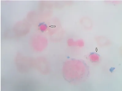
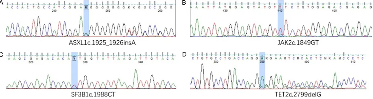
[image:2.612.323.524.73.223.2]Figures 1 and 2). Extracellular iron (Figure 3): ++, I: 1%, II 3%, III 9%, IV 21%, Ringed sidero-blasts 59%. Mature red blood cells are of unequal size and different shapes (Figure 4) with an extremely light staining in central pale area. BM biopsy: hypercellularity, basically no adipose tissue, polymorphous megakaryocytes and dysmegakaryopoiesis like that of ET, exten-sive fibrosis about 1/2 of the total area. Cy- togenetic exam: 46,XY [20]; BM-PCR: JAK2 (V6- 17F) mutation: positive. BCR-ABL, CALR, MPL: negative. PDGFRα/β: negative. Next-generation sequencing in bone marrow sample (Figure 5): Figure 1. Dyserythropoiesis with megaloblastic
[image:2.612.325.524.275.425.2]changes Bone marrow, May-Grünwald-Giemsa (MGG) staining, 100×.
Figure 2. Ring sideroblasts. Bone marrow, Perls’ staining, 100×.
[image:2.612.89.288.286.434.2]MDS/MPN-RS-T with repeated pleural effusion
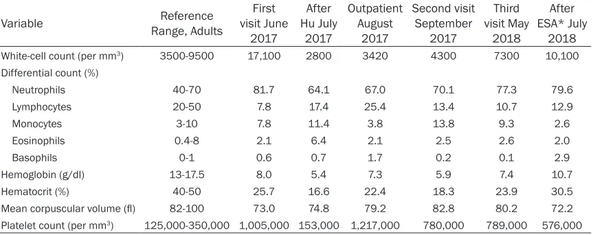
a missense mutation in SF3B1 gene: c.1988C>T (heterozygous, frequency 43.11%); a missense mutation in JAK2 gene: c.1849G>T (heterozy-gous, frequency 60.56%); a frameshift muta-tion in TET2 gene: c.2799delG (p.Gly934Glufs- Ter19) (heterozygous, frequency 72.92%); a fr- ameshift mutation in ASXL1 gene: c. 1925_ 1926insA (p.Gly643ArgfsTer15), four mutati- ons are all related with myeloid hematopathy. After a definitive diagnosis of MDS/MPN-RS-T, the patient mainly accepted supportive treat-ments including transfusion support, cytore-ductive therapy (hydroxyurea for dynamically adjusted doses) and antiplatelet therapy. In- itially, aspirin was not provided because of his persistent positive fecal occult blood. One month later, he was given aspirin 100 mg daily after fecal occult blood turn negative. After a while, the WBC and PLT were gradually declined, but Hb had no significant change under the cir-cumstances of no use of erythropoiesis stimu-lating agents (ESA) at the beginning. Surpri- singly, a significant improvement of Hb was observed when he accepted ESA (40,000 Unit ih biw) since May 2018. The CBC results are shown in Table 1.
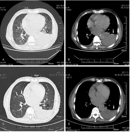
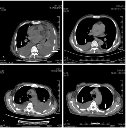
Notably, the patient had no oppression in chest or any other respiratory symptoms, but had a low breathing tone in lower left lung at his first visit. Computed tomography (CT) of the chest showed: bilateral pleural effusion, especially noticeable on the left. Thoracocentesis and chest drainage was adopted by once central venous catheter. For a time, the hydrothorax
were mitigated (Figure 6). However, the hydro-thorax and chest distress appeared repeatedly in October 2017 and May 2018 (Figure 7). Every time after thoracocentesis and chest drainage, the patient got much better. However, several month later, the pleural effusion devel-oped again. Flow cytometry of hydrothorax showed as (Figure 8): Lymphocytes account for about 80~90% of total cells. Exfoliocytology examination of the effusion: mainly lympho-cytes with several histolympho-cytes and mesothelial cells and the final pathological diagnosis of the effusion (many times): No tumor cells were seen (Figure 9). Next-generation sequencing in pleural effusion sample: negative. No gene mutation was detected.
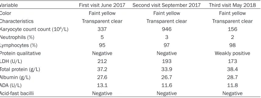
Three routine examinations of pleural fluid showed (Table 2) Acid-fast bacilli, ADA and T-Spot in hydrothorax were all negative. After the last visit, the patient did not back to hospi-tal. According to the telephone follow-up, the old man reports a basically normal life.
Discussion
[image:4.612.92.530.86.257.2]Pleural effusions are rarely observed in patients with hematological malignancy such as acute myelogenous leukemia (AML), acute lympho-cytic leukemia (ALL) and MDS/MPN. Therefore, the underlying etiology of pleural effusions has not been well studied. Reviewing past litera-ture, the etiology of pleural effusions in hema-tological malignancy including: 1) Infections. In a retrospective review [3] of a 10-year series cases of 111 patients with pleural effusion in
Table 1. CBC* results of the patient in different stages
Variable Range, AdultsReference visit June First 2017 After Hu July 2017 Outpatient August 2017 Second visit September 2017 Third visit May 2018 After ESA* July 2018 White-cell count (per mm3) 3500-9500 17,100 2800 3420 4300 7300 10,100
Differential count (%)
Neutrophils 40-70 81.7 64.1 67.0 70.1 77.3 79.6
Lymphocytes 20-50 7.8 17.4 25.4 13.4 10.7 12.9
Monocytes 3-10 7.8 11.4 3.8 13.8 9.3 2.6
Eosinophils 0.4-8 2.1 6.4 2.1 2.5 2.6 2.0
Basophils 0-1 0.6 0.7 1.7 0.2 0.1 2.9
Hemoglobin (g/dl) 13-17.5 8.0 5.4 7.3 5.9 7.4 10.7
Hematocrit (%) 40-50 25.7 16.6 22.4 18.3 23.9 30.5
Mean corpuscular volume (fl) 82-100 73.0 74.8 79.2 82.8 80.2 72.2
Platelet count (per mm3) 125,000-350,000 1,005,000 153,000 1,217,000 780,000 789,000 576,000
Figure 6. Computed tomography (CT) of the chest in June 2017 at the first hospitalization. (A) Lung window and (B) mediastinal window before thoracentesis and drainage (the arrows: left pleural effusion). (C) Lung window and (D) mediastinal window after thoracentesis and drainage (the arrows: pleural effusion was reduced).
leukemia and MDS from 1997 to 2007, the most frequent cause was infection (47%) fol-lowed by malignancy (36%). Similarly, earlier autopsy series for acute leukemia also des- cribe parapneumonic effusions as the most common cause [4]. Overall, infection was pre-dominant cause of pleural effusions followed by 2) Hematologic Malignancy. Malignant pleu-ral effusions (MPEs) may occur at any time dur-ing the course of hematologic malignancies and may signal the presence of disease or indi-cate relapse [5, 6]. Lymphomas are the most common hematologic malignancies associated with pleural effusions, and may occur in 30% of
MDS/MPN-RS-T with repeated pleural effusion
associated with pleural effusion [10]. Similarly, other agents used in the treatment of leukemia and MDS such as fludarabine, cyclophospha-mide and decitabine etc. may also cause pleu-ral effusions [11]. In this case, the patient didn’t receive any treatment before and had no evi-dence of infection. Subsequently, extramedul-lary infiltration was our first suspicion at one time. But the pleural effusion had no evidence of malignancy considering the effusion was nonspecific lymphocyte dominated transudate (since this disease is a lesion of erythrocytes and megakaryocytes), and the exfoliocytology
[image:6.612.89.524.70.514.2]and molecular biology were all negative, as well as many times of the routine examinations on effusion. Combining all the results, it was be- lieved that the effusion had no connection with hematologic malignancy. Attention was also paid to other rare reasons as below 4) Immune mechanisms. Autoimmune manifestations as- sociated with myelodysplasia are known as “autoimmune paraneoplastic syndrome” who- se clinical presentation can include pleural effusion. Autoimmune disorders were diag-nosed in 30 patients (7.4%) of 221 patients treated for MDS reported by Enright [12]. It Figure 7. Mediastinal window of the chest CT scans at the second and third hospitalization. (A) Mediastinal window
before and after (B) thoracentesis and drainage at the second hospitalization (pleural effusion was significantly
et Syndrome. Sweet syndrome is characteriz- ed by skin lesions with red or purple papules. may well be more frequent by an incidence of
[image:7.612.95.517.73.652.2]10 to 13% reported by Saif et al [13]. 5) Swe-
Figure 8. Flow cytometry of cells in pleural effusion. The figure showed that T lymphocytes account for about
MDS/MPN-RS-T with repeated pleural effusion
Extracutaneous manifestations are possible with pulmonary involvement. In a series of 79 patients with Sweet syndrome, Cohen report- ed 9% with MDS and 7% with CML in chronic or acceleration phase for 85% of the associat-ed blood diseases [14]. 6) Hypereosinophilic Syndrome. Matsushima reported three cases of patients presenting this type of MDS with the same chromosomal abnormality der(1q;7p), bone marrow hypereosinophilia and pulmonary involvement [15]. In this case, the patient did not have the above comorbidity, and did not have 7) Pulmonary alveolar proteinosis or 8) Organizing pneumonia. On the other hand, Pulmonary Arterial Hypertension (PAH) is a common complication of myeloproliferative neoplasia, which is reported a poor prognosis [16]. The patient did have a PAH, but a sole PAH still could not well explain the intermittently
asymmetric, lymphocyte dominated pleural effusion. In clinical, 9) Systemic Causes (throm-bosis, liver dysfunction, cardiomyopathy, renal insufficiency, autoimmune conditions, volume overload) are common, which should be care-fully excluded. As a result, the patient still was negative in systemic causes. Therefore, a defi-nite pathogeny for pleural effusion was not found. Therefore, pleural effusion as a possible specific manifestation in MDS/MPN-RS-T was questioned. Although pleural effusions in he- matologic malignancies do not necessarily por-tend poor prognosis [17], further prospective study is needed.
[image:8.612.90.523.70.255.2]In terms of management, no formal guidelines for this disease exist [18] since it is extremely rare. Empirically, the management of MDS/ MPN-RS-T is largely supportive. During his hos-Figure 9. Exfoliative cells of pleural effusion. (A: Wright stain 40×, B: Wright Giemisa staining 100×). The cells in pleural effusion are mainly lymphocytes with several histocytes and mesothelial cells (the arrows: lymphocytes).
Table 2. Routine examinations of pleural fluid during three hospital stays
Variable First visit June 2017 Second visit September 2017 Third visit May 2018
Color Faint yellow Faint yellow Faint yellow
Characteristics Transparent clear Transparent clear Transparent clear
Karyocyte count count (106/L) 337 946 156
Neutrophils (%) 5 3 2
Lymphocytes (%) 95 97 98
Protein qualitative Negative Negative Weakly positive
LDH (U/L) 212 193 173
Total protein (g/L) 37.2 33.9 38.4
Albumin (g/L) 27.6 26.7 28.7
ADA (U/L) 13.1 11.6 11.8
[image:8.612.94.523.318.479.2]pitalization, supportive care like transfusion and drainage procedure for pleural effusion using indwelling pleural catheters was provid-ed. Similar to lower risk MDS, the use of eryth-ropoiesis stimulating agents (ESA) is instituted early on in patients with anemia. In the begin-ning, the patient did not use ESA for some per-sonal reason even we strongly recommend when his serum EPO showing 67.4 mIu/ml. Aspirin therapy is reasonable in MDS/MPN-RS-T, especially in low risk patients with the presence of JAK2V617F [18, 19]. Afterwards, antiplatelet therapy (aspirin dynamically adjust-ed doses) was providadjust-ed. Hydroxyurea is a com-mon drug for cytoreductive therapy. But the value of cytoreductive therapy is debatable because it might exacerbate anemia. On the other hand, maybe patients should accept cyto-reductive therapy when compelled by the pres-ence of multiple risk factors for thrombosis [20]. Considering platelet count 1005~2200× 109/L (normal range, 125-350×109/L) and ot-
her conditions, the Hu therapy was chosen and adjusted dynamically according to the CBC results. According to recent research, Lena- lidomide showed a good clinical activity in this rare disorder [21]. However, economic reasons limited upfront use of it in this patient. Under our strong recommendation, he take the ESA therapy. Surprisingly, his Hb up to highest 90-110 g/L from the lowest 50-65 g/L. At the time of writing, the current state of the patient is stable and capable of living independently. Overall, our patient’s case illustrates that the asymmetric, lymphocyte dominated pleural ef- fusion could possibly be an exceptionally clini-cal manifestations in patient with MDS/MPN-RS-T. Importantly, for super-aged patient, sup-portive care and the use of ESA can be suc-cessful especially when serum EPO <500 mIu/ ml. Therapeutic options should be personalized to each case since there is not yet a standard-ized treatment of this disease.
Acknowledgements
We thank all the volunteers who took part in this study. Miaoxin Peng and Yipeng Ling are the co-first authors.
Written informed consent was obtained from the patient for publication of this case report and accompanying images.
Disclosure of conflict of interest
None.
Address correspondence to: Dr. Bing Chen, Depart- ment of Hematology, Nanjing Drum Tower Hospital Clinical College of Nanjing Medical University, No 321, Zhongshan Road, Gulou District, Nanjing 210036, Jiangsu, China. Tel: +86 25 83105211; E-mail: [email protected]; Dr. Yonggong Yang, Department of Hematology, Nanjing Drum Tower Hospital, School of Medicine, Nanjing University, No 321, Zhongshan Road, Gulou District, Nanjing 210036, Jiangsu, China. Tel: +86 25 83106666; E-mail: [email protected]
References
[1] Patnaik MM, Lasho TL, Finke CM, Hanson CA, King RL, Ketterling RP, Gangat N and Tefferi A. Predictors of survival in refractory anemia with ring sideroblasts and thrombocytosis (RARS-T) and the role of next-generation sequencing. Am J Hematol 2016; 91: 492-498.
[2] Jeromin S, Haferlach T, Weissmann S, Megg- endorfer M, Eder C, Nadarajah N, Alpermann T, Kohlmann A, Kern W, Haferlach C and Sch- nittger S. Refractory anemia with ring sidero-blasts and marked thrombocytosis cases har-bor mutations in SF3B1 or other spliceosome genes accompanied by JAK2V617F and ASXL1 mutations. Haematologica 2015; 100: e125. [3] Faiz SA, Bashoura L, Lei X, Sampat KR, Brown
TC, Eapen GA, Morice RC, Ferrajoli A and Jimenez CA. Pleural effusions in patients with acute leukemia and myelodysplastic syndro- me. Leuk Lymphoma 2013; 54: 329-335. [4] Faiz SA, Sahay S and Jimenez CA. Pleural
effu-sions in acute and chronic leukemia and my-elodysplastic syndrome. Curr Opin Pulm Med 2014; 20: 340-6.
[5] Bashoura L, Eapen GA and Faiz SA. Pulmonary manifestations of lymphoma and leukemia. Clin Chest Med 2017; 38: 187-200.
[6] Alexandrakis MG, Passam FH, Kyriakou DS and Bouros D. Pleural effusions in hematologic malignancies. Chest 2004; 125: 1546-1555. [7] Berkman N, Breuer R, Kramer MR and Polliack
A. Pulmonary involvement in lymphoma. Leuk Lymphoma 1996; 20: 229-237.
[8] Bourantas KL, Tsiara S, Panteli A, Milionis C and Christou L. Pleural effusion in chronic my-elomonocytic leukemia. Acta Haematologica 1998; 99: 34-37.
[9] Gong X, Lu X, Fu Y, Wu X, Yan L, Zhang X and Wang L. Cytological features of chronic myelo-monocytic leukaemia in pleural effusion and
lymph node fine needle aspiration.
Cytopath-ology 2010; 21: 411-413.
MDS/MPN-RS-T with repeated pleural effusion
Chomienne C, Chastang C, Degos L, Fenaux P. Incidence, clinical features, and outcome of all trans-retinoic acid syndrome in 413 cases of newly diagnosed acute promyelocytic leuke-mia. The European APL group. Blood 1998; 92: 2712.
[11] Dimopoulou I, Bamias A, Lyberopoulos P and Dimopoulos MA. Pulmonary toxicity from novel antineoplastic agents. Ann Oncol 2006; 17: 372-379.
[12] Enright H, Jacob HS, Vercellotti G, Howe R, Bel-zer M and Miller W. Paraneoplastic autoim-mune phenomena in patients with myelodys-plastic syndromes: response to immunosup- pressive therapy. Br J Haematol 1995; 91: 403-408.
[13] Saif MW, Hopkins JL and Gore SD. Autoim-mune phenomena in patients with myelodys-plastic syndromes and chronic myelomono- cytic leukemia. Leuk Lymphoma 2002; 43: 2083-2092.
[14] Cohen PR, Talpaz M and Kurzrock R. Malig- nancy-associated Sweet’s syndrome: review of the world literature. J Clin Oncol 1988; 6: 1887-1897.
[15] Matsushima T, Murakami H, Kim K, Uchiumi H, Murata N, Tamura JI, Sawamura M, Karasawa M, Naruse T and Tsuchiya J. Steroid-responsive pulmonary disorders associated with myelo-dysplastic syndromes with der(1q;7p) chromo-somal abnormality. Am J Hematol 1995; 50: 110-115.
[16] Lamour C and Bergeron A. Non-infectious pul-monary complications of myelodysplastic syn-dromes and chronic myeloproliferative disor-ders. Rev Mal Respir 2011; 28: e18-e27. [17] Vakil E, Jimenez CA and Faiz SA. Pleural
effu-sions in hematologic malignancies and their management with indwelling pleural cathe-ters. Curr Opin Pulm Med 2018; 24: 384-391. [18] Patnaik MM and Tefferi A. Refractory anemia
with ring sideroblasts (RARS) and RARS with thrombocytosis (RARS-T): 2017 update on
di-agnosis, risk-stratification, and management.
Am J Hematol 2017; 92: 297-310.
[19] Alvarez-Larrán A, Cervantes F, Pereira A, Are- llano-Rodrigo E, Pérez-Andreu V, Hernández-Boluda JC, Ayats R, Salvador C, Muntañola A, Bellosillo B, Vicente V, Hernández-Nieto L, Bur-galeta C, Xicoy B and Besses C. Observation versus antiplatelet therapy as primary prophy-laxis for thrombosis in low-risk essential throm-bocythemia. Blood 2010; 116: 1205.
[20] Tefferi A and Barbui T. Polycythemia vera and essential thrombocythemia: 2015 update on
diagnosis, risk-stratification and management.
Am J Hematol 2015; 90: 162-173.
[21] Huls G, Mulder AB, Rosati S, van de Loosdrecht