ISSN Online: 2165-7416 ISSN Print: 2165-7408
DOI: 10.4236/ojoph.2018.82012 May 11, 2018 84 Open Journal of Ophthalmology
The Influence of Keratometry on Visual and
Refractive Outcomes after Myopic LASER
in Situ
Keratomileusis
Sara Frazão, Pedro Rodrigues, Samuel Alves, Ana Maria Carvalho,
José Maia Sêco, Hugo Nogueira
Instituto de Oftalmologia Dr. Gama Pinto, Lisbon, Portugal
Abstract
To evaluate the effect of preoperative keratometry on visual and refractive outcomes after Myopic LASER in Situ Keratomileusis (LASIK) in eyes with preoperative spherical equivalent (SE) of −6.00D or less. Material and Me-thods: A retrospective study enrolling clinical records of 482 eyes of 275 pa-tients with myopia who underwent LASIK between 2009 and 2016. Subjects were grouped according to the degree of preoperative mean keratometry (Km), into three groups: Group 1 (Flat Cornea): Km ≤ 42.00 diopters (D); Group 2: 42.00 < Km > 46.00D; Group 3 (Steep Cornea): Km ≥ 46.00D. To evaluate the prognostic impact of keratometry in Myopic LASIK, we consi-dered the results measured at 6 months postoperatively, including uncor-rected distance visual acuity (UDVA), postoperative sphere, cylinder, SE and its variation. Results: The mean preoperative SE was −3.91 ± 1.54D, ranging between −0.88 and −6.00D. The percentage of eyes achieving a postoperative SE of ±0.50D was 39.5%, 31.8% and 26% in groups 1, 2 and 3 respectively. Moreover, in group 3, 14.3% of the eyes had a residual SE of −2.00D or great-er, contrasting with the groups 1 and 2 with only 6% - 7%. These results were found to be statistically significant. Concerning UDVA, eyes achieving 20/25 or more were 81.5%, 81.8% and 71.5%, and 20/50 or less were 6.7%, 6.2% and 11.7% in groups 1, 2 and 3 respectively. Conclusions: Myopic eyes with stee-per corneas seem to have greater tendency to undercorrection, also presenting worse visual outcomes.
Keywords
Myopia, Astigmatism, LASER in Situ Keratomileusis, Keratometry, Spherical Equivalent, Flat Cornea, Steep Cornea
How to cite this paper: Frazão, S., Rodri-gues, P., Alves, S., Carvalho, A.M., Sêco, J.M. and Nogueira, H. (2018) The Influence of Keratometry on Visual and Refractive Outcomes after Myopic LASER in Situ Keratomileusis. Open Journal of Ophthal-mology, 8, 84-90.
https://doi.org/10.4236/ojoph.2018.82012
Received: March 27, 2018 Accepted: May 8, 2018 Published: May 11, 2018
Copyright © 2018 by authors and Scientific Research Publishing Inc. This work is licensed under the Creative Commons Attribution International License (CC BY 4.0).
DOI: 10.4236/ojoph.2018.82012 85 Open Journal of Ophthalmolog
1. Introduction
Laser in situ keratomileuis (LASIK) is, nowadays, the most commonly per-formed keratorefractive surgery, with well-established surgical indications, with numerous studies validating its long-term efficacy and safety [1]. Many prog-nostic factors have been investigated concerning their contribution to the pre-dictability of this procedure.
LASIK is based on the modification of corneal curvature, inducing biome-chanical changes with the purpose of altering refractive power [2]. Myopia and myopic astigmatism are corrected by the flattening of its central anterior surface, so that the increase of corneal radius will lower dioptric power with the objective of emmetropization [3]. For this reason, keratometry is a potential prognostic factor, which could in part explain cases of residual postoperative refractive er-ror or unsatisfactory visual outcome. The specific influence of this prognostic factor is contradictory in the literature [4][5]. Studies reported a trend toward undercorrection in flat corneas in eyes with high myopia, whereas, studies ana-lyzing moderate myopia reported better refractive and visual outcomes in such corneas with lower mean keratometry.
The purpose of our study is to evaluate the effect of preoperative mean kera-tometry on refractive and visual outcomes of LASIK in myopic eyes with preo-perative spherical equivalent (SE) of −6.00 or less.
2. Subjects and Methods
The clinical records of patients who underwent LASIK between 2009 and 2016 in Instituto de Oftalmologia Dr. Gama Pinto (IOGP) were retrospectively ana-lyzed. As inclusion criteria, we considered myopic eyes with a preoperative spherical equivalent of −6.00 D or less, without prior history of ocular proce-dures or other known ocular pathology, including keratoconus or high-order aberrations, with a minimum follow-up period of 6 months postoperatively. Due to the retrospective nature of this study, the sample size was limited to the num-ber of subjects respecting inclusion criteria.
LASIK was performed with the Lasersight Laserscan Lsx Excimer Laser
Sys-tem (Lasersight Technologies, Inc), using standard protocol, by four different
refractive surgeons at IOGP. The preoperative keratometry and pachymetry were considered to plan the surgery. The minimum residual stromal bed was planned to be greater than 250 µm to avoid excessive corneal thinning and poss-ible post-LASIK ectasia. Emmetropia was the final goal in all cases. The superior lamellar flaps were created with the microkeratome MORIA One Use-Plus SBK, creating a 6.5 mm optical zone with 8 mm of blend zone, centered with the cen-ter of the pupil. Following ablation, the flap was replaced. Postoperative evalua-tion and measurements included in this study took place at 6 months post-LASIK.
DOI: 10.4236/ojoph.2018.82012 86 Open Journal of Ophthalmolog with Km of 42.00 diopters (D) or less; Group 2, included eyes with Km ranging from 42.00 D to 46.00 D; Group 3 included eyes with steep corneas, with Km of 46.00 D or higher. In order to evaluate the prognostic impact of keratometry in myopic LASIK, we considered the results measured at 6 months postoperatively, including uncorrected distance visual acuity in ETDRS grading system (UDVA), sphere, cylinder, SE and its variation. Corneal keratometry and refractive values of sphere and cylinder were measured by a trained technician with an au-to-refractometer and keratometer. The variation in keratometry (∆K) and spherical equivalent (∆SE) were calculated subtracting the postoperative value to the preoperative measurement.
The statistical analysis was performed with IBM® SPSS® Statistics version 23 software. Descriptive statistics were performed, including means, standard devi-ation (SD), minimum (min) and maximum (max) and percentage of qualitative variables. The Kruskal-Wallis H test was performed, a non-parametric test for independent variables, with a Mann-Whitney U post-hoc test, to compare the keratometry groups (independent factor), with the corresponding SE and UCVA as dependent factors. The results were considered statistically significant if p < 0.05. The Spearman’s correlation test was run to analyze correlation between va-riables.
3. Results
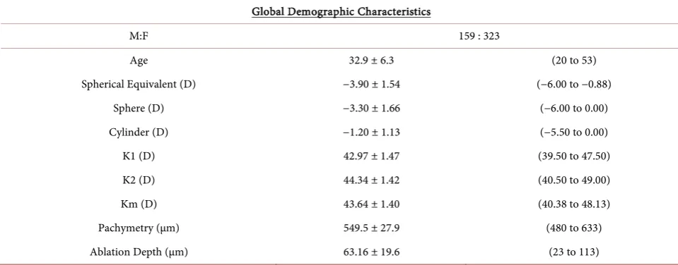
A total of 482 eyes of 275 patients were enrolled in this study, having met all in-clusion criteria. The demographic data and preoperative characteristics are summarized in Table 1.
In our study, the mean age was of 32 ± 6.3 years old, ranging from 20 to 53 years old. The average of SE was −3.90 ± 1.54D, ranging from −6.00 to −0.88D. The mean pachymetry was 549.5 ± 27.9 µm and mean Km was 43.64 ± 1.40D, ranging from 40.38 to 48.13D.
The preoperative characteristics of each Km group are organized in Table 2. Group 1 included a total of 116 eyes; group 2 included 283 eyes and group 3 in-cluded 77 eyes. All three groups had a mean preoperative SE ranging from 3.50 to 4.00 negative diopters. The average preoperative Km was of 40.80 ± 0.47D in group 1, ranging between 40.38 and 42.00D, representing flat corneas; 43.83± 0.69D in group 2, ranging between 42.25 and 45.75D; and 46.79 ± 0.63D in group 3, ranging between 46.00 and 48.13D, representing steep corneas. The av-erage pachymetry in group 1 was of 552.43 ± 32.27 µm, 547.29 ± 26.22 µm in group 2 and 553.5 ± 26.5 µm in group 3.
The mean ablation depth in group 1 was 57.60 ± 19.55 µm, contrasting with 66.35 ± 19.0 µm in group 3 which was considerably higher. This difference might be due to the slightly lower preoperative sphere and SE values in group 1, comparing to the other groups.
DOI: 10.4236/ojoph.2018.82012 87 Open Journal of Ophthalmolog
Table 1. Global demographic characteristics (N = 482 eyes).
Global Demographic Characteristics
M:F 159 : 323
Age 32.9 ± 6.3 (20 to 53)
Spherical Equivalent (D) −3.90 ± 1.54 (−6.00 to −0.88)
Sphere (D) −3.30 ± 1.66 (−6.00 to 0.00)
Cylinder (D) −1.20 ± 1.13 (−5.50 to 0.00)
K1 (D) 42.97 ± 1.47 (39.50 to 47.50)
K2 (D) 44.34 ± 1.42 (40.50 to 49.00)
Km (D) 43.64 ± 1.40 (40.38 to 48.13)
Pachymetry (µm) 549.5 ± 27.9 (480 to 633)
Ablation Depth (µm) 63.16 ± 19.6 (23 to 113)
Table 2. Demographic characteristics in preoperative period divided by groups of mean keratometry (mean ± standard deviation
(minimum to maximum)).
Group 1 - Flat Cornea Km ≤ 42.00D
(N: 116)
Group 2 42.00D < Km > 46.00D
(N: 283)
Group 3 - Steep Cornea Km ≥ 46.00D
(N: 77)
Parameter Mean ± SD Min to max Mean ± SD Min to max Mean ± SD Min to max Spherical Equivalent (D) −3.47 ± 1.54 (−6.00 to −1.00) −4.06 ± 1.55 (−6.00 to −1.00) −3.99 ± 1.38 (−6.00 to −1.75)
Sphere (D) −2.71 ± 1.67 (−6.00 to 0.00) −3.53 ± 1.60 (−6.00 to 0.00) −3.33 ± 1.61 (−6.00 to −0.50) Cylinder (D) −1.51 ± 1.29 (−5.00 to 0.00) −1.06 ± 1.01 (−5.50 to 0.00) −1.25 ± 1.21 (−4.50 to 0.00)
K1 (D) 40.15 ± 0.75 (39.50 to 42.00) 43.20 ± 0.84 (40.75 to 45.00) 44.92 ± 0.99 (42.50 to 47.50) K2 (D) 42.67 ± 0.77 (40.50 to 45.00) 44.45 ± 0.76 (42.75 to 46.50) 47.91 ± 0.71 (46.00 to 49.00) Mean K (D) 40.80 ± 0.47 (40.38 to 42.00) 43.83 ± 0.69 (42.25 to 45.75) 46.79 ± 0.63 (46.00 to 48.13) Pachymetry (µm) 552.43 ± 32.27 (480 to 633) 547.29 ± 26.22 (492 to 622) 553.5 ± 26.5 (505 to 624) Ablation Depth (µm) 57.60 ± 19.55 (23 to 112) 64.61 ± 19.48 (23 to 113) 66.35 ± 19.0 (23 to 101)
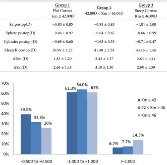
The refractive and keratometric outcomes at 6 months postoperatively are summarized in Table 3, according to Km group. The mean postoperative Km was of 39.99 ± 1.25D in group 1, 41.40 ± 1.54D in group 2 and 43.16 ± 1.46D in group 3. Concerning keratometry variation and SE variation, group 3, representing steep corneas, seemed to have a greater difference between preo-perative and postopreo-perative values, with a ∆SE of 2.98 ± 1.39D and a ∆Km of 2.63 ± 1.34 D, therefore a greater surgical effect, when compared with group 1, representing flat corneas with a ∆SE of 2.66 ± 1.43D and a ∆Km of 1.83 ± 1.20D. This difference was statistically significant (p < 0.008).
[image:4.595.57.540.323.515.2]DOI: 10.4236/ojoph.2018.82012 88 Open Journal of Ophthalmolog
Table 3. Results and variable variations at 6 months postoperatively (mean ± standard
deviation).
Group 1 Flat Cornea Km ≤ 42.00D
Group 2 42.00D < Km > 46.00D
Group 3 Steep Cornea Km ≥ 46.00D SE postop(D) −0.80 ± 0.85 −0.95 ± 0.85 −1.01 ± 1.00 Sphere postop(D) −0.46 ± 0.92 −0.64 ± 0.87 −0.66 ± 0.99 Cylinder postop (D) −0.69 ± 0.60 −0.63 ± 0.55 −0.71 ± 0.47 Mean K postop (D) 39.99 ± 1.25 41.40 ± 1.54 43.16 ± 1.46
∆Km (D) 1.83 ± 1.20 2.43 ± 1.37 2.63 ± 1.34
∆SE (D) 2.66 ± 1.43 3.10 ± 1.50 2.98 ± 1.39
Figure 1. Distribution of postoperative SE refraction by Km group.
only 6.7% and 7.7% of eyes respectively, were left with this degree of undercor-rection. The percentage of eyes achieving a postoperative SE between −0.50 and +0.50D, therefore achieving surgical success, was of 39.5%, 31.8% and 26% in groups 1, 2 and 3 respectively.
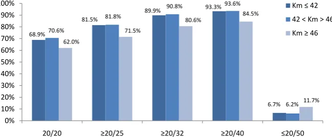
Considering visual outcomes, steep corneas also seemed to have worse UDVA comparing to eyes in the other groups. Figure 2 shows the cumulative UDVA among the keratometry groups, where slightly more than 81% of subjects on both group 1 and 2 achieved 20/25 vision or more, whereas only 71.5% of sub-jects in group 3 achieved the same vision. Additionally, almost 11.7% of eyes in group 3 were left with 20/50 vision or worse, contrasting with 6.7% and 6.2% in groups 1 and 2 respectively.
4. Discussion
The influence of keratometry on refractive outcomes of myopic LASIK has al-ready been debated in various studies, although with contradictory results. Rao
et al. [6] reported a trend toward undercorrection in flat corneas in eyes with
DOI: 10.4236/ojoph.2018.82012 89 Open Journal of Ophthalmolog
Figure 2. Distribution of Cumulative UDVA among keratometry groups.
reported tendency for undercorrection in flat corneas in myopic eyes with preo-perative SE ranging from −6.00 to −12.00D. Contrasting with these studies,
Christiansen et al. [4] studied moderate myopia, with preoperative SE ranging
from −2.00 to −5.99D, reporting better refractive and visual outcomes in flat corneas. The difference between these contrasting results lies in the different grades of myopia analyzed in each study. Studies reporting tendency for under-correction in flat corneas analyzed subjects with high myopia, who were origi-nally excluded from our study due to the fact that eyes with high myopia do not have surgical indication for LASIK in our institution.
Our results seem to be concordant with Christiansen et al.[4] findings con-cerning refractive and visual outcomes after LASIK, with steeper corneas show-ing a greater tendency toward refractive undercorrection and worse visual out-comes. We found a significant correlation between the variation of keratometry and spherical equivalent after LASIK in the evaluated population.
In our study, all three groups were homogeneous concerning preoperative SE and pachymetry. The mean ablation depth performed was greater in steeper corneas comparing to the other groups, showing greater variation of Km after LASIK, with an apparent greater surgical effect. However, although steep cor-neas suffered a greater corneal applanation, this was not reflected on SE varia-tion in the same magnitude. Despite the greater ablavaria-tion depth and surgical ef-fect in steeper corneas, these showed a greater tendency for undercorrection concerning SE and worse visual outcomes comparing to other degrees of preo-perative keratometry. Christiansen et al. proposes more aggressive laser ablation in eyes with Km ≥ 46.00D.
One of our study’s limitations is its retrospective nature, with a short fol-low-up period especially due to disparity in clinical records. It would be inter-esting to project a prospective longitudinal study, with a larger population, di-viding the sample per myopia grades, so as to categorize the influence of kera-tometry in different grades of preoperative myopia. This would certainly be helpful in clinical practice, to optimize the procedure and maximize the success of this refractive surgery.
In conclusion, myopic eyes with steeper corneas seem to have greater tenden-cy to undercorrection, also presenting worse visual outcomes.
68.9%
81.5%
89.9% 93.3%
6.7% 70.6%
81.8%
90.8% 93.6%
6.2% 62.0%
71.5%
80.6% 84.5%
11.7%
0% 10% 20% 30% 40% 50% 60% 70% 80% 90% 100%
20/20 ≥20/25 ≥20/32 ≥20/40 ≤20/50
Km ≤ 42
42 < Km > 46
DOI: 10.4236/ojoph.2018.82012 90 Open Journal of Ophthalmolog
References
[1] Schallhorn, S.C., et al. (2015) Effect of Postoperative Keratometry on Quality of Vi-sion in the Postoperative Period after Myopic Wavefront-Guided Laser in Situ Ke-ratomileusis. Journal of Cataract & Refractive Surgery, 41, 2715-2723.
https://doi.org/10.1016/j.jcrs.2015.06.034
[2] Seven, I., et al. (2016) Comparison of Patient-Specific Computational Modeling Predictions and Clinical Outcomes of LASIK for Myopia. Investigative Ophthal-mology & Visual Science, 57, 6287-6297. https://doi.org/10.1167/iovs.16-19948 [3] Kim, G., et al. (2014) Change in Keratometry after Myopic Laser in Situ
Keratomi-leusis and Photorefractive Keratectomy. Journal of Cataract & Refractive Surgery, 40, 564-574. https://doi.org/10.1016/j.jcrs.2013.09.016
[4] Christiansen, S., et al. (2012) The Effect of Preoperative Keratometry on Visual Outcomes after Moderate Myopic LASIK. Clinical Ophthalmology, 6, 459-464. [5] Mostafa, E.M., et al. (2015) Effect of Flat Cornea on Visual Outcome after LASIK.
Journal of Ophthalmology, 2015, 7. https://doi.org/10.1155/2015/794854
[6] Rao, S.K., et al. (2001) Effect of Preoperative Keratometry on Refractive Outcomes after LASIK. Journal of Cataract & Refractive Surgery, 27, 297-302.
https://doi.org/10.1016/S0886-3350(00)00746-X
[7] Perez-Santonja, J.J., et al. (1997) Laser in Situ Keratomileusis to Correct High Myo-pia. Journal of Cataract & Refractive Surgery, 23, 372-385.