
Original Article
Long non-coding RNA Hottip
modulates high-glucose-induced
inflammation and ECM accumulation
through miR-455-3p/WNT2B in mouse mesangial cells
Xiang-Jun Zhu, Zhaung Gong, Shu-Juan Li, Hai-Ping Jia, Da-Lin Li
Department of Nephrology, Yancheng City No. 1 People’s Hospital, Yancheng, Jiangsu, China
Received December 25, 2018; Accepted January 20, 2019; Epub July 1, 2019; Published July 15, 2019
Abstract: Long non-coding RNAs (lncRNAs) play important roles in the pathogenesis of various diseases, including diabetic nephropathy (DN). However, the detailed mechanism is still largely unknown. High-glucose treated SV40-MES13 cells was used to mimic diabetic nephropathy in vitro. qRT-PCR was introduced to measure Hottip, collagen
type I (Col. I), collagen type IV (Col. IV), fibronectin (FN), PAI-1, miR-455-3p and Wnt2B, IL-6, TNF-α mRNA level. Ellisa was used to examine the expression level of IL-6, TNF-α in the cell culture medium. Western blotting was employed to detect the protein level of Col. I, Col. IV, FN, PAI-1, Wnt2B, β-catenin and cyclin D1. Cell viability was examined
by MTT assay, luciferase reporter assay were used to determine the relationship between Hottip, miR-455-3p and
Wnt2B. In the results, Hottip and Wnt2B was upregulated in db/db DN mice and high-glucose treated mouse me -sangial cells (MMCs) while miR-455-3p was downregulated. High glucose treatment could enhance cell
prolifera-tion, and inflammaprolifera-tion, increase fibrosis-related protein expression and active Wnt2B/β-catenin/cyclin D1 pathway,
while Hottip silencing reversed all the effects caused by high-glucose treatment. miR-455-3p was a sponge target of
Hottip while Wnt2B was a downstream target of miR-445-3p. miR-445-3p inhibitor could suppress the effect of Hot
-tip knockdown in cell proliferation, inflammation and fibrosis-related protein expression. Our data supported lncRNA Hottip/miR-455-3p/Wnt2B axis plays an important role in cell proliferation, inflammation, and extracellular matrix
(ECM) accumulation in diabetic nephropathy.
Keywords: Hottip, diabetic nephropathy, miR-455-3p, renal fibrosis, Wnt2B
Introduction
Diabetic nephropathy (DN), a chronic kidney disease, is the main cause of end-stage renal disease in diabetic patients worldwide [1-5]. About 30%-40% diabetes patients will suffer diabetic nephropathy [6]. The main pathologic characteristics of DN are increased extracellu-lar matrix formation, podocyte loss, increased thickness of basement membrane, glomerular and tubular cell injury which lead to albumin-uria, glomerular matrix accumulation, glomeru-lar hypertrophy and kidney fibrosis or failure [7-10]. DN is a complicated pathologic process which involves many different molecules, and cells [11]. For example, immune inflammatory response [12-15], epithelial-mesenchymal tran- sition [16], apoptosis [17, 18]. Emerging
evi-dence demonstrated that epigenetics is also involved in DN. High glucose stimulation may lead to epigenetic modifications such as DNA methylation [19], histone modification [20, 21], chromatin remodeling [22], and non-coding RNA regulation [23-25].
expression of lncRNAs is related with various diseases, including DN [29]. However, the mechanism of lncRNA in DN is still largely unknown.
In present work, we validated that Hottip was upregulated in db/db DN mice and high-glucose treated mouse mesangial cells (MMCs). Then we hypothesized Hottip was involved in the pathogenesis of diabetic nephropathy and fur-ther study revealed that hottip/miR-455-3p/ Wnt2B played an important role in cell prolifer -ation, apoptosis, and fibrosis-protein expres -sion in DN which provided a novel view of DN pathogenesis.
Materials and methods
Animal model
Diabetic mouse model C57BL/KsJ-db/db and matched normal db/m mice were obtained from the Laboratory Animal Services Center, Yancheng City No. 1 People’s Hospital. All mice were kept in the animal facility of Yancheng City No. 1 People’s Hospital following the animal protocol. The animal experiment was approved by the Ethics Committee of the Animal Resea- rch Institute of Yancheng City No. 1 People’s Hospital and was performed in accordance with the guidelines of the National Institutes of Health Guide for the Care and Use of Laboratory Animals. The kidney cortex was extracted from the kidney tissues of the db/db and db/m mice for analysis.
Cell culture
Mouse mesangial cell lines (MMCs, SV40-MES13) were purchased from American Type Culture Collection (Manassas, VA). SV40-MES13 was cultured in Dulbecco’s modified eagle medium (DMEM) medium containing with 2 mM glutamine, 50 mM β-mercaptoethanol, 20% fetal bovine serum (FBS), 0.1% penicillin/ streptomycin antibiotics. SV40-MES13 cells were treated with low glucose (LG) or high glu-cose (HG), in low-gluglu-cose group, SV40-MES13 were stimulated with 5.5 mM glucose together with 19.5 mM mannitol; and in high-glucose group, MCs were stimulated with 25 mM glu-cose. High glucose treatment was performed to mimic the DN cells. HEK293T cells were cul -tured with DMEM medium containing 10% FBS and 0.1% penicillin/streptomycin antibiotics.
SV40-MES13 and HEK293T cells were cultured in humidified 95% air with 5% carbon dioxide at 37°C and the medium was changed every 2 days.
Quantitative real-time PCR (qRT-PCR)
Total RNA was extracted from mouse model and MMCs using Trizol reagent. For miRNA, cDNA was reversely transcribed by miScript II RT Kit (Qiagen). For mRNA and lncRNAs, cDNA was reversely transcribed by Reverse Tran- scription Reagents (Applied Biosystems). Hot-tip, miR-455-3p, WNT2B, Col. I, Col. IV, FN, and PAI-1 expression levels were measured by SYBR Green (Promega, Madison, WI, USA) according to the manufacturer’s protocol. QuantStudio™ 3 Real-Time PCR Systems (Ther-moFisher, USA) was used to detect the fluores -cence and the relative expression was calcu-lated by 2-ΔΔCt method. U6 and 18s rRNA were used to normalize the expression. Primer sequences are: mouse Hottip, forward 5’-GC- ACCATTCACTCACACTCCTG-3’, reverse 5’-CAAA- ACGGAATGCAACAGTGGA-3’; mouse WNT2B, forward 5’-CCGACGTGTCCCCATCTTC-3’, rever- se 5’-GCCCCTATGTACCACCAGGA-3’; mmu-miR-455-3p, forward 5’-ACACTCCAGCTGGGGCAG- TCCACGGGCATATACAC-3’; mouse Col. I (COL-1A1), forward 5’-GCTCCTCTTAGGGGCCACT-3’, reverse 5’-CCACGTCTCACCATTGGGG-3’; mou- se Col. IV (COL4A1), forward 5’-CTGGCACAA-AAGGGACGAG-3’, reverse 5’-ACGTGGCCGAGA- ATTTCACC-3’; mouse Fibronectin (FN, FN1), for -ward 5’-GATGTCCGAACAGCTATTTACCA-3’, rever- se 5’-CCTTGCGACTTCAGCCACT-3’; mouse PAI- 1 (Serpine1), forward 5’-TTCAGCCCTTGCTTGC- CTC-3’, reverse 5’-ACACTTTTACTCCGAAGTCG- GT-3’; mouse IL-6, TGATGGATGCTTCCA AACTG, GAGCATTGGAAGTTGGGGTA; mouse TNF-α, AC-TGAACTTCGGGGTGATTG, GCTTGGTGGTTTGCT- ACGAC; mouse 18S rRNA, forward 5’-GGC- CCTGTAATTGGAATGAGTC-3’, reverse 5’-CCAA-GATCCAACTACGAGCTT-3’; mouse U6 forward 5’-GTTGACATCCGTAAAGACC-3’, reverse 5’-GG- AGCCAGGGCAGTAA-3’.
Transient transfection
USA) according to the manufacturer’s instru- ctions.
Western blot analysis
Total protein was extracted from MMCs using RIPA lysis buffer. Equal protein samples were loaded and separated with 8%-10% sodium dodecyl sulfate polyacrylamide-gel electropho-resis and then transferred to polyvinylidene difluoride (PVDF) membranes (Bio-Rad, Her-cules, CA, USA). After blocking with 5% dried non-fat milk in TBS for 1 hr at room tempera -ture, the membranes were incubated with dif-ferent primary antibodies including and anti-Col. I (ab34710, 1:1000), anti-anti-Col. IV (ab6586, 1:1000), anti-fibronectin (anti-FN, ab2413, 1:1000), PAI-1 (ab66705, 1:1000), anti-Wnt2B (ab50575, 1:1000), anti-β catenin (ab32572, 1:1000), anti-cyclin D1 (ab16663, 1:1000) and anti-GAPDH (CST#5174, 1:1000) at 4°C overnight. On second day, the mem -branes were washed with TBS-T 3 times for 10 min each time and incubated with HRP-conjugated IgG secondary antibody at room temperature for 1 hr. After washing with TBST three times, the protein signals were detected using PierceTM ECL western blotting substrate
(ThermoFisher Scientific).
Luciferase reporter assay
Hottip-WT, Hottip-MUT, WT and WNT2B-MUT were constructed into pGL3 vector and cotransfected with miR-455-3p into HEK293T cells by Lipofectamine 2000 (Invitrogen). After 48 hrs, the luciferase activity was measured using dual-luciferase reporter system (Promega, Madison, WI, USA).
µL MTT solvent (4 mM HCl, 0.1% NP40 in iso-propanol) was added into each well and incu-bated for 2 h at 37°C. Then we measured the absorbance at 490 nanometers using the Microplate Reader (MG LABTECH, Durham, NC, USA).
Enzyme-linked immunosorbent assay (ELISA)
The levels of tumor necrosis factor alpha (TNF-α) and interleukin-6 (IL-6) were detected by ELISA in culture medium. ELISA kits for TNF-α and IL-6 (BD Biosciences, San Jose, CA) were used according to the manufacturers’ instruc-tions. Results were read at an optical density of 450 nm using a Spectra Max Plus plate reader (Molecular Devices, Sunnyvale, CA).
Statistical analysis
Student t-test was used for statistical analysis. Statistical significance means P value < 0.05 or as indicated. All data were displayed as mean ± SD (standard deviation) which was analyzed by GraphPad Prism 7.0 (GraphPad Software, San Diego, CA, USA).
Results
Hottip was upregulated in DN mice and high-glucose treated MMCs
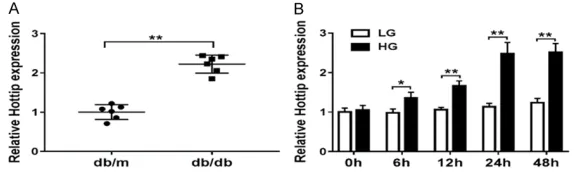
To check the expression of Hottip in diabetic nephropathy, we firstly extracted the kidney cortex from db/db DN mice and db/m non-DN mice. qRT-PCR results demonstrated that Hottip was increased in db/db DN mice about 2 fold compared with db/m non-DN mice (Figure 1A). To mimic DN in vitro, we used low glucose (LG, 5 mM) and high glucose (HG, 25 mM) to treat SV40-MES13 which is one mouse mesan-Figure 1. Hottip expression in DN mice and glucose treated MMCs. A. The
expression of lncRNA Hottip in kidney cortex of db/db and db/m mouse.
B. Mouse mesangial cells (MMCs, SV40-MES13) were treated with
high-glucose (25 mM) and low-high-glucose (5 mM) for 0, 6, 12, 24 and 48 hrs and the expression of Hottip was measured by qRT-PCR. *P < 0.05. **P < 0.01.
Cell viability
MTT assay kit (Sigma-Aldrich) was used to measure the cell viability. The transfected cells were seeded into 96-well plates (Corning Costar, Cor- ning, NY, USA) in a number of 2 × 10³. On different time
[image:3.612.88.377.73.159.2]gial cell line (MMCs) for 0 h, 6 h, 12 h, 24 h and 48 h. Hottip expression increased in a time-dependent manner significantly (Figure 1B). Hottip knockdown inhibited cell proliferation and inflammation in high-glucose treated MMCs
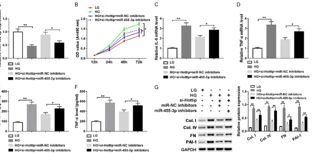
To explore the function of Hottip, we used small interfering RNA (siRNA) to knock down Hottip in SV40-MES13 cells and treated with high glucose (HG, 25 mM) (Figure 2A). The prolifera-tion was inhibited significantly by Hottip knock -down compared with control (Figure 2B). Then, RT-PCR results showed that HG could induce inflammation in SV40-MES13, which was inhib -ited by Hottip knockdown group compared with si-NC control group (Figure 2C and 2D). Next, we examined IL-6, and TNF-α expression in cell culture medium by ELISA and found that high glucose treatment increased IL-6, TNF-α expression which was decreased by Hottip knockdown (Figure 2E and 2F). The results sug-gested that Hottip knockdown could inhibit cell proliferation and inflammation in a DN model in vitro.
Hottip knockdown suppressed the fibrosis-related protein expression and extracellular matrix accumulation in high-glucose treated MMCs
Fibrosis is one pathologic basis of extracellular matrix accumulation and mesangial cell prolif-eration. So we checked the expression of fibro -sis-related proteins including collagen type I (Col. I), collagen type IV (Col. IV), fibronectin (FN) and PAI-1. The mRNA level of these four pro-teins were upregulated in db/db DN mice sig-nificantly (Figure 3A). Meanwhile, in vitro the mRNA (Figure 3B) and protein level (Figure 3C) of Col. I, Col. IV, FN and PAI-1 were increased in high glucose treated SV40-MES13 cells. But, Hottip knockdown could partially inhibit their expression (Figure 3C and 3D). These results demonstrated that Hottip silencing suppressed the fibrosis-related protein expression in vitro.
miR-455-3p acted as a target of Hottip
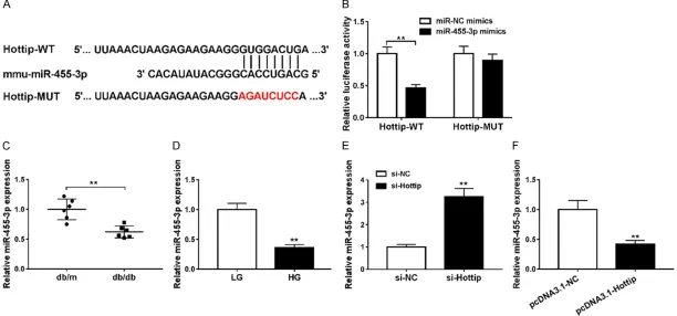
To further understand the function of Hottip, we found that miR-455-3p was a sponge target miRNA of Hottip by LncBase (Figure 4A). Then Figure 2. Hottip knockdown inhibited cell proliferation and inflammation in glucose treated SV40-MES13 cells.
SV40-MES13 cells were transfected with si-Hottip and treated with high glucose (HG) for 24 hr. (A) The expression of
lagen type I (Col. I), collagen type IV (Col. IV), fibronectin (FN) and PAI-1 were all upregulated after miR-455-3p inhibitor transfection (Figure 5G). Therefore, these data indicated that Hottip regulated cell proliferation, inflammation and fibrosis-protein expression by modulating miR-455-3p in MMCs.
Wnt2B is a target of miR-455-3p
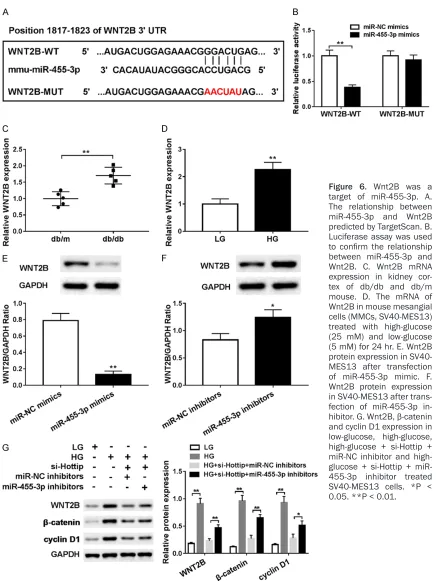
MicroRNAs usually play functions by binding 3’UTR of specific mRNA to regulate the tran -scription or mediate the degradation. So we used TargetScan to predict the target of miR-455-3p and found that WNT2B contains com -plementary sequence (Figure 6A). The lucifer-ase activity was decrelucifer-ased when WNT2B-WT and miR-455-3p mimics were cotransfected into HEK293T (Figure 6B). Then qRT-PCR dem-onstrated that WNT2B was upregulated in db/ db DN mice in vivo (Figure 6C) and high glucose (HG) treated SV40-MES13 cells in vitro (Figure Figure 3. Hottip knockdown suppressed the fibrosis-related protein genera
-tion and extracellular matrix (ECM) accumula-tion in SV40-MES13 cells. A.
The mRNA level of fibrosis-related protein including Col. I, Col. IV, FN and PAI-1 in kidney cortex of db/db and db/m mouse. B. The mRNA level of fibrosis-related protein including Col. I, Col. IV, FN and PAI-1 in mouse me -sangial cells (MMCs, SV40-MES13) treated with high-glucose (25 mM) and
low-glucose (5 mM) for 24 hr. C. Fibrosis-related protein expression in
SV40-MES13 after Hottip knockdown and HG treatment. D. Grey scale analysis of
fibrosis-related proteins. *P < 0.05. **P < 0.01.
we constructed luciferase reporter vector con-taining wild-type Hottip (Hottip-WT) and mutant Hottip (Hottip-MUT). The luciferase activity was decreased when the Hottip-WT and miR-455-3p mimics were cotransfected into HEK293T cells but not Hottip-MUT (Figure 4B). Then, qRT-PCR results showed that miR-455-3p was downregulated in db/db DN mice in vivo (Figure 4C) and high glucose (HG) treated SV40-MES13 cells in vitro (Figure 4D). In addition, knockdown of Hottip induced the expression of miR-455-3p (Figure 4E) while overexpression of Hottip inhibited miR-455-3p expression (Figure 4F). These results indicated that miR-455-3p was a target of Hottip and regulated by Hottip.
Hottip/miR-455-3p regulated cell proliferation, inflammation and ECM accumulation in MMCs
Next, we introduced a rescue experiment using miR-455-3p inhibitor. We treated SV40-MES13
col-Figure 4. mmu-miR-455-3p is a target of Hottip. A. The relationship between Hottip and miR-455-3p was predicted by LncBase. B. Luciferase assay confirmed the
relationship between Hottip and miR-455-3p. C. miR-455-3p expression in kidney cortex of db/db and db/m mouse. D. miR-455-3p expression in mouse mesangial
cells (MMCs, SV40-MES13) treated with high-glucose (25 mM) and low-glucose (5 mM) for 24 hr. E. miR-455-3p expression after Hottip silencing. F. miR-455-3p
[image:6.792.90.702.75.361.2]Figure 5. Hottip/miR-455-3p regulates cell proliferation, inflammation, and ECM accumulation in SV40-MES13 cells. SV40-MES13 cells were divided into 4 groups
and treated with 1) low glucose (5 mM), 2) high glucose (25 mM); 3) si-Hottip + miR-NC inhibitor + high glucose; 4) si-Hottip + miR-455-3p inhibitor + high glucose.
6D). Meanwhile, we found that WNT2B protein level was downregulated by overexpression miR-445-3p (Figure 6E) and upregulated by transfection with miR-445-3p inhibitor (Figure 6F).
[image:8.612.90.526.66.647.2]Wnt2B is a key regulator in Wnt signaling path -way of which β-catenin and cyclin D1 are down -stream target. So next, we checked the expres-sion of Wnt2B, β-catenin and cyclin D1. After high glucose treatment, all of Wnt2B, β-catenin Figure 6. Wnt2B was a
target of miR-455-3p. A. The relationship between
miR-455-3p and Wnt2B predicted by TargetScan. B.
Luciferase assay was used
to confirm the relationship
between miR-455-3p and
Wnt2B. C. Wnt2B mRNA
expression in kidney cor-tex of db/db and db/m mouse. D. The mRNA of
Wnt2B in mouse mesangial
cells (MMCs, SV40-MES13) treated with high-glucose (25 mM) and low-glucose
(5 mM) for 24 hr. E. Wnt2B
protein expression in SV40-MES13 after transfection
of miR-455-3p mimic. F. Wnt2B protein expression
in SV40-MES13 after trans-fection of miR-455-3p
in-hibitor. G. Wnt2B, β-catenin
and cyclin D1 expressions were upregulated significantly, but Hottip silencing could inhibit their expression which could be reversed by miR-455-3p inhibitor (Figure 6G). These data indicated that Wnt2B was a target of miR-455-3p and Wnt pathway was involved in DN.
Discussion
Diabetic nephropathy (DN) is a microvascular complication in diabetic patients and is a leading cause of chronic renal failure [30]. Mutifactors are involved in the pathogenesis of DN. In this study, we used the db/db DN mice [31] model and high-glucose treated mouse mesangial cell line (MMCs, SV40-MES13) [32] to study the underlying molecular mechanism of DN.
Long non-coding RNAs draw a lot attention in recent years. Although lncRNAs are not capable of protein coding, they may regulate the expres-sion of genes at transcriptional and posttran-scriptional level in DN. For example, long non-coding RNA metastasis associated lung ade- nocarcinoma transcript 1 (MALAT1) is increased in kidney cortex in streptozocin-induced diabet-ic nephropathy in mdiabet-ice and in cultured mouse podocytes stimulated with high glucose, knock-down of MALAT1 partially restored the function of podocytes [33]. CYP4B1-PS1-001 is signifi -cantly decreased in DN and overexpression inhibits the proliferation and fibrosis of mesan -gial cells [34]. ENSMUST00000147869 overex-pression could reverse the proliferation and fibrosis-related protein expression in mesangial cells [35]. Long non-coding RNA taurine-upreg-ulated gene 1 (Tug1) overexpression is associ-ated with improvements in mitochondrial bio-energetics in podocytes [36] and alleviates extracellular matrix accumulation by regulating miR-377 in mesangial cells [37]. In our study, we found long non-coding RNA Hottip was upregulated in kidney cortex in db/db DN mice and high glucose treated MMCs SV40-MES13 cells. We further found knockdown of Hottip could inhibit cell proliferation and inflammation as well as decrease fibrosis-related protein in high-glucose treated MMCs by regulating miR-455-3p and Wnt2b.
Extracellular matrix includes collagen, laminin, fibronectin and proteoglycans. Mesangial ma-trix changes include disrupted expression of collagen I, collagen IV, fibronectin, PAI-1 and
others [38, 39]. Recent studies indicated that some signaling pathways or molecules are involved in the regulation of ECM in DN. For example, Notch1 and Jagged1 are significantly increased in tubular epithelial cells in DN patients [40]. HG treatment can activate the PI3K/Akt/mTOR pathway and subsequently promotes the expression of ECM laminin-β1. TGF-β, Col. IV and NF-κB expression were mark -edly decreased in db/db mice treated withTLR4 inhibitor [41]. In addition, various investigations suggested that Wnt/β-catenin pathway is rele -vant to the evolution of renal fibrosis because its inhibition reduces ECM expression [42-44]. Wnt signaling pathways include a group of highly conserved signal transduction pathways which may regulate various biologic processes in different tissues including kidney. Wnt path -ways regulate cell-to-cell interactions in physi-ologic and pathphysi-ologic conditions, such as inflammation, angiogenesis, and fibrosis [45]. Wnt signaling pathways have been character -ized into two major transduction cascades: the Wnt/β-catenin-dependent or canonical path -way and the β-catenin-independent or non-canonical pathway [46]. Canonical Wnt path -way involves the accumulation of β-catenin but this is not true of the non-canonical pathway. Disrupted Wnt pathways are related to various diseases such as cancer [47], neurodegenera-tion [48], and cardiometabolic disorders [49]. Previous studies demonstrated that Wnt family members including Wnt2b, 4, 5b, 6, 7b, 9b and 11 have been expressed in kidney ontogeny [50] while 6, 7b, 9b, 11 are expressed in the early stage of nephrogenesis. In our study, we found Wnt2B was upregulated in db/db DN mice and in the high-glucose treated mouse mesangial cells (MMCs) as well as β-catenin and cyclin D1. In addition, Hottip knockdown decreased their expression while miR-455-3p inhibitor increased the expression of Wnt2B, β-catenin and cyclin D1. This means the canon -ical Wnt signaling pathway was involved in DN.
as activated the Wnt2B/β-catenin/cyclin D1 pathway. However, knockdown of Hottip par-tially reversed the effects caused by HG treat-ment in MMCs which provides a novel and promising target in DN treatment.
Disclosure of conflict of interest
None.
Address correspondence to: Dr. Da-Lin Li, Depart- ment of Nephrology, Yancheng City No. 1 People’s Hospital, 66#, South Renmin Road, Yancheng, Jiangsu, China. Tel: +86-515-88592872; E-mail: [email protected]
References
[1] Barutta F, Mastrocola R, Bellini S, Bruno G and
Gruden G. Cannabinoid receptors in diabetic kidney disease. Curr Diabetes Reports 2018; 18: 9.
[2] Packham DK, Alves TP, Dwyer JP, Atkins R, de Zeeuw D, Cooper M, Shahinfar S, Lewis JB,
Lambers Heerspink HJ. Relative incidence of ESRD versus cardiovascular mortality in pro-teinuric type 2 diabetes and nephropathy: re-sults from the DIAMETRIC (diabetes mellitus
treatment for renal insufficiency consortium) database. Am J Kidney Dis 2012; 59: 75-83.
[3] Afifi A, El Setouhy M, El Sharkawy M, Ali M, Ahmed H, El-Menshawy O, Masoud W. Diabetic
nephropathy as a cause of end-stage renal dis-ease in egypt: a six-year study. East Mediterr Health J 2004; 10: 620-626.
[4] Hadjadj S, Cariou B, Fumeron F, Gand E, Char
-pentier G, Roussel R, Kasmi AA, Gautier JF, Mo
-hammedi K and Gourdy P. Death, end-stage
renal disease and renal function decline in
pa-tients with diabetic nephropathy in French co -horts of type 1 and type 2 diabetes. Diabetolo-gia 2016; 59: 208-216.
[5] Chandie Shaw PK, Vandenbroucke JP, Tjandra YI, Rosendaal FR, Rosman JB, Geerlings W, de Charro FT, van Es LA. Increased end-stage dia -betic nephropathy in indo-asian immigrants liv-ing in the netherlands. Diabetologia 2002; 45: 337-341.
[6] Reddy MA, Park JT and Natarajan R. Epigenetic
modifications in the pathogenesis of diabetic
nephropathy. Semin Nephrol 2013; 33: 341-353.
[7] Hovind P, Rossing P, Tarnow L, Smidt UM and Parving HH. Progression of diabetic
nephropa-thy. Kidney Int 2001; 59: 702-709.
[8] Parving HH. Diabetic nephropathy: prevention
and treatment. Kidney Int 2001; 60:
2041-2055.
[9] Wang YQ, Fan CC, Chen BP and Shi J. Resistin-like molecule beta (RELM-β) regulates prolifer
-ation of human diabetic nephropathy mesan-gial cells via mitogen-activated protein kinases
(MAPK) signaling pathway. Med Sci Monit
2017; 23: 3897-3903.
[10] Zhang X, Song S and Luo H. Regulation of podocyte lesions in diabetic nephropathy via miR-34a in the notch signaling pathway. Medi-cine 2016; 95: e5050.
[11] Sun YM, Su Y, Li J and Wang LF. Recent ad -vances in understanding the biochemical and molecular mechanism of diabetic
nephropa-thy. Biochem Biophys Res Commun 2013;
433: 359-361.
[12] Duransalgado MB and Rubioguerra AF. Dia
-betic nephropathy and inflammation. World J
Diabetes 2014; 5: 393.
[13] Kanasaki K, Taduri G and Koya D. Diabetic ne
-phropathy: the role of inflammation in fibro
-blast activation and kidney fibrosis. Front En -docrinol 2013; 4: 7.
[14] Sun L and Kanwar YS. Relevance of TNF-α in the context of other inflammatory cytokines in the progression of diabetic nephropathy. Kid -ney Int 2015; 88: 662-665.
[15] Wada J and Makino H. Inflammation and the
pathogenesis of diabetic nephropathy. Clin Sci 2013; 124: 139-152.
[16] Ivonne L and Gunter W. Epithelial-to-mesen -chymal transition in diabetic nephropathy: fact
or fiction? Cells 2015; 4: 631-652.
[17] Inagi R, Ishimoto Y and Nangaku M. Proteosta-sis in endoplasmic reticulum--new mecha-nisms in kidney disease. Nat Rev Nephrol 2014; 10: 369-378.
[18] Cao AL, Wang L, Chen X, Wang YM, Guo HJ, Chu S, Liu C, Zhang XM and Peng W. Ursode -oxycholic acid and 4-phenylbutyrate prevent endoplasmic reticulum stress-induced podo-cyte apoptosis in diabetic nephropathy. Lab Investig 2016; 96: 610.
[19] Sapienza C, Lee J, Powell J, Erinle O, Yafai F,
Reichert J, Siraj ES and Madaio M. DNA
meth-ylation profiling identifies epigenetic differenc -es between diabet-es patients with ESRD and diabetes patients without nephropathy. Epi-genetics 2011; 6: 20-28.
[20] Li X, Li C and Sun G. Histone acetylation and its
modifiers in the pathogenesis of diabetic ne -phropathy. J Diabetes Res 2016; 2016: 1-11. [21] Sun G, Cui W, Guo Q and Miao L. Histone lysine
methylation in diabetic nephropathy. J Diabe-tes Res 2015; 2014: 654148.
[22] Badal SS, Yin W, Long J, Corcoran DL, Chang BH, Luan DT, Kanwar YS, Overbeek PA and Danesh FR. miR-93 regulates Msk2-mediated
chromatin remodelling in diabetic nephropa-thy. Nat Commun 2016; 7: 12076.
knockdown prevents progression of diabetic nephropathy. Sci Rep 2016; 6: 26072. [24] He F, Peng F, Xia X, Zhao C, Luo Q, Guan W, Li
Z, Yu X and Huang F. MiR-135a promotes renal fibrosis in diabetic nephropathy by regulating
TRPC1. Diabetologia 2014; 57: 1726-1736. [25] Puthanveetil P, Chen S, Feng B, Gautam A and
Chakrabarti S. Long non-coding RNA MALAT1
regulates hyperglycaemia induced inflamma -tory process in the endothelial cells. J Cell Mol Med 2015; 19: 1418.
[26] Balakirev ES and Ayala FJ. Pseudogenes: are they “junk” or functional DNA? Ann Rev Genet
2002; 37: 123-151.
[27] Yang F, Yi F, Zheng Z, Ling Z, Ding J, Guo J, Mao W, Wang X, Wang X and Ding X. Characteriza -tion of a carcinogenesis-associated long
non-coding RNA. Rna Biol 2012; 9: 110-116.
[28] Monika H and Tony G. Long non-coding RNAs in cancer and development: where do we go
from here? Int J Mol Sci 2015; 16: 1395-1405.
[29] Allison SJ. Diabetic nephropathy: a lncRNA and miRNA megacluster in diabetic nephropathy. Nat Rev Nephrol 2016; 12: 713.
[30] Kanwar YS, Wada J, Sun L, Xie P, Wallner EI, Chen S, Chugh S and Danesh FR. Diabetic ne -phropathy: mechanisms of renal disease
pro-gression. Exp Biol Med 2008; 233: 4-11.
[31] Cohen MP, Sharma K, Jin Y, Hud E, Wu VY, To
-maszewski J and Ziyadeh FN. Prevention of di -abetic nephropathy in db/db mice with glycat-ed albumin antagonists. A novel treatment strategy. J Clin Investig 1995; 95: 2338-2345. [32] Yang Y, Chen G, Cheng X, Teng Z, Cai X, Yang J,
Sun X, Lu W, Wang X and Yao Y. Therapeutic potential of digitoflavone on diabetic nephrop -athy: nuclear factor erythroid 2-related factor
2-dependent anti-oxidant and anti-inflamma -tory effect. Sci Rep 2015; 5: 12377.
[33] Hu M, Wang R, Li X, Fan M, Lin J, Zhen J, Chen
L and Lv Z. Lnc RNA MALAT 1 is dysregulated in diabetic nephropathy and involved in high glu-cose-induced podocyte injury via its interplay
with β-catenin. J Cell Mol Med 2017; 21:
2732-2747.
[34] Min W, Wang S, Di Y, Qin Y and Lu W. A novel long non-coding RNA CYP4B1-PS1-001 regu
-lates proliferation and fibrosis in diabetic ne -phropathy. Mol Cell Endocrinol 2016; 426: 136-145.
[35] Wang M, Yao D, Wang S, Yan Q and Lu W. Long
non-coding RNA ENSMUST00000147869
pro-tects mesangial cells from proliferation and fi -brosis induced by diabetic nephropathy. Endo-crine 2016; 54: 1-12.
[36] Long J, Badal SS, Ye Z, Wang Y, Ayanga BA, Gal
-van DL, Green NH, Chang BH, Overbeek PA and Danesh FR. Long noncoding RNA Tug1
regulates mitochondrial bioenergetics in dia-betic nephropathy. J Clin Investig 2016; 126: 4205-4218.
[37] Duan LJ, Ding M, Hou LJ, Cui YT, Li CJ and Yu DM. Long noncoding RNA TUG1 alleviates ex-tracellular matrix accumulation via mediating
microRNA-377 targeting of PPARγ in diabetic nephropathy. Biochem Biophys Res Commun
2017; 484: 598-604.
[38] Mason RM and Wahab NA. Extracellular matrix
metabolism in diabetic nephropathy. J Am Soc Nephrol 2003; 14: 1358-1373.
[39] Kolset SO, Reinholt FP and Jenssen T. Diabetic
nephropathy and extracellular matrix. J Histo-chem CytoHisto-chem 2012; 60: 976.
[40] Bielesz B, Sirin Y, Han S, Niranjan T, Gruenwald A, Ahn S, Kato H, Pullman J, Gessler M and
Haase VH. Epithelial Notch signaling regulates
interstitial fibrosis development in the kidneys
of mice and humans. J Clin Investig 2010; 120: 4040-4054.
[41] Cha JJ, Hyun YY, Lee MH, Kim JE, Nam DH, Song HK, Kang YS, Lee JE, Kim HW and Han JY.
Renal protective effects of toll-like receptor 4 signaling blockade in type 2 diabetic mice. En-docrinol 2013; 154: 2144-2155.
[42] Hwang I, Seo EY and Ha H. Wnt/β-catenin sig -naling: a novel target for therapeutic
interven-tion of fibrotic kidney disease. Arch Pharm Res
2009; 32: 1653-1662.
[43] Cisternas P, Vio CP and Inestrosa NC. Role of
wnt signaling in tissue fibrosis, lessons from
skeletal muscle and kidney. Curr Mol Med 2014; 14: 510-522.
[44] He W, Dai C, Li Y, Zeng G, Monga SP and Liu Y. Wnt/β-catenin signaling promotes renal inter
-stitial fibrosis. J Am Soc Nephrol 2009; 20:
765-776.
[45] Macdonald BT, Tamai K and He X.
Wnt/beta-catenin signaling: components, mechanisms, and diseases. Dev Cell 2009; 17: 9-26. [46] Rao TP and Kühl M. An updated overview on
wnt signaling pathways: a prelude for more. Circ Res 2010; 106: 1798-1806.
[47] Anastas JN and Moon RT. WNT signalling path -ways as therapeutic targets in cancer. Nat Rev Cancer 2013; 13: 11-26.
[48] Berwick DC and Harvey K. The importance of
wnt signalling for neurodegeneration in
parkin-son’s disease. Biochem Soc Trans 2012; 40:
1123-1128.
[49] Schinner S. Wnt-signalling and the metabolic
syndrome. Horm Metab Res 2009; 41: 159-163.
[50] Pulkkinen K, Murugan S and Vainio S. Wnt sig
![keylogger[kissmefox] pdf](data:image/gif;base64,R0lGODlhAQABAIAAAP///wAAACH5BAEAAAAALAAAAAABAAEAAAICRAEAOw==)