Nijmegen
The following full text is a publisher's version.
For additional information about this publication click this link.
http://hdl.handle.net/2066/189883
Please be advised that this information was generated on 2018-04-11 and may be subject to
change.
Visual discrimination of screen-detected
persistent from transient subsolid nodules:
An observer study
Kaman Chung1*, Francesco Ciompi1, Ernst T. Scholten1, Jin Mo Goo2, Mathias Prokop1, Colin Jacobs1, Bram van Ginneken1, Cornelia M. Schaefer-Prokop1,3
1 Department of Radiology and Nuclear Medicine, Radboud University Medical Center, Nijmegen, the Netherlands, 2 Department of Radiology, Seoul National University College of Medicine, and Institute of Radiation Medicine, Seoul National University Medical Research Center, Seoul, Korea, 3 Department of Radiology, Meander Medical Center, Amersfoort, the Netherlands
Abstract
Purpose
To evaluate whether, and to which extent, experienced radiologists are able to visually cor-rectly differentiate transient from persistent subsolid nodules from a single CT examination alone and to determine CT morphological features to make this differentiation.
Materials and methods
We selected 86 transient and 135 persistent subsolid nodules from the National Lung Screening Trial (NLST) database. Four experienced radiologists visually assessed a prede-fined list of morphological features and gave a final judgment on a continuous scale (0– 100). To assess observer performance, area under the receiver operating characteristic (ROC) curve was calculated. Statistical differences of morphological features between tran-sient and persistent lesions were calculated using Chi-square. Inter-observer agreement of morphological features was evaluated by percentage agreement.
Results
Forty-nine lesions were excluded by at least 2 observers, leaving 172 lesions for analysis. On average observers were able to differentiate transient from persistent subsolid nodules
10 mm with an area under the curve of 0.75 (95% CI 0.67–0.82). Nodule type, lesion mar-gin, presence of a well-defined border, and pleural retraction showed significant differences between transient and persistent lesions in two observers. Average pair-wise percentage agreement for these features was 81%, 64%, 47% and 89% respectively. Agreement for other morphological features varied from 53% to 95%.
Conclusion
The visual capacity of experienced radiologists to differentiate persistent and transient sub-solid nodules is moderate in subsub-solid nodules larger than 10 mm. Performance of the visual a1111111111 a1111111111 a1111111111 a1111111111 a1111111111 OPEN ACCESS
Citation: Chung K, Ciompi F, Scholten ET, Goo JM,
Prokop M, Jacobs C, et al. (2018) Visual discrimination of screen-detected persistent from transient subsolid nodules: An observer study. PLoS ONE 13(2): e0191874.https://doi.org/ 10.1371/journal.pone.0191874
Editor: Gayle E. Woloschak, Northwestern
University Feinberg School of Medicine, UNITED STATES
Received: March 22, 2017 Accepted: January 12, 2018 Published: February 13, 2018
Copyright:©2018 Chung et al. This is an open access article distributed under the terms of the
Creative Commons Attribution License, which permits unrestricted use, distribution, and reproduction in any medium, provided the original author and source are credited.
Data Availability Statement: “CT data are from the
NLST study whose workers may be contacted at
https://biometry.nci.nih.gov/cdas/nlst/.”
Funding: (Dutch Cancer Society (KWF
Kankerbestrijding) grant number: KUN 2013−6110
https://www.kwf.nl/english/pages/default.aspx"The funders had no role in study design, data collection and analysis, decision to publish, or preparation of the manuscript."
assessment of CT morphology alone is not sufficient to generally abandon a short-term fol-low-up for subsolid nodules.
Introduction
Results of lung cancer screening Computed Tomography (CT) studies revealed the importance of subsolid nodules as potential early adenocarcinomas. In the Early Lung Cancer Action Proj-ect (ELCAP) study the prevalence of subsolid nodules was found to be lower compared to solid nodules. However, subsolid nodules demonstrated a higher malignancy rate in the
detected subsolid nodules of 34% (15/44) compared to 7% (14/189) for solid nodules [1].
Another study evaluating a group of clinically and screen-detected lesions even reported 81%
(43/53) of resected subsolid nodules to be (pre)malignant [2].
The most frequent benign disease causing subsolid nodules is a focal infection [3,4]. Other
more rare underlying benign diseases are a focal organizing pneumonia or focal fibrosis [5,6].
Subsolid nodules caused by infection will eventually disappear. Differentiation of transience versus persistence of subsolid nodules thus represents the first diagnostic task to discriminate between benign and potentially malignant lesions, and a short-term three months follow-up
has been recommended by the Fleischner Society and the British Thoracic Society [7,8]. The
percentage of subsolid nodules detected in screening studies varied from 2% to 20% of all
base-line screen-detected non-calcified nodules [1,9,10]. Prospective discrimination of transient
from persistent lesions would therefore contribute to the reduction of follow-up CTs. Previous studies on this subject evaluated the contribution of texture analysis and clinical features, but
did not assess human observer performance [11–13].
The only other morphological feature used for risk prediction of subsolid nodules besides
persistence and lesion growth, is nodule size and the presence/size of a solid component [10,
14]. For solid nodules spiculation is an important predictor of malignancy in a recently
pub-lished (screening) risk model [10]. However, for subsolid nodules no additional morphological
features have been established. Defining morphological features for transient and persistent subsolid nodules would be a valuable first step.
The purpose of this study was therefore to evaluate whether and to which extent experi-enced radiologists would be able to differentiate transient from persistent subsolid nodules from a single CT examination by visual analysis alone. Secondly, we aimed to identify which morphological features are used by the radiologists to make this differentiation.
Materials and methods
Study population
We recruited subsolid nodules from the National Lung Screening Trial (NLST). The NLST was approved by the institutional board at each participating medical institution and
partici-pants provided written informed consent before randomization [15]. In total the NLST had
26,722 participants. Of those, 3194 participants had at least one subsolid nodule annotated by the NLST screening radiologist in any of the 3 screening rounds. Nine participants did not have any scans available, leaving 3185 participants for further analysis.
For this observer study, we used baseline (year 0) subsolid nodules only. The NLST annota-tions did not contain year-to-year linking between the same lesions, therefore we re-annotated all lesions by using information from the NLST database (slice number, nodule type, lobe loca-tion, size). Annotations were done by two medical students and one medical researcher using
Competing interests: "The authors have declared
in-house software (CIRRUS Lung Screening, Diagnostic Image Analysis Group, Radboud Uni-versity Medical Center, Nijmegen, the Netherlands). A subsolid nodule was defined as tran-sient if the nodule had disappeared on follow-up CT. A subsolid nodule was defined as persistent if the nodule remained visible on follow-up CT.
Subsequently we only selected CTs with a slice thickness of2 mm, to ensure the quality
of the coronal and sagittal projections of the lesions. As morphology is more difficult to assess
in smaller lesions and thicker slices, we only selected lesions10 mm (rounded average
diam-eter) in this observer study. In total 232 subsolid lesions were eligible for our study. Eleven lesions (11/232, 4.7%) could not be located on the scans. Thus, our final dataset for the observer study contained 221 subsolid lesions.
Observer study
All study lesions were independently evaluated by four experienced radiologists (ETS, CSP,
MP, and JMG). All of them had>15 years of experience in reading chest CTs and had
exten-sive experience with evaluating screen-detected nodules. Nodules were presented in a random order to each observer. Observers were asked to score the morphological nodule features using a predefined list. The list of morphological features as well as the definitions can be found in
Table 1. In addition, they were asked to estimate the probability that the lesion was persistent on a scale between 0 and 100, with 0 representing certainly transient and 100 representing cer-tainly persistent.
Reading methodology
A reading workstation designed to optimize workflow and to document the scoring data was used (CIRRUS Observer, Diagnostic Image Analysis Group, Radboud University Medical Center, Nijmegen, the Netherlands). After opening a case, a magnified axial view of the nodule under evaluation was centered in the middle of the display. Coronal/sagittal projections were
available on the right side of the screen (Fig 1). The position of the magnified view was
indi-cated by center lines on the smaller views. Using this set-up, observers did not have to search for the lesion. For all cases, the full 3D CT dataset was available for evaluation. All views could be (de)magnified. A standard lung window with a width of 1500 HU and a center of -650 HU was used as a default, but could be adjusted if necessary. Nodule diameters could be measured manually using electronic calipers.
The morphological features to be scored were listed on the left side of the monitor display. Scoring had to be completed before the next lesion could be displayed. Observers were allowed to place any comments if needed. No specifications with respect to comments were made pro-spectively. Lesions indicated in the comments for exclusion by 2 or more observers were omit-ted for further analysis. No information about follow-up appearance, persistence or any other outcome was provided.
Statistical analysis
Receiver Operating Characteristic (ROC) analysis was performed for each observer. Areas
under the curve (Az) and 95% confidence intervals (CI) were calculated to determine the
abil-ity to differentiate between transient and persistent lesions. We did not take into account within-participant correlation of participants with multiple nodules, because multiple nodules
in a participant were considered as multiple independent nodules [16,17]. Univariate analysis
(Chi-square) per observer was used to assess whether a certain morphological feature was
scored significantly different in transient or persistent nodules. P-values<0.05 were
by calculating percentage agreement for each pair of observers. Statistical analyses were per-formed using SPSS, version 20.0 (SPSS, Chicago, Ill).
Results
Study group
Of all lesions 61% (135/221) were persistent. The median average diameter of persistent lesions was 12.0 mm (IQR 10.0–15.0 mm). Accordingly, 39% (86/221) were transient and had a median average diameter of 12.0 mm (IQR 11.0–16.5 mm). Forty-nine lesions were excluded from further data analysis because at least 2 of the 4 observers had made the comment that the opacification under review, which had been marked as subsolid nodule in the NLST database, would in fact not represent a nodular (subsolid) opacification when taking all three planes into consideration. Comments leading to exclusion were non-nodular (N = 11), solid lesion (N = 8), wall of emphysema (N = 3), apical scarring, (N = 7), fibrosis (N = 5) and (plate-like)
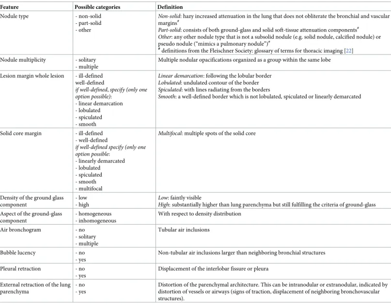
Table 1. List of morphological features scored by each observer. For all features one category had to be chosen obligatorily.
Feature Possible categories Definition
Nodule type - non-solid - part-solid - other
Non-solid: hazy increased attenuation in the lung that does not obliterate the bronchial and vascular margins#
Part-solid: consists of both ground-glass and solid soft-tissue attenuation components# Other: any other nodule type that is not a subsolid nodule (e.g. solid nodule, calcified nodule) or pseudo nodule (“mimics a pulmonary nodule”)#
#
definitions from the Fleischner Society: glossary of terms for thoracic imaging [22] Nodule multiplicity - solitary
- multiple
Multiple nodular opacifications organized as a group within the same lobe Lesion margin whole lesion - ill-defined
well-defined
if well-defined,specify (only one option possible):
- linear demarcation - lobulated - spiculated - smooth
Linear demarcation: following the lobular border Lobulated: undulated contour of the border Spiculated: with lines radiating from the borders
Smooth: a well-defined border which is not lobulated, spiculated or linearly demarcated
Solid core margin - ill-defined - well-defined
if well-defined specify (only one option possible: - linearly demarcated - lobulated - spiculated - smooth - multifocal
Multifocal: multiple spots of the solid core
Density of the ground glass component
- low - high
Low: faintly visible
High: substantially higher than lung parenchyma but still fulfilling the criteria of ground-glass Aspect of the ground-glass
component
- homogeneous - inhomogeneous
With respect to density distribution Air bronchogram - no
- solitary - multiple
Tubular air inclusions
Bubble lucency - no - yes
Non-tubular air inclusions larger than neighboring bronchial structures Pleural retraction - no
- yes
Displacement of the interlobar fissure or pleura External retraction of the lung
parenchyma
- no - yes
Distortion of the parenchymal architecture. This can be intranodular or extranodular, indicated by distortion of vessels or airways (signs of traction, displacement of neighboring bronchovascular structures).
atelectasis (N = 15). Thus the final study group consisted of 172 subsolid lesions (101 persis-tent, 71 transient).
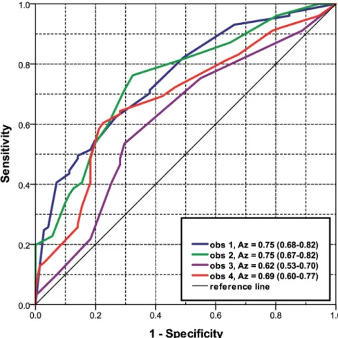
Discrimination of persistent from transient nodules
Observers 1 to 4 separately achieved an Azfor discriminating persistent from transient
subso-lid nodules of 0.75 (95% CI 0.68–0.82), 0.75 (95% CI 0.67–0.82), 0.62 (95% CI 0.53–0.70) and
0.69 (95% CI 0.60–0.77), respectively (Fig 2).
Considering the score of 50 as a threshold for discriminating between transience (scores 0–50) and persistence (scores 51–100), the four observers correctly identified 58/71 (82%), 63/ 71 (89%), 51/71 (72%) and 55/71 (77%) transient lesions. The observers correctly identified 52/101 (51%), 37/101 (37%), 47/101 (47%) and 61/101 (60%) persistent nodules, respectively.
Taking the same thresholds for transience (0–50) and persistence (51–100), all four observ-ers agreed on the same classification in 105 of the 172 nodules (61%). 68 of these 105 nodules (65%) were correctly classified, 37 of the 105 nodules (35%) were misclassified by all four observers. Thirty of the correctly classified nodules were persistent and 38 were transient. Figs
3and4show examples of correctly and incorrectly identified lesions for which all or the
majority of observers agreed on the classification.
Fig 1. Reading workstation. The morphological features to be scored are listed on the left side of the monitor display. Lower-left corner has two text fields to enter the
probability (0–100) and any comments. A magnified axial view of the nodule under evaluation is centered in the middle of the display. Coronal/sagittal projections are available on the right side of the screen, display size of the three projections was interchangeable. Processing tools such as windowing and magnification as well the full 3D CT dataset were available at any time.
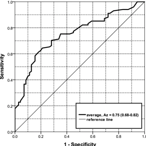
Averaging the scores of the four observers resulted in an Azof 0.75 (95% CI 0.68–0.82) (Fig
5). Using the average scores a sensitivity of>90% for persistent lesions was only achieved at
the expense of a specificity of<30% (e.g., sensitivity/specificity is 91% / 28%).
Morphology assessment: Univariate analysis
Morphological features that showed significant difference between transient and persistent in
at least 2 observers are listed inTable 2. At a significance level of p<0.05, nodule type and
lesion margin were scored significantly different by 2 observers (p = 0.016 and p = 0.025, p = 0.001 and p = 0.044 respectively). Part-solid nodules were more often seen in persistent lesions compared to transient lesions in all observers, reaching statistical significance in two of them (p = 0.016 and p = 0.025). The subcategory of a well-defined border yielded significant
difference in 2 observers (p<0.001 and p = 0.001). Linear demarcation following the lobular
border was the only feature in this category to be seen more often in transient lesions in three observers. Lobulated, spiculated and smooth borders were scored more often in persistent lesions. Pleural retraction was observed more frequently in persistent than transient lesions reaching significance in two observers (p = 0.006, p = 0.037).
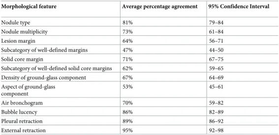
Inter-reader variability of morphology
Average pair-wise percentage agreement was highest in external retraction, pleural retraction and bubble lucency (95%, 89%, and 86% respectively). Nodule type had an agreement of 81%,
Fig 2. Receiver Operating Characteristic (ROC) curves for observer 1, 2, 3 and 4 to predict the persistence of the subsolid lesions10 mm. Az(Areas Under the Curve) and 95% confidence interval in parenthesis, obs = observer.
followed by nodule multiplicity (73%), solid core margin (71%) and presence of an air-bronch-ogram (70%). Lower agreement was found in density of ground-glass component (67%), lesion margin (64%) and the subcategory of a well-defined solid core margin (62%). Lowest agree-ment was found for aspect of ground-glass component (53%) and the subcategory of a well-defined lesion margin (47%). The average pair-wise agreement and the 95% confidence
inter-vals can be found inTable 3.
Fig 3. (a) Correctly identified transient lesion with a probability score of40 by all four observers. (b) Correctly identified persistent lesion with a probability score of80 by all four observers. (c) Incorrectly identified lesion by majority of observers: transient lesion, but scored as persistent (probability score60). (d) Incorrectly identified lesion by majority of observers: persistent lesion, but scored as transient (probability score40).
https://doi.org/10.1371/journal.pone.0191874.g003
Fig 4. (a) A transient lesion with disagreement (2 versus 2) among observers. (b) A persistent lesion with
disagreement (2 versus 2) among observers.
Fig 5. Receiver Operating Characteristic (ROC) curves for the average of all four observers. Az(Area Under the Curve) and 95% confidence interval in parenthesis.
https://doi.org/10.1371/journal.pone.0191874.g005
Table 2. Univariate analyses. Table describes morphological features with at least 2 observers in which the feature is seen significantly different between transient (T)
and persistent (P) subsolid nodules using Chi-square. The total number of included nodules after exclusion is 172.
T P P-value Obs. 1 T P P-value Obs. 2 T P P-value Obs. 3 T P P-Value Obs. 4 Nodule type non-solid 46 49 45 47 43 43 49 49 part-solid 23 52 P = 0.016 25 53 P = 0.081 27 55 P = 0.064 21 48 P = 0.025 other 2 0 1 1 1 3 1 4 Lesion Margin ill-defined 29 30 P = 0.130 55 51 P<0.001 37 68 P = 0.044 38 39 P = 0.053 well-defined 30 71 16 50 34 33 33 62 If well-defined N = 112 N = 66 N = 67 N = 96 linearly demarcated 27 13 1 3 18 9 23 16 lobulated 1 15 P<0.001 8 28 P = 0.813 0 1 P = 0.063 7 20 P = 0.001 spiculated 5 14 1 4 0 2 3 16 smooth 8 29 7 14 15 22 1 10 Pleural Retraction no 66 78 P = 0.006 71 96 P = 0.057 67 95 P = 0.933 68 87 P = 0.037 yes 5 23 0 5 4 6 3 14 https://doi.org/10.1371/journal.pone.0191874.t002
Discussion
The most frequent cause of transient subsolid nodules is focal infection. A persistent subsolid nodule, however, is potentially malignant and requires follow-up or alternative diagnostic work-up. A prospective estimation of whether the lesion would be persistent or transient would aid in reducing unnecessary follow-ups. This is the first study assessing the performance of human visual analysis for predicting the likelihood of persistence in subsolid nodules. Results of our study indicate that experienced radiologists are at best only moderately able
(average Azof all readings 0.75) to visually differentiate transient from persistent character in
subsolid nodules10 mm. In addition the individual performance among the observers
var-ied substantially with Azvalues ranging from 0.62 to 0.75. Given the variability among the
observers, the moderate agreement and the imperfect performance of experienced radiologists, human visual analysis alone has to be considered insufficient to reproducibly predict if a subsolid nodule is persistent or transient. In that respect our results confirm published
management strategies [7,8] that recommend a 3-month follow-up CT for clarification of
persistency.
A study by Lee HJ et al. [18] evaluated the performance of radiologists predicting benign
and malignant subsolid nodules, a differentiation that might be less complex, since persistent lesions can be both benign and malignant and malignant lesions may expose more suggestive features. However, even with the availability of several clinical parameters (age, sex, pack years,
history of lung cancer) and knowledge of predefined predictive CT information, an average Az
value of 0.77 for non-solid and of 0.76 for part-solid nodules were achieved, thus in fact com-parable to our results.
Secondly, we found that none of the morphology features yielded significant discrimination in all four observers. Most promising features were nodule type, lesion margin, presence of a well-defined lesion margin and pleural traction. The average pair-wise percentage agreement was relatively high in nodule type and pleural retraction (81% and 89% respectively). A consid-erably lower agreement, however, was found for features that had to be rated qualitatively such as lesion margin in general or the subcategory of a well-defined lesion margin (63% and 47%, respectively), indicating that these features do not appear to be sufficiently definable by visual analysis to serve as a broadly applicable criterion within a screening process.
Table 3. Average pair-wise percentage agreement of the morphological features.
Morphological feature Average percentage agreement 95% Confidence Interval
Nodule type 81% 79–84
Nodule multiplicity 73% 61–84
Lesion margin 64% 56–71
Subcategory of well-defined margins 47% 44–50
Solid core margin 71% 67–75
Subcategory of well-defined solid core margins 62% 59–65
Density of ground-glass component 67% 64–69
Aspect of ground-glass component 53% 45–61 Air bronchogram 70% 59–82 Bubble lucency 86% 82–89 Pleural retraction 89% 86–92 External retraction 95% 92–98 https://doi.org/10.1371/journal.pone.0191874.t003
Interestingly however, when looking at the subcategory of a well-defined border, three observers scored linearly demarcated border more frequently in transient lesions (27/40, 18/ 27, and 23/39) compared to persistent lesions. We did not prospectively define whether the lin-ear demarcation following the lobular border had to be present in several projections, which most likely contributed to the fact that one observer scored the feature only 4 times. The
find-ing of linear demarcation shows similarity with a findfind-ing reported by Felix et al. [13]. Their
study described a polygonal shape (defined “as a lesion with linear or concave margins at every corner”) as indicative for a transient lesion. Furthermore, they found that transient subsolid nodules were more frequently lobulated than persistent nodules. The finding of lobulation
being predictive for transience reported by Felix et al. [13] is in contradiction to the other
study by Lee SM et al. [19], who reported lobulation as indicative for malignancy. Similarly we
found that 74% to 100% of the lobulated lesions were found to be persistent (15/16, 28/36, 1/1, 20/27 respectively).
In this study we selected the subsolid nodules following the nodule type annotations of the NLST database. Previous studies have shown that the agreement among radiologists is only
moderate with regards to the differentiation of part-solid, non-solid and solid nodules [20,21].
Therefore we decided to exclude all lesions that were considered not a subsolid nodule by at least 2 of the 4 experienced observers in our study, as indicated in their comments. We did so, to increase accuracy and reliability of the observer data.
Our study has some limitations. First, our study did not include any elaborate texture or quantitative analysis. Visual CT features in combination with elaborate objectively quantifiable measures might not only improve performance but also achieve a higher reproducibility.
Sec-ond, we selected lesions10 mm only, taking into account the fact that the majority of the
NLST CTs has not been reconstructed with 1 mm slice thickness, thus not providing isotropic high resolution image quality in all three projections. The level of performance and reader agreement we found, confirms the notion that visual assessment of morphological features in
lesions<10mm will be even more difficult and less reliable. Last, the CT examinations of the
NLST trial have been obtained with different scanners and variable slice thickness. Though
only scans with a slice thickness of2 mm were included, the diverging image quality might
have influenced the visual assessment of the nodules.
In conclusion, experienced radiologists are moderately able to determine persistent and
transient nodule character in lesions10 mm visually. There are morphological features
indicative for the discrimination of persistent and transient nodules, but none of them yielded significant discrimination in all four observers. Our results show that performance of the visual assessment of CT morphology alone is not sufficient to generally abandon a short-term follow-up and inter-reader variability plays a substantial role even among highly experienced
observers.
Supporting information
S1 File. Main data file. Data file containing observers’ scores (probability that the lesion was
persistent on a scale between 0–100 and morphology) and the ground truth for each case. (SAV)
Acknowledgments
The authors thank the National Cancer Institute for access to NCI’s data collected by the National Lung Screening Trial (project no. NLST-74). The statements contained herein are solely those of the authors and do not represent or imply concurrence or endorsement by NCI.
Author Contributions
Conceptualization: Kaman Chung, Francesco Ciompi, Ernst T. Scholten, Jin Mo Goo,
Mathias Prokop, Colin Jacobs, Bram van Ginneken, Cornelia M. Schaefer-Prokop.
Data curation: Kaman Chung, Francesco Ciompi, Ernst T. Scholten, Colin Jacobs. Formal analysis: Kaman Chung, Francesco Ciompi, Colin Jacobs, Cornelia M.
Schaefer-Prokop.
Funding acquisition: Cornelia M. Schaefer-Prokop.
Investigation: Kaman Chung, Francesco Ciompi, Bram van Ginneken.
Methodology: Kaman Chung, Francesco Ciompi, Ernst T. Scholten, Jin Mo Goo, Mathias
Prokop, Colin Jacobs, Bram van Ginneken, Cornelia M. Schaefer-Prokop.
Project administration: Kaman Chung.
Resources: Kaman Chung, Francesco Ciompi, Ernst T. Scholten, Jin Mo Goo, Mathias
Pro-kop, Colin Jacobs, Bram van Ginneken.
Software: Francesco Ciompi, Colin Jacobs.
Supervision: Bram van Ginneken, Cornelia M. Schaefer-Prokop. Visualization: Kaman Chung, Francesco Ciompi.
Writing – original draft: Kaman Chung, Cornelia M. Schaefer-Prokop.
Writing – review & editing: Kaman Chung, Francesco Ciompi, Ernst T. Scholten, Jin Mo
Goo, Mathias Prokop, Colin Jacobs, Bram van Ginneken, Cornelia M. Schaefer-Prokop.
References
1. Henschke CI, Yankelevitz DF, Mirtcheva R, McGuinness G, McCauley D, Miettinen OS. CT screen-ing for lung cancer: frequency and significance of part-solid and nonsolid nodules. AJR Am J Roent-genol. 2002; 178(5):1053–7. Epub 2002/04/18.https://doi.org/10.2214/ajr.178.5.1781053PMID: 11959700.
2. Kim HY, Shim YM, Lee KS, Han J, Yi CA, Kim YK. Persistent pulmonary nodular ground-glass opacity at thin-section CT: histopathologic comparisons. Radiology. 2007; 245(1):267–75. Epub 2007/09/22. https://doi.org/10.1148/radiol.2451061682PMID:17885195.
3. Godoy MC, Truong MT, Sabloff B, Naidich DP. Subsolid pulmonary nodule management and lung ade-nocarcinoma classification: state of the art and future trends. Semin Roentgenol. 2013; 48(4):295–307. Epub 2013/09/17.https://doi.org/10.1053/j.ro.2013.03.013PMID:24034262.
4. Oh JY, Kwon SY, Yoon HI, Lee SM, Yim JJ, Lee JH, et al. Clinical significance of a solitary ground-glass opacity (GGO) lesion of the lung detected by chest CT. Lung Cancer. 2007; 55(1):67–73. Epub 2006/ 11/10.https://doi.org/10.1016/j.lungcan.2006.09.009PMID:17092604.
5. Yang PS, Lee KS, Han J, Kim EA, Kim TS, Choo IW. Focal organizing pneumonia: CT and pathologic findings. J Korean Med Sci. 2001; 16(5):573–8. Epub 2001/10/20.https://doi.org/10.3346/jkms.2001. 16.5.573PMID:11641525; PubMed Central PMCID: PMC3057585.
6. Park CM, Goo JM, Lee HJ, Lee CH, Chun EJ, Im JG. Nodular ground-glass opacity at thin-section CT: histologic correlation and evaluation of change at follow-up. Radiographics. 2007; 27(2):391–408. Epub 2007/03/22.https://doi.org/10.1148/rg.272065061PMID:17374860.
7. Naidich DP, Bankier AA, MacMahon H, Schaefer-Prokop CM, Pistolesi M, Goo JM, et al. Recommen-dations for the management of subsolid pulmonary nodules detected at CT: a statement from the Fleischner Society. Radiology. 2013; 266(1):304–17. Epub 2012/10/17.https://doi.org/10.1148/radiol. 12120628PMID:23070270.
8. Callister ME, Baldwin DR, Akram AR, Barnard S, Cane P, Draffan J, et al. British Thoracic Society guidelines for the investigation and management of pulmonary nodules. Thorax. 2015; 70 Suppl 2:ii1– ii54. Epub 2015/06/18.https://doi.org/10.1136/thoraxjnl-2015-207168PMID:26082159.
9. van Klaveren RJ, Oudkerk M, Prokop M, Scholten ET, Nackaerts K, Vernhout R, et al. Management of lung nodules detected by volume CT scanning. N Engl J Med. 2009; 361(23):2221–9. Epub 2009/12/ 04.https://doi.org/10.1056/NEJMoa0906085PMID:19955524.
10. McWilliams A, Tammemagi MC, Mayo JR, Roberts H, Liu G, Soghrati K, et al. Probability of cancer in pulmonary nodules detected on first screening CT. N Engl J Med. 2013; 369(10):910–9. Epub 2013/09/ 06.https://doi.org/10.1056/NEJMoa1214726PMID:24004118; PubMed Central PMCID:
PMC3951177.
11. Lee SH, Lee SM, Goo JM, Kim KG, Kim YJ, Park CM. Usefulness of texture analysis in differentiating transient from persistent part-solid nodules(PSNs): a retrospective study. PloS one. 2014; 9(1):e85167. Epub 2014/01/15.https://doi.org/10.1371/journal.pone.0085167PMID:24416357; PubMed Central PMCID: PMC3885675.
12. Lee SM, Park CM, Goo JM, Lee CH, Lee HJ, Kim KG, et al. Transient part-solid nodules detected at screening thin-section CT for lung cancer: comparison with persistent part-solid nodules. Radiology. 2010; 255(1):242–51. Epub 2010/02/23.https://doi.org/10.1148/radiol.09090547PMID:20173104. 13. Felix L, Serra-Tosio G, Lantuejoul S, Timsit JF, Moro-Sibilot D, Brambilla C, et al. CT characteristics of
resolving ground-glass opacities in a lung cancer screening programme. Eur J Radiol. 2011; 77(3):410– 6. Epub 2009/10/07.https://doi.org/10.1016/j.ejrad.2009.09.008PMID:19804950.
14. American College of Radiology. Lung CT Screening Reporting and Data System (Lung-RADS): Avail-able at:http://www.acr.org/Quality-Safety/Resources/LungRADS; [May 27, 2015].
15. Aberle DR, Berg CD, Black WC, Church TR, Fagerstrom RM, Galen B, et al. The National Lung Screen-ing Trial: overview and study design. Radiology. 2011; 258(1):243–53. Epub 2010/11/04.https://doi. org/10.1148/radiol.10091808PMID:21045183; PubMed Central PMCID: PMC3009383.
16. Goo JM, Park CM, Lee HJ. Ground-glass nodules on chest CT as imaging biomarkers in the manage-ment of lung adenocarcinoma. AJR Am J Roentgenol. 2011; 196(3):533–43. Epub 2011/02/24.https:// doi.org/10.2214/AJR.10.5813PMID:21343494.
17. Vazquez M, Carter D, Brambilla E, Gazdar A, Noguchi M, Travis WD, et al. Solitary and multiple resected adenocarcinomas after CT screening for lung cancer: histopathologic features and their prog-nostic implications. Lung Cancer. 2009; 64(2):148–54. Epub 2008/10/28.https://doi.org/10.1016/j. lungcan.2008.08.009PMID:18951650; PubMed Central PMCID: PMC2849638.
18. Lee HJ, Goo JM, Lee CH, Park CM, Kim KG, Park EA, et al. Predictive CT findings of malignancy in ground-glass nodules on thin-section chest CT: the effects on radiologist performance. Eur Radiol. 2009; 19(3):552–60. Epub 2008/10/18.https://doi.org/10.1007/s00330-008-1188-2PMID:18925404. 19. Lee SM, Park CM, Goo JM, Lee HJ, Wi JY, Kang CH. Invasive pulmonary adenocarcinomas versus
preinvasive lesions appearing as ground-glass nodules: differentiation by using CT features. Radiology. 2013; 268(1):265–73. Epub 2013/03/08.https://doi.org/10.1148/radiol.13120949PMID:23468575. 20. van Riel SJ, Sanchez CI, Bankier AA, Naidich DP, Verschakelen J, Scholten ET, et al. Observer
Vari-ability for Classification of Pulmonary Nodules on Low-Dose CT Images and Its Effect on Nodule Man-agement. Radiology. 2015; 277(3):863–71. Epub 2015/05/29.https://doi.org/10.1148/radiol. 2015142700PMID:26020438.
21. Ridge CA, Yildirim A, Boiselle PM, Franquet T, Schaefer-Prokop CM, Tack D, et al. Differentiating between Subsolid and Solid Pulmonary Nodules at CT: Inter- and Intraobserver Agreement between Experienced Thoracic Radiologists. Radiology. 2015:150714. Epub 2015/10/13.https://doi.org/10. 1148/radiol.2015150714PMID:26458208.
22. Hansell DM, Bankier AA, MacMahon H, McLoud TC, Muller NL, Remy J. Fleischner Society: glossary of terms for thoracic imaging. Radiology. 2008; 246(3):697–722. Epub 2008/01/16.https://doi.org/10. 1148/radiol.2462070712PMID:18195376.