ISSN 2287-6782 (Online)
Korean J Endocrine Surg 2014;14:1-6
Endocrine Surgery
분화 갑상선암에서 측경부 림프절 절제술의 범위
연세대학교 의과대학 외과학교실, 내분비연구소이초록ㆍ남기현
Lateral Neck Node Dissection in Differentiated Thyroid
Carcinoma
Cervical lymph node metastasis is common in patients with differentiated thyroid
carcinoma (DTC). Lateral neck node metastases are a significant consideration in surgical
management of patients with DTC. However, the optimal extent of therapeutic lateral neck
dissection remains controversial. Optimizing the surgical extent of lymph node dissection
is fundamental to balancing the surgical morbidity and oncological benefits in DTC
patients with lateral neck metastasis. Consideration of the individualized appropriate
surgical extent of lateral lymph node is important in treatment of DTC patients.
Key Words:
Differentiated thyroid carcinoma, Lateral node metastasis, Neck dissection
중심 단어
:
분화갑상선암, 측경부 림프절 전이, 경부 곽청술
Cho Rok Lee, Kee-Hyun Nam
Department of Surgery, Yonsei University College of Medicine, Institution of Endocrine Research, Seoul, Korea
Received March 22, 2014, Revised March 22, 2014, Accepted March 25, 2014 Correspondence: Kee-Hyun Nam
Department of Surgery, Yonsei University College of Medicine, 250 Seongsanno, Seodaemun-gu, Seoul 120-752, Korea Tel: +82-2-2228-2100
Fax: +82-2-313-8289 E-mail: [email protected]
Copyright © 2014 Korean Association of Thyroid and Endocrine Surgeons; KATES. All Rights Reserved.
cc This is an Open Access article distributed under the terms of the Creative Commons Attribution Non-Commercial License (http://creativecommons.org/licenses/by-nc/3.0) which permits unrestricted non-commercial use, distribution, and reproduction in any medium, provided the original work is properly cited.
서 론
갑상선암은 내분비 종양중에 가장 흔히 발견되는 암으로,
2011년 12월 29일 보건복지부 중앙 암 등록본부가 발표한 통계
에 의하면 2005년 이후부터는 국내 여성암 중 가장 호발하는 암
종이다. 국내 갑상선암의 아형을 보면, 95%이 분화갑상선암인
유두암으로 지난 몇 년간 그 빈도가 매우 빠른 속도로 증가하고
있다. 분화갑상선암의 우선적인 치료는 수술이나 갑사선절제 혹
은 림프절 절제의 수술범위에 대해서는 현재까지 논란이 계속되
고 있다. 이는 분화갑상선암의 진행속도가 느리고, 진행되는 양
상이 병리학적인 구분에 따라 다르며, 요오드의 섭취율이 치료
에 결정적인 역할을 하는 암의 특징 때문이라고 볼 수 있겠다.
갑상선 유두암의 경부 림프절 전이가 임상적으로 예후에 미치
는 영향에 대해서는 아직도 논란의 여지가 있다.(
1
) 육안으로 관
찰되는(macroscopic) 림프절 전이가 재발의 위험을 높인다는
것은 여러 연구에서 입증이 되었으나, 미세 림프절
전이(micro-scopic)가 재발율과 생존율에 미치는 영향에 대해서는 아직 논
란이 되고 있다.(
2-6
) 그러나 이러한 논란에도 불구하고 현재 분
화갑상선암의 치료방침은 전세계적으로 일반화된 추세를 따르
는 경향을 보이고 있으며, 이러한 경향은 여러 가지 분화갑상선
암에서 측경부 림프절 전이에 대한 외과적 치료방침에서도 나타
나고 있다. 분화갑상선암의 아형에 따른 림프관 배액경로와 림
프절 전이 빈도, 진단 당시 전이 림프절의 위치 등이 측경부 림프
절의 절제범위를 결정하는 중요한 지표가 된다.
본 론
1) 갑상선의 림프관 배액
갑상선의 여포는 림프관을 싸여 있고 림프관들은 갑상선내의
림프절과 갑상선 피막 주변 림프절과 치밀한 연결을 갖고 있다.
일반적으로 갑상선의 림프절 전이 경로는 먼저 중앙구획 림프절
군으로 가고, 다음으로 내경정맥 림프절군과 후경삼각 림프절군
으로 배액된다. 주요 림프관은 갑상선 혈관의 주행을 따라 상방,
측방, 하방의 세 방향으로 주행 경로를 갖는데, 갑상선의 상부는
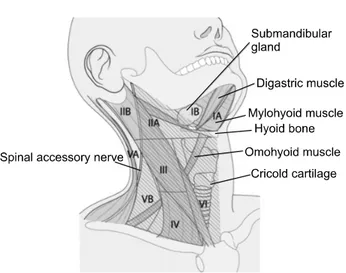
Lymph node group Description Submental group
(sublevel IA)
Lymph nodes within the triangular boundary of the anterior belly of the diagastric muscles and the hyoid bone.
Submandibular group (sublevel IB)
Lymph nodes within the boundaries of the anterior and posterior bellies of the digastrics muscle and the body of the mandible. The submandibular gland is included in the specimen when the lymph nodes within this triangle are removed.
Upper jugular group (sublevels IIA and IIB)
Lymph nodes located around the upper third of the internal jugular vein and adjacent spinal accessory nerve, extending from the level of the carotid bifurcation (surgical landmark) or hyoid bone (clinical landmark) to the skull base. The posterior boundary is the posterior border of the sternocleidomastoid muscle and the anterior boundary is the lateral border of the sternohyoid muscle.
Middle jugular group (level III)
Lymph nodes located around the middle third of the internal jugular vein, extending from the carotid bifurcation superiorly to the omohyoid muscle (surgical landmark), or to the cricothyroid notch (clinical landmark) inferiorly. The posterior boundary is the posterior border of the sternocleidomastoid muscle, and the anterior boundary is the lateral border of the sternohyoid muscle.
Lower jugular group (level IV)
Lymph nodes located around the lower third of the internal jugular vein, extending from the omohyoid muscle superiorly to the clavicle inferiorly. The posterior boundary is the posterior border of the sternocleidomastoid muscle, and the anterior boundary is the lateral border of the sternohyoid muscle. Posterior triangle
group (sublevels VA and VB)
This group is composed predominantly of the lymph nodes located along the lower half of the spinal accessory nerve and the transverse cervical artery.
Anterior compartement group (level VI)
Lymph nodes in this compartment include the pre-and paratracheal nodes, precricoid (Delphian) node, and the perithyroidal nodes including the lymph nodes aling the recurrent laryngeal nerves. The lateral boundaries are the common carotid arteries, the superior boundary is the hyoid bone, and the inferior boundary is the suprasternal notch.
Table 1. Definition of the lateral neck anatomy (11)
Fig. 1. Nodal levels with corresponding anatomic landmarks.(48)
상갑상선 혈관을 향해 연결되며, 측방으로는 중앙구획 림프절을
거쳐 중갑상선 정맥을 따라 중, 하부 내경정맥 림프절군으로 연
결된다. 하부의 림프 경로는 전기관(pretracheal), 측방기관
(paratracheal) 림프절과 하부 내경정맥 림프절로 연결되고, 갑
상선 협부는 주로 전후두 림프절(prelaryngeal node), 전기관
림프절, 상부 내경정맥 림프절로 연결된다. 갑상선에서 전상격
동 림프절(anterior superior mediastinal node)이나 후인두 림
프절(retropharyngeal node)로는 흔히 연결되지만, 하악 림프
절(submandibular node)이나 턱밑 림프절(suprahyoid node)
로의 연결은 흔하지 않다. 갑상선 피막 주변 림프절, 전기관, 전
후두 림프절을 통해서 반대편(contralateral side)으로도 연결
될 수 있지만 빈도는 낮은 것으로 알려져 있다.(
7-10
)
2) 경부 림프절 분류
경부 림프절은 편측에 평균 75개 가량 있으며 주로 내경정맥
과 척추 부신경(spinal accessory)을 따라 분포하며 일부는 이
두 림프관쇄(lymphatic chanin)를 서로 연결하는 횡림프관쇄
가 분포한다. 이러한 림프계(lymphatic system)는 경부의 주요
장기를 둘러싸고 있는 경부 근막과 근막 사이에 지방조직과 함께
존재한다. 2002년 미국 이비인후과 및 두경부 외과 위원회
(Committee for Head and Neck Surgery and Oncology of
the American Academy of Otolaryngology-Head and Neck
surgery)에서는 경부 림프절 분류와 그에 따른 경부 림프절 곽청
술의 분류를 개정하였다. 측경부 림프절은 제 1림프군부터 제5
림프군까지 분류하였다. 제1림프절군은 이하(submental) 및 악
하군(submandibular group)을 의미하며, 제2, 3, 4 림프절군은
상, 중, 하 경부군(jugular group)을, 제5림프절군은 후삼각군
(posterior triangular group)을 의미한다. 제6림프절군은 전경
부군(anterior compartment group)을 의미한다. 또한 제1, 2,
5 림프절군은 림프절 전이의 위험도에 따라 다시 Ia, Ib, IIa, IIb,
Va, Vb 림프절로 세분화 했다(Table 1, Fig. 1).(
11
)
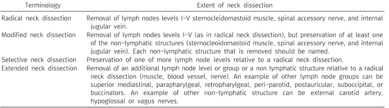
Terminology Extent of neck dissection
Radical neck dissection Removal of lymph nodes levels I-V sternocleidomastoid muscle, spinal accessory nerve, and internal jugular vein.
Modified neck dissection Removal of lymph nodes levels I-V (as in radical neck dissection), but preservation of at least one of the non-lymphatic structures (sternocleoidomastoid muscle, spinal accessory nerve, and internal jugular vein). Each non-lymphatic structure that is removed should be named.
Selective neck dissection Preservation of one of more lymph node levels relative to a radical neck dissection.
Extended neck dissection Removal of an additional lymph node level or group or a non lymphatic structure relative to a radical neck dissection (muscle, blood vessel, nerve). An example of other lymph node groups can be superior mediastinal, parapharylgeal, retropharylgeal, peri-parotid, postauricular, suboccipital, or buccinators. An example of other non-lymphatic structure can be external carotid artery, hypoglossal or vagus nerves.
Table 2. Classification of neck dissections (8)
3) 분화 갑상선암에서 림프절 전이의 경로
분화 갑상선암중에서 갑상선 유두암은 주변 림프절의 전이가
잘 되는 것으로 알려져 있다. 여포암에서는 림프절 전이가 흔하
지 않으며, 림프절 전이를 동반하는 여포암의 경우는 유두암의
여포형 변이에 대해 고려해 봐야 한다. 갑상선암의 림프절 전이
는 중앙구획 림프절로부터 시작되어 측경부로 전이되는데 주로
중, 하부 내경정맥 림프절로 전이된다.(
12
) 림프절 전이가 흔하
게 동반되는 유두상 갑상선암에서 림프절 전이가 있는 환자의
80%는 중앙구획과 중, 하부 내경정맥 림프절에 전이를 보이고,
상 내경정맥 주위나 후경삼각 부위에는 낮은 빈도를 보인다. 하
악 림프절이나 턱밑 림프절로의 전이는 림프절 전이가 아주 심한
경우를 제외하고는 드물고, 상격동 림프절로의 전이도 가능하지
만 비교적 드문 것으로 알려져 있다. 갑상선암의 전이는 주로 동
측의 림프절로 일어나지만 반대측으로 전이되는 경우도 드물게
있는데, 반대측 내경정맥 림프절군까지 전이되는 경우에서 종격
동 림프절 전이를 동반하는 경우가 많은 것으로 알려져 있
다.(
13-16
)
유두 갑상선암에서 림프절 전이의 유무는 원발암의 크기와 연
관이 있다는 보고가 있지만, 초기에도 림프절 전이가 흔히 발견
되며, 직경 1 cm 미만의 미세 유두암의 경우에도 림프절 전이가
발견된다는 여러 보고가 있다.(
7,12,13,17
) 분화 갑상선암(유두
암, 여포암)에서 경부 림프절 전이는 매우 흔하게 발견되고 있으
며, 유두암의 경우는 초진시 중심 림프절 전이가 30%에서 80%,
측경부 림프절 전이가 10
∼
30%까지 발견되는 것으로 보고되고
있다.(
1,18,19
) 따라서 유두상 갑상선암은 처음 치료 당시 이미
주변 림프절(regional lymph node) 전이 가능성을 염두에 두고
치료방침을 정해야 한다.
원격전이가 없는 갑상선 내부의 여포암(intrathyroidal
follicular carcinoma)의 경우 림프절 전이는 거의 없는 것으로
알려져 있지만, 갑상선 외부 연부조직의 침범(extrathyroidal
extension)이 있는 경우에는 주변 림프절 전이가 발생할 수 있
다.(
20
) 임상적으로 여포암의 림프절 전이가 나타나는 경우는
10% 미만으로, 여포암에서 림프절의 전이는 질병이 진행된 병
기임을 시사하는 하나의 증거가 된다.(
21
)
4) 측경부 림프절 절제술의 분류
측경부 림프절 절제 수술로는 육안적으로 전이가 의심되는 림
프절 만을 딸기 따는 식(berry picking)으로 제거하는 방법은 높
은 재발률을 보이므로 암 수술에서는 적용되지 않으며,일반적으
로 전형적 경부 곽청술(classic radical neck dissection)이 원칙
이며, 다음과 같이 근치적 경부 곽청술(radical neck
dis-section), 변형 근치적 경부 곽청술(modified radical neck
dissection), 선택적 경부 곽청술(selective neck dissection) 및
확대 근치적 경부 곽청술(extended radical neck dissection)
로 분류한다(
8,11
).
근치적 경부 곽청술은 제1림프군부터 제5림프군까지 림프군
의 제거 및 내경정맥, 척추 부신경, 흉쇄유돌근을 제거하는 수술
법을 말한다. 변형 근치적 경부 곽청술은 포괄적인 림프군의 제
거는 동일하나 척추 부신경, 흉쇄유돌근 및 내경정맥 중 하나 이
상을 보존하는 수술이나, 선택적 경부 곽청술은 제1림프군부터
제5림프군까지 중 하나 이상의 경부 림프군을 보존하면서 척추
부신경, 흉쇄유돌근 및 내경정맥을 보존하는 수술법이다. 확대
근치적 경부 곽청술을 근치적 경부 곽청술에서 하나 이상의 림프
군이나 구조물을 추가적으로 절제하는 수술이다(Table 2).
1906년 Crile이 두경부 암의 치료를 위해 전통적인 근치적 경
부 림프절 곽청술을 최초로 시행한 이후로 경부 림프절로 전이된
두경부 암에서는 근치적 경부 림프절 곽청술이 표준술식으로 자
리 잡았다.(
22
) 근치적 경부 림프절 곽청술은 미관상, 신체 기능
상 결함을 남기지만 두경부암에서 뛰어난 국소 제어와 완치의 확
률을 높이는 것으로 알려져 있다.(
23-25
)그러나 양호한 예후를
보이는 분화갑상선암의 측경부 림프절 전이에서는 근치적 경부
림프절 곽청술이 표준술식으로 적용되지는 않는다.
분화갑상선암에서는 변형 근치적 경부 림프절 곽청술이 기능
적인 림프절 절제술로 주로 이용되며, 이름처럼 근육, 신경, 혈관
등의 중요 구조물의 기능을 보존하면서 경부의 림프절을 총괄적
으로 제거할 수 있는 술식이다.(
24,26,27
) 일반적으로 분화갑상
선암에서 제1림프절까지 전이되는 경우는 매우 드물기 때문에
대개의 경우 변형 근치적 림프절 곽청술은 제2 림프절부터 제5
림프절까지의 림프절을 제거하는 것을 의미한다.(
28
)
5. 분화갑상선암에서 측경부 림프절의 절제범위
분화갑상선암의 림프절 전이가 예후에 어떤 영향을 미치는지
는 암의 종류별로 차이가 있다. 유두암에서는 진단 당시의 림프
절 전이는 재발에는 영향을 미치나 생존율에는 영향을 미치지 않
는다는 것이다. 여포암 환자에서 주변 림프절 전이의 발생 빈도
는 높지 않으나, 림프절 전이가 동반되어 있는 경우의 원발 종양
은 더욱 공격적인 성향을 보인다.(
17,19,29
) 따라서 여포암은 임
상적으로 림프절 전이가 확인되거나 갑상선피막 침범이 있는 경
우가 아니라면 중앙구획 혹은 측경부 림프절 절제술은 필요하지
않다.
현재 대다수의 외과의들은 유두암에서 예방적인 측경부 림프
절 곽청술은 필요하지 않다는데 동의 한다. 또한 광범위한 측경
부 림프절 전이를 보이는 유두암 환자에서 변형 근치적 경부 림
프절 곽청술을 시행하는 것이 제한적인 림프절 생검만 시행하는
것에 비해 암의 재발율을 낮추는 효과적인 방법이라는 의견에는
이견이 없으나, 측경부 림프절 절제 범위가 생존율과 어떤 관계
가 있는지는 여전히 논란의 여지가 되고 있다.(
29-31
) 한 연구에
서는 갑상선 유두암에서 발견되는 림프절 전이가 재발뿐 아니라
생존율에도 영향을 미치며, 이런 경우에서 좀 더 광범위한 치료
가 예후에는 크게 영향을 미치지 못한다고 한다.(
32
) 따라서 유
두암이 측경부 림프절 전이를 보인 경우, 두경부암에서 적용되
는 근치적 경부 곽청술을 일괄적으로 적용한다는 것은 합리적이
지 못한 생각이며, 오히려 합병증만 증가시킬 가능성이 있다는
의견들이 많다.
측경부 림프절 절제술 후의 합병증은 다른 수술 후 합병증과
마찬가지로 삶의 질에 영향을 미친다. 대표적으로 생길 수 있는
합병증으로는 척추 부신경이 손상되었을 때 나타나는 어깨 증후
군(shoulder syndrome)을 들 수 있다. 이는 승모근(trapezius
muscle)의 마비로 인해 어깨 쳐짐, 팔의 외전의 감소, 통증 등의
증상이 나타내는 것으로, 승모근의 마비는 기능의 손상 뿐 아니
라 외관상의 문제 또한 일으킬 수 있다. 모든 술자가 수술 중 척추
부신경을 보존하기 위해 노력을 하지만, 대개 제2B 림프군
(sublevel IIB) 또는 제5 림프군(level V)을 제거하는 과정에서
의도치 않은 기구에 의한 열손상, 견인 등에 의해 발생하는 합병
증으로 알려져 있다.(
33
) 심지어는 척추 부신경이 완벽하게 보존
된다고 하더라도 제2B 림프군(level IIB)을 제거하는 과정에서
수술 중 견인에 의해 척추 부신경이 손상을 입는 경우 근치적 경
부 곽청술을 받은 환자의 79%, 변형된 근치적 경부 곽청술을 받
은 환자의 65%, 선택적 경부 곽청술을 받은 환자의 52%에서 수
술 후 어깨 통증이 동반 되었다는 연구 결과가 있다.(
34,35
)
(1) 제2B 림프군 절제술(Level IIB dissection)에 관한 이견
측경부 림프절 전이가 있는 유두암 환자에서 제2B 림프군
(sublevels IIA & IIB)의 전이 빈도와 경향에 관한 연구들에 따르
면, 제2A 림프군(level IIA) 전이 혹은 다중 군(multi-level) 림프
절 전이가 발견될 때 제2B림프군(level IIB)의 전이를 의심할 수
있다는 결론을 얻었다. 따라서 수술 후 합병증 등을 고려하여, 측
경부 전이가 발견된 모든 환자에서 반드시 변형 근치적 측제2A
와 제2B 림프군을 모두 포함한 측경부 림프절 곽청술을 시행하
기 보다는, 제2A 림프군 및 다수의 측경부 림프절 전이가 발견될
때에 적극적인 변형된 근치적 림프절 곽청술을 시행할 것을 권유
하고 있다.(
36-39
)
(2) 제5 림프군 절제술(Level V dissection)에 관한 이견
측경부 림프절 곽청술의 범위에 있어 제5림프군 절제술 또한
제2B림프군 절제와 함께 많은 이견이 있다. 제5림프절의 전이율
은 약 16%에서 60%까지 보고되고 있다.(
28,36,39-41
) 제5림프
군 절제술도 척추 부신경의 손상으로 인한 수술후 합병증을 유발
시키는 주요 술식으로 인식되고 있다. Kupferman 등은 제2림프
군부터 제5림프군까지 변형된 근치적 림프절 곽청술을 시행한
환자의 약 30%에서 수술 후 일시적인 척추 부신경의 마비증상이
나타남을 보고했다.(
42
) 또한 Inoue 등과 Terrell 등은 제5림프
절(level V)을 제거하는 술식과 보존하는 술식의 비교로 척추 부
신경 기능 보존과 관련된 술 후 합병증이 삶의 질에 미치는 영향
에 대한 연구를 통해 척추 부신경 기능의 보존을 주장하였다.
(
43,44
)
또한 많은 연구들이 갑상선 유두암의 측경부 전이가 있을 때
선택적 경부 곽청술만 시행해도 충분하다는 견해의 결과를 발표
했다. Turanli 등이 변형 근치적 경부 곽청술 및 선택적 경부 곽
청술을 시행한 61명을 대상으로 조사한 결과 수술의 범위에 따
른 차이가 생존율(disease free survival, overall survival) 및 국
소 재발률에는 영향을 미치지 않는 것을 발표했고,(
45
) Kandil
등도 측경부로 전이된 갑상선암 환자에서 근치적 경부 곽청술을
받은 환자군이 선택적 경부 곽청술을을 시행한 환자군에 비해 생
존율에 차이가 없음을 보고하였다.(
46
) Caron 등은 106명의 갑
상선 유두암 환자에서 제1 림프군 및 5림프군(level 1 and V) 전
이의 비율이 매우 낮으므로 임상 또는 영상결과에서 전이가 의심
되는 경우에만 제거술을 시행할 것을 보고하였다.(
47
) 따라서 제
5림프절도 제2B 림프절과 마찬가지로 술 전 초음파에서 림프절
전이의 소견이 보이거나, 다수의 림프절 전이가 있는 경우에만
시행할 것을 주장하고 있다.(
28,36,39-41
)
대다수의 연구에서 림프절 전이의 유무가 예후에 악영향을 미
치지 않는다는 결론을 내리고 있지만, 림프절 전이는 더욱 진행
된 갑상선 암과 연관이 되어 있다는 것에 대해서는 이견이 없을
것이다. 따라서 갑상선암의 명백한 광범위 측경부 림프절 전이
의 증거가 있다면 합병증의 비율을 높일 가능성이 있어도 반드시
변형된 근치적 림프절 곽청술을 시행하는 것이 올바른 치료 방법
이나, 일부 제한적인 림프절 전이를 보이는 환자에서는 제 2A,
제 3 및 제 4 림프군만을 내경 정맥 림프절 곽청술과 같은 선택적
림프절 곽청술을 시행하는 방법도 고려해 볼 수 있을 것이다.
결 론
측경부 림프절 전이를 보이는 분화 갑상선암 환자에서 측경부
림프절 절제 범위는 진단시 암 병기, 림프절 전이의 정도, 및 수술
후 삶의 질에 영향을 주는 합병증 가능성 등과 같은 환자 관련 요
인들을 모두 고려하여 결정해야 하며, 외과의사의 경험과 판단
도 반드시 고려해야 한다. 분화갑상선암이 광범위한 측경부 림
프절 전이가 있다면 합병증의 가능성이 있어도 변형 근치적 림프
절 곽청술을 시행하는 것이 올바른 치료 방법이나, 일부 제한적
인 림프절 전이를 보이는 환자에서는 분화 갑상선암의 양호한 예
후와 술 후 삶의 질을 유지할 수 있는 선택적 림프절 곽청술을 시
행하는 방법도 고려해 볼 수 있을 것이다.
REFERENCES
1. Stack BC Jr, Ferris RL, Goldenberg D, Haymart M, Shaha A, Sheth S, et al; American Thyroid Association Surgical Affairs Committee. American Thyroid Association consensus review and statement regarding the anatomy, terminology, and ration-ale for lateral neck dissection in differentiated thyroid cancer. Thyroid 2012;22:501-8.
2. Lundgren CI, Hall P, Dickman PW, Zedenius J. Clinically sig-nificant prognostic factors for differentiated thyroid carcino-ma: a population-based, nested case-control study. Cancer 2006;106:524-31.
3. Podnos YD, Smith D, Wagman LD, Ellenhorn JD. The im-plication of lymph node metastasis on survival in patients with well-differentiated thyroid cancer. Am Surg 2005;71:731-4. 4. Leboulleux S, Rubino C, Baudin E, Caillou B, Hartl DM, Bidart
JM, et al. Prognostic factors for persistent or recurrent disease of papillary thyroid carcinoma with neck lymph node meta-stases and/or tumor extension beyond the thyroid capsule at
initial diagnosis. J Clin Endocrinol Metab 2005;90:5723-9. 5. Mazzaferri EL, Young RL. Papillary thyroid carcinoma: a 10 year
follow-up report of the impact of therapy in 576 patients. Am J Med 1981;70:511-8.
6. Loh KC, Greenspan FS, Gee L, Miller TR, Yeo PP. Pathological tumor-node-metastasis (pTNM) staging for papillary and fol-licular thyroid carcinomas: a retrospective analysis of 700 patients. J Clin Endocrinol Metab 1997;82:3553-62.
7. Noguchi S, Noguchi A, Murakami N. Papillary carcinoma of the thyroid. I. Developing pattern of metastasis. Cancer 1970;26: 1053-60.
8. Ferlito A, Robbins KT, Silver CE, Hasegawa Y, Rinaldo A. Classification of neck dissections: an evolving system. Auris Nasus Larynx 2009;36:127-34.
9. Smith R, Taylor SM, Trites JR, Smith A. Patterns of lymph node metastases to the submuscular recess. J Otolaryngol 2007;36: 203-7.
10. Committee on Classification of Regional Lymph Nodes of Japan Society of Clinical Oncology. Classification of regional lymph nodes in Japan. Int J Clin Oncol 2003;8:248-75.
11. Robbins KT, Clayman G, Levine PA, Medina J, Sessions R, Shaha A, et al; American Head and Neck Society; American Academy of Otolaryngology--Head and Neck Surgery. Neck dissection classification update: revisions proposed by the American Head and Neck Society and the American Academy of Otolaryngol-ogy-Head and Neck Surgery. Arch Otolaryngol Head Neck Surg 2002;128:751-8.
12. Noguchi S, Murakami N. The value of lymph-node dissection in patients with differentiated thyroid cancer. Surg Clin North Am 1987;67:251-61.
13. Scheumann GF, Gimm O, Wegener G, Hundeshagen H, Dralle H. Prognostic significance and surgical management of locore-gional lymph node metastases in papillary thyroid cancer. World J Surg 1994;18:559-67.
14. Frankenthaler RA, Sellin RV, Cangir A, Goepfert H. Lymph node metastasis from papillary-follicular thyroid carcinoma in young patients. Am J Surg 1990;160:341-3.
15. Dralle H, Damm I, Scheumann GF, Kotzerke J, Kupsch E, Geerlings H, et al. Compartment-oriented microdissection of regional lymph nodes in medullary thyroid carcinoma. Surg Today 1994;24:112-21.
16. Dralle H, Machens A. Surgical management of the lateral neck compartment for metastatic thyroid cancer. Curr Opin Oncol 2013;25:20-6.
17. Hay ID, Grant CS, van Heerden JA, Goellner JR, Ebersold JR, Bergstralh EJ. Papillary thyroid microcarcinoma: a study of 535 cases observed in a 50-year period. Surgery 1992;112:1139-46. 18. Shaha AR, Shah JP, Loree TR. Patterns of nodal and distant
metastasis based on histologic varieties in differentiated carci-noma of the thyroid. Am J Surg 1996;172:692-4.
19. DeGroot LJ. Long-term impact of initial and surgical therapy on papillary and follicular thyroid cancer. Am J Med 1994;97: 499-500.
20. Cady B, Sedgwick CE, Meissner WA, Bookwalter JR, Romagosa V, Werber J. Changing clinical, pathologic, therapeutic, and survival patterns in differentiated thyroid carcinoma. Ann Surg
1976;184:541-53.
21. Young RL, Mazzaferri EL, Rahe AJ, Dorfman SG. Pure follicular thyroid carcinoma: impact of therapy in 214 patients. J Nucl Med 1980;21:733-7.
22. Crile G. Landmark article Dec 1, 1906: Excision of cancer of the head and neck. With special reference to the plan of dissection based on one hundred and thirty-two operations. By George Crile. JAMA 1987;258:3286-93.
23. Sako K, Marchetta FC, Razack MS, Shedd DP. Modified radical neck dissection for metastatic carcinoma of the thyroid. A reappraisal. Am J Surg 1985;150:500-2.
24. Marchetta FC, Sako K, Matsuura H. Modified neck dissection for carcinoma of the thyroid gland. Am J Surg 1970;120:452-5. 25. McConahey WM, Hay ID, Woolner LB, van Heerden JA, Taylor
WF. Papillary thyroid cancer treated at the Mayo Clinic, 1946 through 1970: initial manifestations, pathologic findings, ther-apy, and outcome. Mayo Clin Proc 1986;61:978-96.
26. Attie JN. Modified neck dissection in treatment of thyroid can-cer: a safe procedure. Eur J Cancer Clin Oncol 1988;24:315-24. 27. Attie JN, Khafif RA, Steckler RM. Elective neck dissection in
papillary carcinoma of the thyroid. Am J Surg 1971;122:464-71. 28. Kupferman ME, Weinstock YE, Santillan AA, Mishra A, Roberts
D, Clayman GL, et al. Predictors of level V metastasis in well-differentiated thyroid cancer. Head Neck 2008;30: 1469-74.
29. Conzo G, Docimo G, Mauriello C, Gambardella C, Esposito D, Cavallo F, et al. The current status of lymph node dissection in the treatment of papillary thyroid cancer. A literature review. Clin Ter 2013;164:e343-6.
30. Sugino K, Kure Y, Iwasaki H, Ozaki O, Mimura T, Matsumoto A, et al. Metastases to the regional lymph nodes, lymph node re-currence, and distant metastases in nonadvanced papillary thy-roid carcinoma. Surg Today 1995;25:324-8.
31. Clark OH, Duh QY. Thyroid cancer. Med Clin North Am 1991;75:211-34.
32. Noguchi M, Earashi M, Kitagawa H, Ohta N, Thomas M, Miyazaki I, et al. Papillary thyroid cancer and its surgical. J Surg Oncol 1992;49:140-6.
33. Nahum AM, Mullally W, Marmor L. A syndrome resulting from radical neck dissection. Arch Otolaryngol 1961;74:424-8. 34. Cheng PT, Hao SP, Lin YH, Yeh AR. Objective comparison of
shoulder dysfunction after three neck dissection techniques. Ann Otol Rhinol Laryngol 2000;109:761-6.
35. Dijkstra PU, van Wilgen PC, Buijs RP, Brendeke W, de Goede CJ, Kerst A, et al. Incidence of shoulder pain after neck dissection: a clinical explorative study for risk factors. Head Neck 2001;23:947-53.
36. Lee BJ, Wang SG, Lee JC, Son SM, Kim IJ, Kim YK. Level IIb lymph node metastasis in neck dissection for papillary thyroid carcinoma. Arch Otolaryngol Head Neck Surg 2007;133: 1028-30.
37. Lee J, Sung TY, Nam KH, Chung WY, Soh EY, Park CS. Is level IIb lymph node dissection always necessary in N1b papillary thy-roid carcinoma patients? World J Surg 2008;32:716-21. 38. Koo BS, Yoon YH, Kim JM, Choi EC, Lim YC. Predictive factors
of level IIb lymph node metastasis in patients with papillary thy-roid carcinoma. Ann Surg Oncol 2009;16:1344-7.
39. Roh JL, Kim JM, Park CI. Lateral cervical lymph node metastases from papillary thyroid carcinoma: pattern of nodal metastases and optimal strategy for neck dissection. Ann Surg Oncol 2008;15:1177-82.
40. Kupferman ME, Patterson M, Mandel SJ, LiVolsi V, Weber RS. Patterns of lateral neck metastasis in papillary thyroid carcinoma. Arch Otolaryngol Head Neck Surg 2004;130: 857-60.
41. Yanir Y, Doweck I. Regional metastases in well-differentiated thyroid carcinoma: pattern of spread. Laryngoscope 2008;118: 433-6.
42. Kupferman ME, Patterson DM, Mandel SJ, LiVolsi V, Weber RS. Safety of modified radical neck dissection for differentiated thy-roid carcinoma. Laryngoscope 2004;114:403-6.
43. Inoue H, Nibu K, Saito M, Otsuki N, Ishida H, Onitsuka T, et al. Quality of life after neck dissection. Arch Otolaryngol Head Neck Surg 2006;132:662-6.
44. Terrell JE, Welsh DE, Bradford CR, Chepeha DB, Esclamado RM, Hogikyan ND, et al. Pain, quality of life, and spinal accessory nerve status after neck dissection. Laryngoscope 2000;110: 620-6.
45. Turanli S. Is the type of dissection in lateral neck metastasis for differentiated thyroid carcinoma important? Otolaryngol Head Neck Surg 2007;136:957-60.
46. Kandil E, Friedlander P, Noureldine S, Islam T, Tufano RP. Impact of extensive neck dissection on survival from papillary thyroid cancer. ORL J Otorhinolaryngol Relat Spec 2011;73: 330-5.
47. Caron NR, Tan YY, Ogilvie JB, Triponez F, Reiff ES, Kebebew E, et al. Selective modified radical neck dissection for papillary thyroid cancer-is level I, II and V dissection always necessary? World J Surg 2006;30:833-40.
48. Kang SW, Lee SH, Ryu HR, Lee KY, Jeong JJ, Nam KH, et al. Initial experience with robot-assisted modified radical neck dis-section for the management of thyroid carcinoma with lateral neck node metastasis. Surgery 2010;148:1214-21.