PDF hosted at the Radboud Repository of the Radboud University
Nijmegen
The following full text is a publisher's version.
For additional information about this publication click this link. http://hdl.handle.net/2066/107700
Please be advised that this information was generated on 2018-07-08 and may be subject to change.
4 Dibenzoazacyclooctynes: Synthesis and Bioconjugation
Table of Contents
List of Abbreviations 6
1. Bioconjugation: Unlimited Opportunities 9
1.1 Bioconjugation 10
1.2 Nature inspired modifications 11
1.3 Naturally occurring reactive groups 16
1.4 Labeling of cysteine residues 19
1.5 Bioorthogonal Reactions 23
1.6 Introduction of bioorthogonal functionalities 29
1.7 Azide-related chemistry 34
1.8 Conclusions and outlook 44
1.9 Aim and outline of this thesis 45
1.10 References 47
2. Dibenzoazacyclooctynes for enzyme PEGylation 55
2.1 Introduction 56
2.2 Synthesis 57
2.3 Kinetics 59
2.4 Protein modification 61
2.5 Conclusions and outlook 62
2.6 Acknowledgements 64
2.7 Experimental Section 64
2.8 References 73
3. Synthesis of DIBAC analogues 75
3.1 Introduction 76
3.2 Modifications based on DIBAC and intermediates 76 3.3 DIBAC analogues from substituted iodobenzyl alcohols 78
3.4 Bromination yields anti- and syn-isomers 82
3.5 Double bromide elimination 84
3.6 Base screen towards formation of 68 and 70 86 3.7 DIBAC analogue synthesis via cyclopropenone deprotection 88 3.8 Towards a fluorogenic dibenzoazacyclooctyne 90 3.9 Kinetics of synthesized dibenzoazacyclooctyne analogues 92
3.10 Conclusions and outlook 95
3.11 Acknowledgements 96
3.12 Experimental Section 96
3.13 References 120
4. Introducing functional handles to tumor-targeting 121
single domain antibody A12
4.1 Introduction 122
4.2 Expressed Protein Ligation 124
4.3 Affinity studies 127
4.4 Dual labeling using SPAAC and thiol-maleimide ligation 128
4.5 Conclusions 130
4.6 Acknowledgements 130
4.7 Experimental Section 130
5. Biodistribution studies with tumor-targeting 137 polymersomes
5.1 Introduction 138
5.2 Preparation of targeting PEG-b-PS polymersomes 140 5.3 Affinity experiments with A12-functionalized polymersomes 142 5.4 Preparation of targeting PEG-b-PB polymersomes 143 5.5 Affinity studies of A12-functionalized PEG-b-PB polymersomes 145
5.6 Conclusions and outlook 146
5.7 Acknowledgements 147
5.8 Experimental Section 147
5.9 References 152
6. Formation of an enzyme-metal catalyst hybrid system 155
using Strain-Promoted Azide Alkyne Cycloaddition
6.1 Introduction 156
6.2 A hybrid catalyst using DIBAC-pincer: attempts to conjugate 158 Pincer-ligands to CalB
6.3 A hybrid catalyst via the dibenzocyclooctadiyne approach: 160 Cyclooctyne introduction in CalB
6.4 A hybrid catalyst via the dibenzocyclooctadiyne approach: 165 N3-catalysts
6.5 Catalysis with hybrid system 166
6.6 Conclusions and outlook 169
6.7 Acknowledgments 170
6.8 Experimental Section 170
6.9 References 176
7. Towards identification of glycosylation sites on 179
mucin-type glycoproteins
7.1 Introduction 180
7.2 Deglycosylation of glycoproteins 182
7.3 Labeling studies 187
7.4 Lysate enrichment and deglycosylation 191
7.5 Conclusions and outlook 193
7.6 Acknowledgements 194
7.7 Experimental Section 194
7.8 References 199
8. Conclusions and Perspective 201
8.1 Bioorthogonal ligation reactions 202
8.2 New bioorthogonal reactions and reactive probes 203
8.3 Outlook for biological research 204
8.4 References 204
Summary 206
Samenvatting 210
6 Dibenzoazacyclooctynes: Synthesis and Bioconjugation
List of Abbreviations
AIBN Ar b BARAC BCN Boc br BSA CalB calcd CBD Cbz CDR CI CoumBARAC CuAAC d dba DBU DIBAC DIBAL-H DIBO DIBODY DiFMU DIFO DIMAC DKR DLS DMAP DMF DMSO DNA DTPA DTT e.g. EDC EDG EDTA EI ELISA ELP EM EPL equiv ESI Et et al. EtOAc EWG Fmoc FRET g GalNAc GalNAz GlcNAc GlcNAz h HRMS HRP 2,2'-azabisisobutyronitile argon block biarylazacyclooctynone 9-hydroxymethylbicyclo[6.1.0]nonyne tert-butoxycarbonyl broad (in NMR) bovine serum albumin Candida Antarctica Lipase B calculatedchitin binding domain benzyloxycarbonyl
complementarity determining regions chemical ionization
coumarinbiarylazacyclooctynones
Cu(I)-catalyzed Azide Alkyne Cycloaddition doublet (in NMR) dibenzylidene acetone 1,8-diazabicyclo[5.4.0]undec-7-ene dibenzoazacyclooctyne diisobutylaluminum hydride dibenzocyclooctynol dibenzocyclooctadiyne 6,8-Difluoro-7-Hydroxy-4-Methylcoumarin difluorocyclooctyne dimethoxyazacyclooctyne dynamic kinetic resolution dynamic light scattering 4-dimethylaminopyridine N,N-dimethylformamide dimethylsulfoxide deoxyribonucleic acid diethylenetriaminepentaacetic acid dithiothreitol exempli gratia 1-Ethyl-3-(3-dimethylaminopropyl)carbodiimide electron donating group
ethylenediaminetetraacetic acid electron impact
enzyme-linked immuno sorbent assay elastin-like polypeptide
electron microscopy expressed protein ligation equivalents
electronspray ionization ethyl
et alia
ethyl acetate
electron withdrawing group 9-fluorenylmethoxycarbonyl Förster resonance energy transfer gram N-acetyl galactosamine N-azidoacetylgalactosamine N-acetyl glucosamine N-azidoacetylglucosamine hour(s)
high resolution mass spectrum Horse Radish Peroxidase
HWE Hz ICPMS IMPACT IR J k KHMDS KOtBu KR L LC LDA Lys m m M MALDI-TOF mCPBA Me MES MESNA min MOFO MS MWCO NaHMDS NBS n-BuLi NHS NMR ºC PB PBS PEG ppm PS q r.t. RF RNA s SATA SDS SDS-Page SPAAC t t TBS TCEP TEM TFMSA THF TLC TMB TMS μ UV VHH VSV WHO Horner-Wadsworth-Emmons hertz
inductively coupled plasma-mass spectrometry
Intein-Mediated Purification with an Affinity Chitin-binding Tag infrared
coupling constant (in NMR) kilo potassium bis(trimethylsilyl)amide potassium tert-butoxide kinetic resolution liter(s) liquid chromatography lithium diisopropylamide lysozyme milli multiplet (in NMR) molar
matrix-assisted laser desorption ionization time-of-flight meta-chloroperoxybenzoic acid methyl morpholineethanesulfonic acid sodium 2-sulfanylethanesulfonate minutes monofluorocyclooctyne mass spectrometry molecular weight cut off sodium bis(trimethylsilyl)amide
N-bromosuccinimide
n-butyllithium
N-hydroxysuccinimide nuclear magnetic resonance degrees Celsius (Centigrade) polybutadiene
phosphate buffered saline polyethyleneglycol parts per million polystyrene quartet (in NMR) room temperature retention factor ribonucleic acid singlet (in NMR) N-Succinimidyl S-Acetylthioacetate sodium dodecyl sulfate
sodium dodecyl sulfate polyacrylamide gel electrophoresis Strain-Promoted Azide Alkyne Cycloaddition
tert
triplet (in NMR)
tert-butyldimethylsilyl
tris(2-carboxyethyl)phosphine tunneling electron microscopy trifluoromethane sulfonic acid tertahydrofuran
thin layer chromatography 3,3’,5,5’-tetramethylbenzidine trimethylsilyl
micro ultraviolet
heavy chain-only antibody vesicular stomatitis virus World Health Organization
Part of this chapter was or will be published:
Marjoke F. Debets, Criest W. J. van der Doelen, Floris P. J. T. Rutjes, Floris L. van Delft, ChemBioChem 2010, 11, 1168-1184.
Marjoke F. Debets, Sander S. van Berkel, Jan Dommerholt, Ton J. Dirks, Floris P J. T. Rutjes, Floris L. van Delft, Accounts Chem. Res. 2011, 44, 805-815.
Marjoke F. Debets, Jan C. M. van Hest, Floris P. J. T. Rutjes, submitted
This chapter will provide a literature overview of methods used for protein modification, which is a field that has largely evolved in the last 15 years. In the 1990s, protein modification was either achieved via natural amino acid residue labeling, or by using biology inspired methods like Sortase or expressed protein ligation.
Ever since, many new reactions have been developed, including those that do not involve naturally
occurring functional handles. Especially the
development of these so-called bioorthogonal
ligations have significantly contributed to the study of biological processes.
1
Bioconjugation:
10 Dibenzoazacyclooctynes: Synthesis and Bioconjugation
1.1 Bioconjugation
Bioconjugation is the process of linking two or more molecules, of which at least one is a biomolecule, together. In this manner, the properties of the individual components are combined.1 Chemically, clearly, examples of reactions ligating two or more molecules have been numerous for decades. However, biomolecule labeling poses large limitations on reaction conditions, e.g. solvent use, concentration, and available reactive groups.
In general, biological systems flourish best in an aqueous environment. This means that the reagents will need to be stable in aqueous systems, and the reaction needs to proceed in water. In addition, biomolecules are only present in low concentrations, let alone the concentration of the reactive group in these biomolecules. Therefore, reaction speed needs to be high in order to obtain significant modification over a convenient time span.
Apart from these requirements, in proteins only limited reactive groups are available, i.e. amines, thiols, phenols and indoles. This implies that for modification of proteins suitable reaction partners will have to be identified that react selectively with either of these functional handles. Alternatively, new, non-biologically present, labels can be introduced into the biological system. With this realization, the development of bioorthogonal reactions hit the road. For a reaction to be bioorthogonal, apart from the requirements as mentioned for bioconjugation, it needs to be selective for its target, and not give cross-reactivity with any of the functional groups present in biological systems.
Apart from expanding the scope of biomolecule modification, bioorthogonal ligation also allows for the selective and controlled introduction of labels, not only on single proteins, but also on cell-surfaces, in living cells, and even in live animals. These features open many opportunities for studying biological processes, which are not possible when using for example lysine or cysteine labeling.
Although the complete genome of many organisms has been elucidated, many biological processes like post-translational modifications, protein-protein interactions, and protein translocation are not yet understood. With new bioorthogonal ligation reactions it has been possible to e.g. study protein formation during embryogenesis,2 image tumors,3 and track glycan formation and translocation.4
In the last decade, research on bioconjugation has grown at an incredible pace, and thousands of papers have been published on new ligation methods or applications thereof. In this chapter an overview is given of the different ligation strategies available for protein modification. The aim is to give an impression of different types of ligation strategies and how they can be used in biological research. This chapter will not provide a complete overview of all methods known, and/or all applications possible, as this would be prohibitively extensive as an introduction to a thesis. Instead, reviews are often cited which give a more elaborate description of certain topics. Due to the extreme expansion of the field
since the start of the research which led to this thesis (november 2008), some research described in this chapter will overlap with research from this thesis.
In this chapter first all different labeling methods are described, i.e. nature inspired modifications (section 1.2), labeling naturally occurring reactive groups
(section 1.3), with a specific section on thiol-mediated ligation (section 1.4), and
bioorthogonal reactions (section 1.5). In all these sections, only direct introduction of a label (e.g. fluorescent dye, liposome, or radiolabel) is described. In case one of the afore mentioned methods is used for the introduction of a non-natural functional handle (instead of a label) this is described in section 1.6, which elaborates on the different methods to introduce these reactive partners. Lastly, as azide-related chemistry is currently the most-studied bioorthogonal reaction,
section 1.7 describes the different azide-reactive probes developed and highlights
a few applications of these reactions.
1.2 Nature inspired modifications
During evolution, nature has developed a wide range of tools for fast and selective modifications of biomolecules. For example, nature uses enzymes to cleave proteins, ligate proteins, or functionalize proteins. In addition, nature uses certain sequences which are used for either recognition of substrates or for post-translational modifications. Modifying these techniques as used in nature such that they can be performed outside living systems in a controlled fashion, can result in methods for labeling of specific biomolecules. In recent research, some of the known biological tools have found application for protein modification and cell surface modification.5
1.2.1 Sortase ligations
Sortases are enzymes produced by Gram-positive bacteria which covalently attach cell surface proteins to the cell wall.6 In protein modifications, the most commonly used Sortase is Sortase A (SrtA), mainly because bioactive and water-soluble variants of SrtA are widely available.7 When SrtA recognizes a small so-called “sorting signal”, i.e. LPXTG, the active cysteine residue of SrtA cleaves the bond between glycine and threonine, leaving an activated thio-ester. Next, the N-terminus of a peptide or protein can attack on the activated ester, thereby forming the desired conjugate, as depicted in Scheme 1.8 This method then, in general, leads to C-terminally functionalized proteins, albeit also methods resulting in N-terminal functionalization have been reported.9, 10 These sortases have, among others, been used for the preparation of peptide, protein-protein, peptide-nucleic acid, protein-lipid, and protein-glycopeptide conjugates.6, 7 Recently, it was also shown that SrtA can be used to ligate and cyclize proteins in different cellular compartments.11
In addition to these numerous ligations, SrtA has been used to site-specifically label living cells. Ploegh et al. showed that surface protein CD40L, containing a C-terminal LPETG-tag, could be expressed on the surface of HEK-cells.12 When
12 Dibenzoazacyclooctynes: Synthesis and Bioconjugation
these cells were subsequently incubated in SrtA containing medium, together with biotinylated-pentaglycine, cells were equipped with a biotin label. Nagamune and co-workers later extended this methodology by reporting the N-terminal labeling of enhanced cyan fluorescent protein (ECFP), which was targeted to the cell-surface of HEK cells.
Scheme 1. Mechanism for Sortase-mediated ligation. 1.2.2 Transglutaminase
Transglutaminases (TGases) are enzymes causing protein cross-linking and introduction of primary alkylamines. The enzymes are involved in, for example, blood coagulation, apoptosis, epidermal differentiation, and reproductive processes.13 The cysteine in the active site of TGases can attack on the carboxyamide of the glutamine side chain. Attack of the ε-amine of lysines, or small alkylamines, on the resulting thio-ester then results in protein-crosslinkage or introduction of a small alkyl-spacer to proteins, as shown in Scheme 2.13 In addition, under certain conditions, also water or small alcohols can attack on the thio-ester, forming free acids or esters, respectively.
Scheme 2. Mechanism for transglutaminase-mediated ligations.
To be able to use TGases for protein labeling, the protein of interest needs to be equipped with a recognition peptide. To this end, Sato et al. attached a TGase recognition peptide to Interleukin-2 at the N-terminus or the C-terminus. The thus introduced glutamine was now equipped with either a dansyl-group,14 or a PEG-tail.15 In a similar manner, other proteins have also been PEGylated.16 Taki and co-workers, introduced a TGase recognition site to glutathione S-transferase at
either the N-terminus or the C-terminus, and subsequently labeled the protein with fluorescein.17
In spite of the great potential this class of enzymes displays, only limited examples of protein labeling using TGases are known.18 The main problem is that no general consensus is found for the peptide sequence recognized by TGases.19 Therefore, research mainly focusses on the identification of TGase substrates and recognition peptides using biotin-labels or fluorescent tags.
1.2.3 Tags based on biological interactions
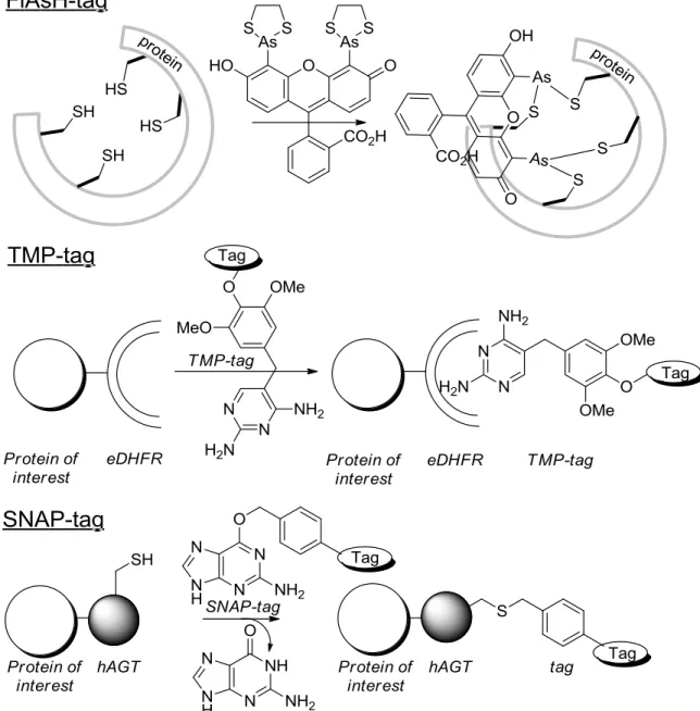
A highly efficient and selective way of labeling proteins is by using protein-based chemical tags.20 These tags are created based on naturally occurring interactions between proteins and small molecules. Common tags are the tetracysteine tag (FlAsH-tag), the trimethoprim (TMP)-tag, and the SNAP-tag.
14 Dibenzoazacyclooctynes: Synthesis and Bioconjugation
The FlAsH-tag is inspired by the binding of trivalent arsenic to paired cysteine-residues of the cofactor lipoic acid, which is largely responsible for the toxic effect of arsenic compounds. In short, the protein of interest is equipped with the FlAsH-tag (a 15 amino acid FlAsH-tag, with a tetracysteine core, CCXXCC).21 Subsequently, the modified protein can be ligated to a fluorogenic bisarsenical fluorescein ligand. Advantage of the tag is its small size. Disadvantages, on the other hand, are that in cysteine-rich environments a high degree of aspecific labeling is obtained, and that the bisarsenical fluorescein ligand is toxic.22 Nonetheless, the FlAsH-tag has successfully been applied for labeling in many types of mammalian cells, bacterial cells, and even in vivo in Drosophilia eyes.22 In addition, the FlAsH tag has also been used for the examination of the trafficking and signaling of proteins in neurons,23 and for FRET studies together with CFP to investigate protein-protein interactions and conformational changes.24, 25 As alternative for the FlAsH-tag, a tetraserine motif was developed which selectively recognizes bis-boronic acids.26
The TMP-tag is based on the interaction of dihydrofolate reductase (DHFR) with antibiotic trimethoprim (TMP), which shows high binding affinity and selectivity. To use this tag for protein labeling, DHFR is attached to the protein of interest; next, the protein can be labeled by the addition of a TMP-fluorophore.27-29 The TMP-tag has been used for the labeling of intracellular proteins in live cells,30, 31 and imaging of the spliceosome.32
Probably the most applied tag in tag-based labeling is the SNAP-tag. For this strategy, a protein is fused with human repair protein O6-alkylguanine-DNA alkyltransferase. This fusion protein then forms a covalent bond with a labeled O6 -benzylguanine.33, 34 The SNAP-tag has been used for the imaging of different proteins in mammalian and yeast cells,33, 34 imaging of the spliceosome,32 the visualization of protein-protein interactions,35 studying of cellular processes like endocytosis36 and HIV-cell interactions,37 and for the imaging of tumor xenographts.38
1.2.4 Expressed protein ligation
A crucial process in protein synthesis is post-translational modification. A prominent example is the exclusion of a precisely defined part of the protein (the so-called intein) and the subsequent conjugation of the N-extein and C-extein.39, 40 With few exceptions, there are no limitations to the amino acids present in the exteins, however, inteins are characterized by several sequence constraints, e.g. an N-terminal and C-terminal serine, cysteine or threonine, and an asparagine at the C-terminal end of the intein.40 At this moment, over 500 different inteins have been identified.41 One of the sequential similarities between almost all inteins is the cysteine, threonine, or serine at the N-terminal site of the intein. The thiol or alcohol moiety of the N-terminal residue attacks on the amide bond, thereby forming an activated thio-ester or ester bond. Subsequently, the thiol or alcohol from a Cys/Ser/Thr at the C-terminal site of the intein attacks on this activated ester. A cyclization reaction caused by attack of the amide of the asparagine
residue which is always conserved removes the intein. After an S-N or O-N acyl shift, the two exteins are fused by the formation of a stable amide bond. A schematic representation of this process is shown in Scheme 3.40
Scheme 3. Mechanism for protein splicing. Two exteins are ligated together by removal of an intein.
The implementation of this methodology on the labeling of proteins by introducing different peptides containing for example a fluorescein or biotin was first applied by Muir et al.42 and Xu and co-workers.43 This protein labeling strategy, named expressed protein ligation (EPL), was based on the finding that mutation of the C-terminal site of the intein obstructed protein splicing, but still allowed for thio-ester formation.44, 45 In this methodology, the protein of choice is produced as a fusion protein, C-terminally linked to a modified intein and a binding domain, e.g. a chitin binding binding (CBD). Upon treatment with a small thiol, the fusion protein is released from the intein, and the remaining thio-ester can react with a peptide-like compound containing an N-terminal cysteine in a Native Chemical Ligation (NCL) type reaction,46 as depicted in Scheme 4. Over the last 15 years, many different applications of this EPL technique have been published,47 especially since the constructs became better accessible by the commercial availability of Intein-Mediated Purifcation with an Affinity Chitin-binding Tag (IMPACT) kits.48
Scheme 4. Mechanism for expressed protein ligation.
EPL has widely been used for the introduction of fluorescent probes,49-51 attachment of proteins to solid supports,52 protein cyclization,53 and selective
16 Dibenzoazacyclooctynes: Synthesis and Bioconjugation
isotope-labeling of proteins.54 In addition to this, EPL has also been used to attach proteins to micelles,55 liposomes,56 and magnetic nanoparticles.57 In all these examples the surface of the micelles, liposomes, or nanoparticles was equipped with cysteines. The thiol-groups of these cysteine residues then reacted with the thio-ester of a protein, which was formed by splicing of the intein using a small thiol.
1.3 Naturally occurring reactive groups
Apart from using enzymes and the recognitionpatterns of protein fragments to perform protein modifications, also the side-chains of some natural amino acids contain attractive functional groups to perform ligations. Most prominent in literature are modifications of cysteine-residues, which will be discussed in section 1.4, and lysine-residues.
1.3.1 Lysine labeling
Probably, the most commonly used reagents for lysine modification, and protein modification in general, are NHS-esters, isocyanates and isothiocyanates. These modifications simply require mixing of the protein and the reagent at the right pH (between 8 and 9 for NHS-esters, between 9 and 10 for NCS- and NCO-based ligations). Since many dyes, affinity labels, and metal chelators are available as amine-reactive reagents, labeling of proteins is greatly facilitated in this manner. In addition to these ligation reactions, lysines can also be modified using reductive amination with aldehydes,58 or using sulfonyl chlorides.1, 59, 60 Notably, modifications that apply to the ε-amine of lysines, can also modify the N-terminus.
Although most of these modifications are relatively straightforward, disadvantage is that no selectivity concerning the site of modification and number of modifications is achieved during labeling. In general, if the success of the experiment is independent of the site or degree of modification, lysine-modification is an almost perfect option. On the other hand, when lysines are involved in for example enzyme functioning or epitope binding (e.g. antibodies) a different strategy is required.
1.3.2 Glutamate and aspartate labeling
An alternative to lysine-modification, is the modification of glutamate and aspartate residues. The acid moieties of these residues can be activated by adding EDC, if desired in combination with HOBt, and subsequently reacted with an amine.60 However, also for this method, no selectivity can be obtained as also glutamate and aspartate residues are highly abundant in proteins.
1.3.3 Tyrosine and tryptophan labeling
If more selectivity is required, tyrosine and tryptophan residues are better alternatives, as these are less abundant on protein surfaces.
The tyrosine-residue can be modified using electrophilic aromatic substitution, and metal-catalyzed reactions. Metal-catalyzed labeling can be achieved using Ni(OAc)2 and a labeled phenol, leading to formation of a C-C bond between the two carbon-atoms at the ortho-position with respect to the alcohol-functionalities (not shown).61, 62 Alternatively, Pd(OAc)2 in combination with an allylic ester, carbamate, or carbonate can be used to label the tyrosine alcohol, Scheme 5, route A.63
By means of electrophilic aromatic substitution, iodides64 and nitro groups65 can be introduced, but the most general method involves reaction of the tyrosine alcohol with diazonium salts.66 A wide range of diazonium salts can be prepared by treating an aniline with sodium nitrite under acidic conditions. Treatment of the protein with these diazonium salts results in the modification of a tyrosine residue, ortho to the alcohol functionality, as shown in Scheme 5, route B. This process proceeds in high yield when electron-poor anilines are used as starting material, and results in limited labeling for electon-rich anilines.67, 68
Alternatively, electrophilic aromatic substitution can be achieved using Mannich-type reactions, shown in Scheme 5, route D.69 First, an imine is formed from an aldehyde and an electron-rich amine. The phenyl ring of the tyrosine then attacks on the imine, again introducing the functionality ortho with respect to the alcohol functionality. This reaction proceeds best with electron-rich anilines, making it complementary to the diazonium-coupling reaction. Recently, Barbas et al. showed that a similar reaction can also take place when a cyclic diazodicarboxamide is used, Scheme 5, route C.70
Scheme 5. Methods used for tyrosine labeling.
Among others, these tyrosine-based labeling strategies were used for lipidation (using route A),63 PEGylation (via route B),71 fluorescent labeling (route C),70 or for the introduction of peptides (route D).72
The least abundant amino acid residue in proteins is tryptophan. Francis et al. demonstrated that this residue can be modified using a Rh-carbenoid, which reacts with the indole of tryptophan, yielding a mixture of N-alkyl and 2-alkyl labeled sidechains.73 This labeling was recently shown to be applicable in the
18 Dibenzoazacyclooctynes: Synthesis and Bioconjugation
identification of binders to FynSH3 domain, an important therapeutic target for cancer treatment.74 To achieve this, potential binders were equipped with a rhodium catalyst, followed by the formation of the Rh-carbenoid. In the end, proximity-driven modification led to the ligation of peptide and protein.
1.3.4 Modification of the N-terminus
Probably the most specific labeling targets in a protein are the C-terminus and N-terminus, as of both only one is present in each protein. While for the C-terminus EPL is the only widely applicable strategy, multiple modification strategies of the N-terminus have been reported, most of them involving specific amino acid residues at this terminus. One method being independent of the N-terminal residue is acylation and alkylation (as described for lysine modification). The lower pKa of the N-terminus compared to the lysine side chains, makes selective modification of the N-terminus over the ε-amines possible.75
NCL is, clearly, not only a suitable strategy for C-terminal modification, but can also be used for modification of a protein at the N-terminus, with as requisite that the N-terminal residue is a cysteine. If this is the case, the protein can react with a thio-ester, either formed synthetically or by using EPL.42
Alternatively, the N-terminal cysteine can also be used for formation of a thiazolidine in reaction with an aldehyde. This reaction can also occur with N-terminal serine, threonine, histidine, tryptophan, and asparagine residues, forming different cyclic products, as shown in Scheme 6.76 In addition, the N-terminal tryptophan can be used in a Pictet-Spengler reaction, forming a similar tricyclic structure.77
Scheme 6. N-terminal labeling of Trp, Cys, Ser, Thr, Asn, and His using an aldehyde.
Lastly, modifications of the N-terminus are known which do not directly introduce a label, but introduce a functionalizable handle instead. This handle can then subsequently be used for introduction of a label. This type of modification will be discussed in section 1.6.
1.4 Labeling of cysteine residues
The amino acid least accessible for modification in nature is cysteine, since this residue, in general, occur as di-thiol-bridges.60 Only upon reduction of these disulfide bond, labeling of thiols can be achieved. However, this often has an effect on the tertiary structure. A free thiol can also be introduced using a so-called N-Succinimidyl S-Acetylthioacetate (SATA)-modification.78 This is an NHS-based reagent, introducing a protected thiol to lysine residues, which can, after deprotection, be used for labeling. Clearly, this makes thiol-related labeling as aspecific as other lysine-modifying strategies. Alternatively, an additional cysteine residue can be introduced via site-directed mutagenesis, yielding only one functionalizable amino acid. Disadvantage of this method is that it sometimes leads to unwanted disulfide bond formation and thereby disruption of the tertiary structure.
1.4.1 Traditional thiol-labeling
The most commonly used reagents for the labeling of thiols are maleimides, iodoacetamides, vinyl sulfones, and disulfides, Scheme 7. All except the latter are based on nucleophilic attack of the reactive SH on the activated carbon-atom (resulting in a Michael-type addition or substitution of the iodide). In general, these reactions are selective for thiols, and no labeling of lysines is observed. If necessary, lysine labeling can be further reduced by lowering the pH.
Scheme 7. Traditional thiol-labeling reactions. Thiol-maleimide ligation
The value of the thiol-maleimide ligation (pathway A, Scheme 7) is probably best underscored by the commercial availability of a wide range of reagents (e.g. fluorescent dyes and affinity tags). A nice example of thiol-introduction followed by thiol-maleimide ligation was published by Blankenberg et al.79 They published the introduction of a tag, containing one cysteine, to vascular endothelial growth factor (VEGF). Subsequently, a metal-chelator was ligated to VEGF using
thiol-20 Dibenzoazacyclooctynes: Synthesis and Bioconjugation
maleimide ligation and next labeled with technetium. Ultimately, this conjugate was used for biodistribution studies.
A comparable reaction was published by Finn et al., who showed that oxanorbornadienes as shown in Scheme 8 give a Michael-type addition.80 The thus obtained conjugates undergo a retro-Diels-Alder (rDA) reaction, releasing substituted furans, Scheme 8. In a follow-up paper it was shown that the rate for this rDA cleavage can be controlled by variation of the substituents, making it a potential reaction for drug-delivery.81 Notably, upon prolonged reaction, also lysine-residues were labeled. These lysine-conjugates proved more stable and rDA cleavage was significantly slower.
Scheme 8. Protein labeling using a thiol-oxanorbornadiene ligation, followed by a retro-Diels-Alder reaction.
Labeling using iodoacetamide
Similarly to the maleimides, iodoacetamide reagents (pathway B, Scheme 7) containing metal-chelators82 or dyes83 have been developed. Bruschi et al. prepared two new iodoacetamide dyes and subsequently showed the use of these dyes in the analysis of the oxidation damage in patients with glomerulosclerosis, based on that thiols become unreactive upon oxidation.83 In addition to these labeling applications, iodoacetamide is often used prior to tryptic digest, to prevent refolding of the protein, and it can be used to prevent thiols from giving undesired labeling.84
Thiol-vinyl sulfone labeling
Thiol-vinyl sulfone labeling (pathway C, Scheme 7) is based on a Michael-type addition of the free thiol to the conjugated double bond. Gonzalez et al. used this reaction for the immobilization of vinyl sulfone saccharides on glass plates and for the attachment of neoglycoproteins. Subsequently, the synthesized neoglycopeptides were added to the glass plates and the carbohydrate-binding proteins were identified.85 In addition, this method was used for the PEGylation of proteins,86 and the immobilization of proteins to nanoparticles.87-89
Disulfide-based reagents
Labeling using disulfide reagents (pathway D, Scheme 7) relies on the intrinsic tendency of thiols to form disulfide bonds. Upon addition of a disulfide an exchange takes place, thus forming a mixed disulfide linkage. This type of reaction works best when a symmetrical disulfide is used, or when upon exchange, the
remaining disulfide rearranges to a non-reactive species. Ellmann’s reagent (used to determine the concentration of thiol-functionalities in a sample) is based on this disulfide exchange, as is the introduction of a nitroxide paramagnetic spin label to proteins using S-(2,2,5,5-tetramethyl-2,5-dihydro-1H-pyrrol-3-yl)methyl methane sulfonothioate (MTSL).
A special property of the disulfide bond formation is that the reaction is reversible; upon entering a reductive environment, the bond is cleaved. This feature of the disulfide bond has proven useful in drug delivery studies.90 For example, Zalipsky and co-workers showed that lipopolymers with a disulfide bond between the PEG-part and the lipid-part can be created.91 The liposomes prepared from this type of lipopolymers could be used for the triggered release of doxorubicin.92
1.4.2 Thiol-ene and thiol-yne ligation
The reaction between a free thiol and an alkene (the thiol-ene reaction, Scheme 9) was reported over a century ago by Porter.93 The finding that the thiol-ene reaction proceeds according to a free radical mechanism was reported four decades later by Kharasch et al.94 However, it was not until 2007 that this reaction was first used for the modification of proteins. Kunz et al. reported the preparation of synthetic vaccines by the conjugation of a tumor-associated glycopeptide to BSA.95 The protein was first equipped with either a thiol or an alkene, using an EDC-coupling, thereby labeling the lysines. Next, glycopeptides were prepared with either a thiol or an alkene, and eventually photochemically conjugated to BSA. However, since EDC-coupling was used to introduce one of the functional handles, no specificity was obtained (24 and 25 labels were introduced for both cases). Later, Dondoni and co-workers used the thiol-ene reaction for the glycosylation of BSA.96 Dondoni equipped sugar moieties with an alkene handle, and photochemically ligated these to the free thiols of BSA. In addition to synthesis of glycosylated proteins,97 the thiol-ene ligation was used for the coupling of ubiquitin molecules98 and site-specific protein acetylation.99 With the latter, an excellent mimic for native acetylated residues was formed and it was shown that histones acetylated in this manner were still recognized by histone deacetylases.
Scheme 9. Thiol-ene (left) and thiol-yne (right) ligation.
An alternative for the thiol-ene reaction is the thiol-yne ligation, Scheme 9. Originally, this reaction was reported by Jones et al. in 1949.100 Using this reaction, two thiols can add to a single reagent. However, it was not until 2011 that this was used for the dual modification of BSA, introducing a sugar-moiety and a
22 Dibenzoazacyclooctynes: Synthesis and Bioconjugation
fluorescein.101 Until now, this is the only example known for protein modification. The reaction has been more widely applied in material science for e.g. the creation of dendrimer like structures and surface modifications.102
1.4.3 Thiol bridging reagents
All thiol-related ligations described earlier require either the breaking of a disulfide bridge or the introduction of an additional thiol via site-directed mutagenesis. Disadvantage of the first is that the tertiary structure could potentially be destabilized or disturbed, disadvantage of the latter is that dimerization or disulfide scrambling can occur.103 An alternative was published by Brocchini et al.104, 105 An α,β-unsaturated ketone containing a β’-sulfonyl leaving group was prepared, Scheme 10. Addition to a protein with a reduced disulfide bridge resulted in Michael addition, followed by elimination of the sulfonyl group. Subsequently, the second thiol can react with the reformed α,β-unsaturated ketone, summarized in Scheme 10. In this manner, different proteins were PEGylated, while retaining the stability normally rendered by the disulfide bond. This was confirmed by enzyme activity and immunogenicity studies.104
Scheme 10. PEGylation via cysteine ligation reforming the broken disulfide bridge.
Alternatively, Baker et al. reported that also bromomaleimides can be used for this purpose, allowing for either reformation of the thiol-bridge or introduction of an additional functionality (e.g. a glucose). The distinguishing characteristic of this reaction is that labeling can be reversed by the addition of large excess of glutathione, Scheme 11.106
Scheme 11. Reversible cysteine labeling.
Disadvantage of both methods described above is that reduction of the disulfide bond needs to be performed prior to addition of the labeling agent, which could still cause protein unfolding. To circumvent this, Baker et al. published a maleimide with two thiophenols, which could be added together with the reducing agent.107 Haddleton et al. showed that this reagent was so stable that it could survive atom transfer radical polymerisation (ATRP) conditions. Thus a
maleimide-functionalized poly(methacrylate) was prepared, and used for the conjugation of this polymer to a disulfide bridge containing hormone (salmon calcitonin).108
1.5 Bioorthogonal reactions
Labeling of proteins, by modifications inspired by nature, using chemical tags or enzyme-mediated modification (section 1.2), or by the labeling of naturally occurring amino acids (section 1.3 and 1.4) still suffers from some significant disadvantages. For nature inspired modifications often large reagents and/or the introduction of large recognition parts are required. Alternatively, for the modification of naturally occurring amino acids selectivity is hard to achieve. Even though some amino acids (like tyrosine and cysteine) are low abundant and can be used for selective protein labeling, problems are likely to arise when applying these ligations in bigger biological systems (e.g. in cells or in vivo). In these systems, reagents need to be stable in aqueous environment (unlike diazonium salts or imines), catalyst requirements pose large challenges, and thiol-reactive probes are likely to give cross-reactivity with other proteins and thiol-containing reagents.
To avoid biological interference and allow for live cell and in vivo labeling, in the last decade many groups have focussed on the development of bioorthogonal reactions. These reactions can occur in living systems, and show no cross-reactivity with reagents present in biological systems, like lysines, cysteines, and glutathione.
Since the first modification of a biological system using an unnatural functional group in 1996,109 many new bioorthogonal reactions have been published. Handles which have been shown to be bioorthogonal and applicable in biological system modification are aldehydes, azides, nitrones, nitrile oxides, tetrazines, tetrazoles, quadrocyclanes, and alkenes. All bioorthogonal reactions used in bioconjugation are summarized in Table 1.
Introduction of the functional handles to proteins or cell surfaces will be discussed in section 1.6.
1.5.1 Ligations with ketones and aldehydes
The first non-natural functional handle to be used for the modification of proteins was the carbonyl-functionality (aldehydes and ketones). Although these can react with amine-groups forming imines, in physiological media the carbonyl lies on the favored site of the equilibrium. On the other hand, ketones and aldehydes do form stable conjugation products with alkyloxyamines (entry 1) and hydrazines (entry 2), forming oximes and hydrazones, respectively.110 These reactions have been used for the labeling of proteins109 including in living cells,111 virus particles,68 and on cell surfaces.112
24 Dibenzoazacyclooctynes: Synthesis and Bioconjugation
Table 1. Summary of bioorthogonal reactions used in biological system modification.
Entry Functional
handle Reaction partner Product Catalyst
1 None 2 None 3 Cu(I) or Ru(II)a 4 None 5 None 6b None 7 Cu(I) or Ru (II)c 8 None 9 None 10b None 11b None
12 None 13 None 14 None 15b None 16b UV-light 17 UV-light 18b UV-light 19b none 20 Grubbs’ catalyst 21 Pd(OAc)2(ligand)2 22 Pd(OAc)2(ligand)2
[a] Cu(I) catalysis yields the 1,4-isomer, Ru(II) yields the 1,5-isomer. [b] Both regioisomers are obtained, although only one is shown in the table.
[c] Catalyst is only required when regioselective control is desired, Cu(I) yielding the 1,4 -isomer, Ru(II) yielding the 1,5 isomer.
26 Dibenzoazacyclooctynes: Synthesis and Bioconjugation
1.5.2 Ligations with azides
Currently, azides are the most commonly used bioorthogonal reagent in the modification of biological systems. Its popularity can be explained by the inertness and small size of the azide moiety, the facile introduction and, most important, the unique reactivity. In addition to that, introduction of an azide onto a substrate generally has only minimal effect on its structure.113 Reaction of azides in a Staudinger reduction and the Huisgen 1,3-dipolar cycloaddition were already well established in the previous century, however, they either do not form a bond between the two reaction partners, or require too harsh conditions for protein labeling.
In 2000, Bertozzi et al. developed a Staudinger ligation (entry 4). This reaction is a modification of the long-known Staudinger reduction which was now applied for the formation of an amide bond via rearrangement of the aza-ylide intermediate.114 An alternative was developed by Raines and co-workers, who combined the Staudinger reduction with an NCL (entry 5). 115
A more biocompatible version of the 1,3-Huisgen dipolar cycloaddition was discovered independently by Meldal116 and Sharpless117 who found that the 1,3-dipolar cycloaddition of azides and acetylenes can be catalyzed by sources of copper(I) (entry 3). Copper was found to catalyze the [3+2]-cycloaddition of azides and acetylenes in aqueous solvents at physiological pH and temperature and the reaction is compatible with biomolecules like sugars, proteins and nucleic acids. The resulting 1,4-disubstituted triazole adducts are formed with exclusive regioselectivity and high yield. Although the reaction has found many applications, the required Cu(I) is toxic to cells and binds to for example active sites of enzymes and metal chelators. To avoid the use of this catalyst, Cu-free cycloadditions using strained alkynes were developed.
Already in 1953, Blomquist and Liu published the first synthesis of cyclooctyne and its suspected ring-strain, which became apparent from the reported explosive reaction of cyclooctyne and phenyl azide.118 The structural identity of the resulting liquid was later corroborated as the product of a 1,3-dipolar cycloaddition.119 Ever since, many different cycloalkynes have been reported. It was noted that a cyclooctyne is the smallest cycloalkyne which can be isolated. However, when bigger atoms are added (e.g. Si or S), also cycloheptynes could be isolated, especially when stabilized by the addition of methyl-groups adjacent to the alkyne.120
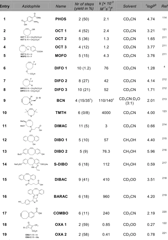
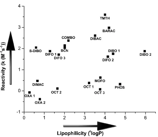
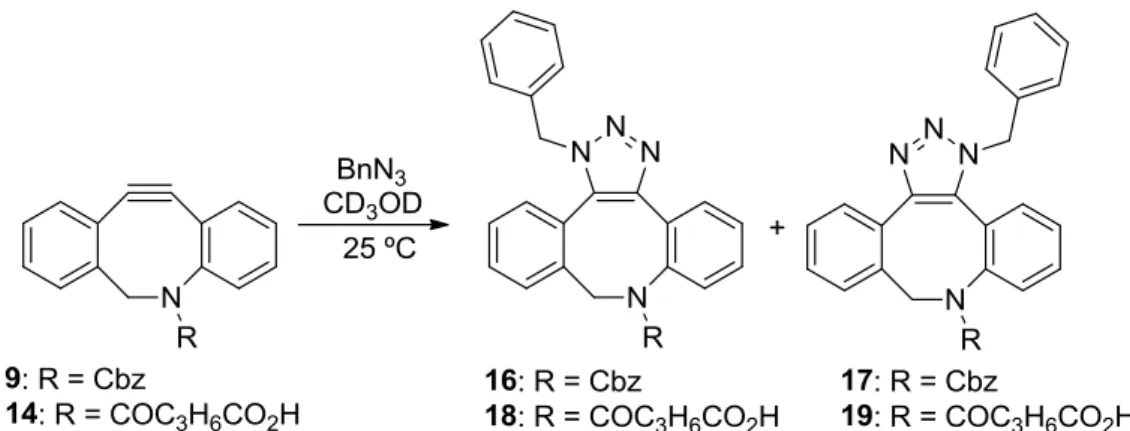
In spite of all this work, mainly performed in the 1960s and 1970s, it was not until 2004 that Bertozzi and co-workers realized the potential of these strained alkynes in the strain-promoted azide alkyne cycloaddition (SPAAC, entry 6).121 Since this first publication many other (dibenzo)cyclooctynes have been developed, and applications of SPAAC are numerous. In addition also oxanorbornadienes122 and thiacycloheptynes123 were shown to react with azides (not shown in Table 1)).
Since the developed reaction partners for azides and examples of bioconjugation with azides are numerous, this will be further elaborated in section 1.7.
1.5.3 Ligations with nitrile oxides and nitrones
Compared to azides, nitrile oxides and nitrones are stronger 1,3-dipoles, making them faster partners in cycloadditions. Unfortunately, this also makes them less stable and therefore more troublesome in biochemical applications. Advantage of using nitrile oxides is that, because of their increased reactivity, they react with simple terminal alkynes without the need of a catalyst (entry 7). This feature has mainly been applied by Heaney et al. for the modification of ribonucleosides.124 In addition, Ravoo and co-workers showed that this reaction can be used for surface modification.125 Carell et al. showed that nitrile oxides can also be used for the reaction with alkenes (entry 8) and norbornenes (entry 9). Here, nucleotides were equipped with norbornenes or alkenes and subsequently used for the synthesis of oligonucleotides, and lastly reacted with in situ formed nitrile-oxides.126, 127 In the study involving alkene-nitrile oxide ligation, not only the alkene-functionality but also alkyne-groups were introduced. It was shown that the alkynes could first be labeled using CuAAC, whereupon the alkenes were labeled with nitrile oxides.127
Most recently it was shown that nitrile oxides also react with cyclooctynes (the so-called Strain-Promoted Alkyne Nitrile Oxide Cycloaddition (SPANOC), entry 10).128, 129 This reaction showed almost a 100-fold rate increase compared to SPAAC, and was used for the modification of oligosaccharides,128 small peptides,129 and DNA modification on solid phase.130
Disadvantage of using nitrile-oxides is that their reactivity also makes them unstable, and in situ preparation directly followed by cycloaddition is required. Nitrones, on the other hand, appeared to be more stable and still react in a fast and selective manner with cyclooctynes in a Strain-Promoted Alkyne Nitrone Cycloaddition (SPANC, entry 11).131, 132
For this reaction, the reactivity largely depended on the nature of the nitrone, with rate constants ranging within 3 orders of magnitude. Cyclic nitrones were found to be most efficient.133, 134 SPANC has found multiple applications in cancer research. Pezacki et al. targeted human breast cancer cells with epidermal growth factor (EGF), which was functionalized with cyclic nitrones using NHS-based chemistry. The cells could subsequently be labeled using a biotin-functionalized cyclooctyne, and imaged with a fluorescent streptavidin.133 Prosperi and co-workers showed that SPANC could be used for the attachment of nitrone-functionalized antibodies to magnetic nanoparticles. After modification, these particles still showed affinity for tumor marker HER2.135
1.5.4 Ligations with tetrazines
An interesting alternative to the 1,3-dipolar cycloadditions as described above, are the tetrazine-based ligations. These reactions are inverse electron-demand Diels-Alder reactions, followed by a retro-Diels-Alder reaction, eliminating N2, shown in Scheme 12. As for the reaction between cyclooctynes and azides, older literature already shows extensive investigations into cycloadditions between tetrazines and strained alkenes and alkynes. In these reports, the effect of
ring-28 Dibenzoazacyclooctynes: Synthesis and Bioconjugation
strain, double bond stereochemistry, and the presence of heteroatoms in rings on the reaction rate have already been extensively investigated.136, 137 In spite of this research, only in 2008 Hilderbrand and Fox almost simultaneously published the modification of proteins and cell surfaces using tetrazine-based ligations (Scheme 12).
Scheme 12. Inverse-electron demand Diels-Alder reaction followed by a retro-Diels-Alder reaction.
Hilderbrand and co-workers demonstrated that norbornenes react with tetrazines (entry 13) with similar rates (1.6-1.9 M-1s-1)138 as the fastest SPAAC reactions (1.5 M-1s-1 being the fastest rate reported).139 This reaction was successfully applied in the labeling of tumor cells.
Fox et al. showed that tetrazines react extremely fast with different trans-cyclooctenes (entry 12, rate constants of 1-22 × 103 M-1s-1) compared to the SPAAC reaction.140, 141 This reaction has for example been applied for the intracellular labeling of anti-cancer drug taxol, and the preparation of 18F-PET tracers.142 Interestingly, the incredible speed of these reactions, also allowed for pretargeting. It was shown that sequential incubation of tumor cells with trans-cyclooctene labeled antibodies, followed by tetrazine-functionalized nanoparticles, led to a higher degree of labeling as for the full antibody-nanoparticle construct.143 Rossin et al. showed the potential of this reaction for in vivo labeling. They pretargeted tumors in mice with trans-cyclooctene functionalized antibodies, and then separately injected an In3+-labeled tetrazine and showed successful tumor labeling.3 Advantage of this last method is that the small tetrazine is cleared within several hours from the blood, whereas the full antibody takes over 24 hours before clearance. This significantly decreases the time between injection and imaging, which especially might turn out useful for fast decaying radioisotopes.
In addition, it was recently shown that tetrazines react with cyclooctynes (entry 14),144 which was applied for protein modification.145, 146 Interestingly, monosubtituted tetrazines do react at much lower rate with dibenzocyclooctynes, compared to cyclooctynes. Disubstituted tetrazines give no cycloaddition product at all with dibenzocyclooctynes.147 Another interesting, newly arisen probe used for tetrazine-based ligation is the cyclopropene (entry 15),148 which already found application in cell surface labeling.148, 149
Notably, not all tetrazines are stable in serum, some degrading in water and/or by the addition of thiols. Hilderbrand et al. prepared a wide range of tetrazines and tested them for reactivity and serum stability.150 In addition, they prepared tetrazines which become fluorescent upon reaction.151
1.5.5 Ligations with tetrazoles
Tetrazoles themselves are no reactive partners in cycloaddition reactions. However, it was shown that upon irradiation with UV-light, nitrogen is excluded and a nitrile imine 1,3-dipole is formed.152 In the absence of a reactive partner for this dipole, formation of a water-adduct was observed, however, in the presence of acrylamides (entry 16),153 norbornenes (entry 17)154 and cyclopropenes (entry 18)155 the nitrile imines rapidly form pyrazolines, as shown in Scheme 13. The reactivity of the 1,3-dipole depends on substituents on the nitrile imine.156 It was shown that this reaction can be used for labeling of proteins, also inside cells,157 for example towards studying perturbing protein localization.158 Notably, the reaction only occurs upon UV irradiation, thereby allowing temporal and spatial control over the ligation.
Scheme 13. Formation of nitrile imines from tetrazoles, followed by a 1,3-dipolar cycloaddition with alkenes.
1.5.6 Other bioorthogonal ligation reactions
In the last couple of years also metal-catalyzed reactions have found application in the modification of proteins. For example, Davis et al. showed that cross-metathesis can be used for protein modification (entry 20). For this modification, an S-allyl cysteine or Se-allyl functionality is required which reacts with allyl-functionalized saccharides or small ethylene glycol tails.159, 160 It was also demonstrated that the Suzuki-Miyaura coupling (entry 22) 161 and the Sonogashira reaction (entry 21)162 can be applied for the labeling of proteins.
A completely new bioorthogonal reaction was recently developed by Sletten and Bertozzi, who sought to develop a new reaction, orthogonal to the known cohort of bioconjugations.163 They encountered that quadrocyclanes have a large ring-strain which makes them likely to react fast with a suitable reaction partner, which was found in Ni(dithiolene)2 complex (entry 19). The obtained cycloaddition product however slowly decomposed, unless a metal-chelator was added. This reaction could be used for protein modification and proved orthogonal to SPAAC and oxime-formation.
1.6 Introduction of bioorthogonal functionalities
The advantage of bioorthogonal chemistry is that selective labeling can be obtained without the interference of biological substrates. This automatically
30 Dibenzoazacyclooctynes: Synthesis and Bioconjugation
implicates that the functional handle will have to be introduced prior to labeling. The methods by which the introduction has been achieved can roughly be divided in three sections. The most straightforward way is by using either nature-inspired methods or by using amino acid modification. In the latter case, no selectivity in labeling site and number of modifications can be achieved. For protein modification, full selectivity can be achieved when non-natural amino acids are incorporated. In this manner, either a natural amino acid is substituted for a non-natural one, or the stop-codon is used for the introduction of a 21st amino acid.164 For cell-surface labeling, a widely applied strategy is by growing cells in the presence of a functionalized sugar-moiety. In this manner, the cell’s metabolism partially incorporates the functionalized sugar instead of the natural version, thereby introducing a functional handle to the glycans.112, 114
1.6.1 Chemical Introduction of handles to proteins
The most straightforward but least selective method for the introduction of handles to proteins has been the modification of lysines. In this manner, for example, tetrazoles,153 nitrones,133 trans-cyclooctenes,3 norbornenes,165 and quadrocyclanes163 were introduced. In addition, it was shown that ε-amines of lysines can be converted into azides, using a diazo-transfer reaction.166 Alternatively, also and tyrosine-residues can be labeled. By cysteine-labeling, trans-cyclooctenes were incorporated using a thiol-maleimide ligation140 and S-allyl-groups required for metathesis were formed via cysteine alkylation.167 Francis et al. showed that ketones could be introduced to virus particles via diazonium-coupling to tyrosine.68 Subsequently, the ketones could be used for oxime formation with labeled hydroxylamines.
A more selective site for modification is the N-terminus. A general method for introduction of an aldehyde at the N-terminus proceeds via either PLP-oxidation or NaIO4 oxidation. Pyridoxal phosphate (PLP) can transform the N-terminal NH2 into an aldehyde, as shown in Scheme 14.168 This reaction works excellent for Ala, Gly, Asp, and Asn, other N-terminal residues could also be modified, only less efficient.169 Using NaIO4, the N-termini of proteins with an N-terminal serine and threonine were altered to an aldehyde.170
Alternatively, aldehydes can also be introduced using formylglycine-generating enzyme (FGE). This enzyme requires CxPxR as recognition domain and subsequently converts the cysteine to formylglycine.171 It was shown that aldehydes can be introduced in E. coli171 and mammalian cells.172 The thus formed aldehydes could be used for protein labeling (using hydroxylamines or hydrazones), however, they could also be used for the formation of nitrones. To this end, the aldehyde was reacted with N-alkylhydroxylamines yielding the nitrone.132
Other handles that were introduced using biology-inspired methods are alkynes, azides, and tetrazoles. Azides and cyclooctynes could be introduced at either the N-terminus or the C-terminus using Sortase (see section 1.2.1).10 C-terminally, a protein was converted to its thio-ester using Sortase, followed by addition of an azide or cyclooctyne containing peptide. For N-terminal modification, the functional handle was attached to a peptide containing the Sortase-recognition sequence. The thio-ester formed by Sortase could then react with the N-terminus of a protein of interest.
Recently, it was also shown that transglutaminases can be used for the introduction of alkynes and azides.173, 174 The handle was attached to an alkylamine, to resemble the lysine side chain, which is essential for efficient recognition. The thus introduced alkyne and azide could subsequently be labeled using either CuAAC or SPAAC. In addition, van Geel et al., showed that this process can also be applied in live cells (HEK and Movas).174
Lastly, also EPL and NCL were used for the introduction of azides and tetrazoles. Tolbert et al. produced proteins with N-terminal cysteines and ligated these via NCL to azide- and alkyne-containing thio-esters, thereby forming N-terminally functionalized proteins.175, Via EPL, azides, alkynes, and tetrazoles could be introduced C-terminally by attaching them to the carboxyl-site of a cysteine (see also Chapter 4).153, 176
1.6.2 Non-natural AA incorporation
The amino acid sequence of natural proteins is encoded for by DNA. Prior to protein synthesis, this DNA is transcribed to mRNA, which can subsequently be read out by different tRNAs leading to sequence-specific protein synthesis. tRNA is highly folded, relatively short (around 80 bases) RNA, which contains an anticodon loop recognizing an mRNA triplet codon. Aminoacyl tRNA synthetase (aaRS) charges the tRNA with the amino acid corresponding to the mRNA codon, at the 3’-terminus. In this manner all 20 amino acids (encoded for by 64 triplet codons) can be incorporated into the protein. However, with interest growing in bioorthogonal functional handles, a need was born for the introduction of non-natural amino acids. Two different methods have been developed for the incorporation of non-natural amino acids. Both methods rely on the mode of action of tRNA.
32 Dibenzoazacyclooctynes: Synthesis and Bioconjugation
Tirrell et al. developed a method known as residue-specific incorporation, which is based on the manipulation of the aminoacylation step (coupling of the amino acid to the tRNA by aaRS).164, 177 For this method an auxotrophic strain, where the
synthetic pathway towards one natural amino acid is blocked, is used. The proteins are then produced in media containing 19 natural amino acids and one unnatural amino acid. Consequently, the unnatural amino acid is now incorporated instead of the lacking natural amino acid. That is, as long as the tRNA synthetase of the natural amino acid also recognizes the unnatural amino acid. To improve the recognition, modifications to the recognition site of the aaRS can be made, however, care must be taken not to lose selectivity. This method has been used for the introduction of azides,178 alkynes, and alkenes179 into proteins. Subsequently, these handles were used for Staudinger ligation,178 CuAAC,180 SPAAC113 and thiol-ene ligation.167 Recently, it was shown that azidohomoalanine and homopropargylglycine can also be metabolically incorporated in live zebrafish. Labeling the obtained functional handles, followed by enrichment or fluorescent imaging (depending on the label introduced), gave insight in protein production over time.180
The group of Schultz pioneered the development of the site-specific introduction of a non-natural amino acid in a specific protein.164, 181 To achieve this, first a selective recognition pattern in the mRNA needed to be identified. This was done by introducing either a four base codon or an amber stop codon (UAG) at the desired modification site. The four base codon method suffers from misreading, causing frame-shift, therefore probably the amber-suppresion method found most application. Since E. coli scarcely uses amber stop codons, introduction of a tRNA which recognizes this stop codon will hardly lead to incorporation of the non-natural amino acid into undesired proteins.
Modification of naturally occurring tRNAs and the corresponding synthetases could lead to the introduction of non-natural amino acids. To this end, in general a tRNA/aaRS couple is taken from a different organism, which is orthogonal to all tRNA/aaRS couples in the organism of interest. Often tRNApyl/PylRS obtained from Methanosarcina mazei is used, which recognizes the amber stop codon.
The crucial factor for success is the modification of the synthetase such that it does not acylate any wildtype tRNA (which would cause introduction of the natural amino acid instead of natural amino acids), nor should it equip the non-natural tRNA with a non-natural amino acid. A general approach to the development of such a synthetase is by generating a library of mutant synthetases and subsequently select for the desired synthetase. Selection can be performed using a positive and negative selection protocol. As positive selection, the synthetase and tRNA together with natural and non-natural amino acids are added to cells containing a plasmid encoding for β-lactamase gene, with UAG-mutations at non-essential sites. Production of this protein will render cells resistant to ampicillin. Therefore, surviving cells were able to incorporate either a natural or non-natural amino acid at the UAG site. As negative selection experiment, only natural amino acids were added. Cells dying under these circumstances contained a
tRNA/tRNA-synthetase pair that selectively incorporated a non-natural amino acid. These cells could be isolated by using an exact replica plate. An alternative, in which the surviving cells contain the tRNA/tRNA-synthetase pair selective for the non-natural amino acid, is when amber stop codons are introduced at non-crucial positions of a toxic gene (e.g. the barnase gene).164 When cells are grown in media containing only natural amino acids, cells surviving under these circumstances contained a tRNA/tRNA-synthetase pair that selectively incorporated a non-natural amino acid. Alternatively, a similar protocol can be designed producing GFP, selecting for fluorescent cells in the positive selection, and for non-fluorescent cells in the negative selection. The development of the tRNA-synthetases is a very laborious process, as for each new non-natural amino acid the synthetase will have to be optimized.
This method has recently found use for the introduction of a wide range of bioorthogonal reaction partners, i.e. alkenes,182 ketones111 (subsequently used for cyclooctyne or azide introduction),183 iodobenzenes,184 tetrazoles,185 tetrazines,186 trans-cyclooctenes,145 norbornenes,154, 187 cyclooctynes,145, 146 and cyclopropenes.155
Chin et al. showed that the amber-suppression technology could also be translated to C. elegans, incorporating non-natural amino acids into a fusion protein of GFP and mCherry. The linker between the two proteins contained a UAG codon, thereby non-natural amino acid incorporation could easily be confirmed by the colocalization of GFP and mCherry-related signals.188 Recently, it was also shown that fruit fly D. melanogaster can incorporate norbornenes using this strategy at different developmental stages, in specific tissues and subsets of cells within these tissues.189
Although the method developed by Schultz et al. allows for the introduction of a wider variety of non-natural amino acids, this method is not the better choice of the two methods per se. The amber suppression codon technology requires the introduction of the amber codon into a specific protein of interest, whereas with the method as developed by Tirrell all proteins containing the substituted amino acid are modified.
The latter method is highly suitable for proteomics and research for which proteins are produced in a certain time span in vitro or in vivo. On the other hand, with this method, multiple handles are introduced in different proteins at multiple sites. For the labeling of a single protein at a single site, the pyrrolysyl tRNA/pyrrolysyl-tRNA synthetase method is the most elegant alternative, which, as was recently shown, can also be used for in vivo labeling.189
1.6.3 Cell-surface labeling
Over 50% of the proteins are post-translationally glycosylated and changes in glycosylation pattern are associated with multiple cancer types and other diseases. However, as glycosylation processes are not template-driven, a high