STRUCTURE AND FUNCTION STUDIES OF MICROBIAL CONJUGATIVE DNA TRANSFER & GI DRUG REACTIVATION PROCESSES
Rebecca Mae Pollet
A dissertation submitted to the faculty at the University of North Carolina at Chapel Hill in partial fulfillment of the requirements for the degree of Doctor of Philosophy in the Biochemistry and
Biophysics Department in the School of Medicine.
Chapel Hill 2016
Approved by: Matthew R Redinbo Kevin Slep
© 2016
ABSTRACT
Rebecca Mae Pollet: Structure and Function Studies of Microbial Conjugative DNA Transfer & GI Drug Reactivation Processes
(Under the direction of Matthew R Redinbo)
suggests that disrupting NES function may be a viable option for disrupting conjugative plasmid transfer.
β-glucuronidase (GUS) enzymes are responsible for the severe drug toxicity associated with the chemotherapy drug irinotecan. Previous characterization of E. coli GUS identified inhibitors whose efficacy is dependent on interaction with an active site loop termed Loop 1. Analysis of the Human Microbiome Project sequencing data establishes a catalog of GUS sequences present in the human gastrointestinal tract. Sequences in this catalog are classified according to the presence and size of an active site loop and we identify at least one sequence from each class for further
To two real life superheroes:
ACKNOWLEDGEMENTS
It has truly taken a village to complete this work. I must begin by thanking our collaborators- Neville Firth (University of Sydney, Australia), Josh Ramsay (Curtin University, Australia), and Anthony Richardson (UNC/University of Pittsburgh) for the NES project and Raad Gharaibeh (UNC-Charlotte) and Tope Keku (UNC) for the GUS project. Their assistance in this work has added more depth than I could ever have hoped to impart alone. The University of North Carolina at Chapel Hill has provided a fantastic training environment and several labs have opened their doors to me during my time here. The labs of Gary Pielak, David Lawrence, Saskia Neher, William Zamboni, Eric Brustad, Bo Li, and Dorothy Erie have shared equipment, knowledge, and a critical eye for evaluating data far beyond what was required of both the students and professors to aid in my research. The students and staff of UNC’s BBSP, IMSD, and TIBBS programs have provided valuable companionship and training throughout my graduate career. I am truly indebted to Ashalla Freeman and the IMSD family for your constant support even when I didn’t know I needed it.
The Redinbo lab has been a wonderful place to work, learn, and grow
accurate representation of the rest of my graduate career. Aadra, Bill, Mike, Sam, and Kristen, thank you for your advice on all aspects of the GUS project and your patience for the many times you have been left waiting for my data, protocol, or time. I can only begin to imagine the great things each of you are going to do and where this project will go. Julianne, our time in the Redinbo lab has been an ever-changing adventure. Thank you for your friendship and for constantly expanding the types of research to which I have been exposed.
I thank all the former members of the Redinbo machine who helped build the lab as well as these projects. I owe special recognition to Monica for her guidance and patience when I was a brand new lab member; Michael Johnson for his mentoring, friendship, and constant support from afar; Krystle for her friendship and advice on research and teaching endeavors; and Laurie for her attention to my kitties and patience as I learned crystallography.
assays that didn’t always replicate the way we expected. You have pushed me to clarify my understanding of assays, our enzymes, and to refine my mentorship skills. Finally, Emma - this work would not have happened without you. You changed the course of my research and I cannot begin to thank you for the countless hours you spent in lab. But more than that, I am so thankful for your friendship; you gave me the true Carolina student experience and always have an open ear to talk about science or our families. Emory is not ready for you.
I am incredibly grateful to Matt for the opportunity to work in his laboratory. Despite our jokes about your time on sabbatical, you have provided support, advice, encouragement, and, perhaps most important, a great example of how to run a
successful laboratory. I greatly respect your love of science and drive to do something of impact rather than what is easy. You are not just my research advisor, you are a mentor for my life and career.
understanding my love of science and never hesitating to ask about new research you read about. Cynthia deserves special thanks for going to coffee shops with me to work, learning enough science to understand what I’m talking about, and always making sure I’m having fun. I’m not sure this dissertation would have been written without your support. Finally, and most importantly, I thank my husband Lucas for his patience, support, and his efforts to learn about my work. Thank you for going to lab to start overnights and telling me when it’s time to stop working. I like doing life with you.
TABLE OF CONTENTS
LIST OF TABLES ... xvi
LIST OF FIGURES ... xvii
CHAPTER 1: INTRODUCTION TO CONJUGATIVE PLASMID TRANSFER OF pSKS41 IN STAPHYLOCOCCUS AUREUS ... 1
Staphylococcus aureus and Antibiotic Resistance ... 1
Conjugative Plasmid Transfer ... 3
Types of Plasmids ... 5
Introduction to S. aureus plasmid pSK41 ... 7
Introduction to Nicking Enzyme of Staphylococcus (NES) ... 8
References ... 15
CHAPTER 2: PROCESSING OF NONCONJUGATIVE RESISTANCE PLASMIDS BY CONJUGATIVE NICKING ENZYME OF STAPHYLOCOCCI ... 18
Introduction ... 18
Materials and Methods ... 21
Cloning, Expression and Purification of the Relaxase Domain of NES ... 21
Cloning, Expression and Purification of Full-Length NES ... 22
DNA Binding Studies ... 23
Structure Modeling ... 26
Plasmid Sequence Analysis ... 26
Bacterial Strains, Plasmids, Growth and Assay Conditions ... 27
Results ... 28
Role of the C-terminal Domain of NES ... 28
Characterization of NES Hairpin Loop 1 and 2 ... 29
Modeling of NES Bound to pSK156 and pCA347 ... 31
Characterization of NES Processing of pSK156 and pCA347 ... 33
Relaxase-in trans Mobilization by pSK41 In Vivo ... 35
Discussion ... 35
References ... 53
CHAPTER 3. INHIBITION OF NICKING ENZYME IN STAPHYLOCOCCI (NES) ... 56
Introduction ... 56
Materials and Methods ... 58
Polyamide Synthesis ... 58
DNA Duplex Melting Temperature Analysis ... 59
Polyamide Inhibition ... 59
Chelator Fragment Library Synthesis ... 60
Chelator Fragment Library Inhibition ... 60
Results ... 61
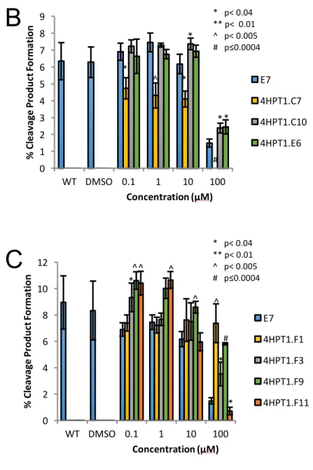
Polyamide Inhibition ... 61
HPT1 Library... 65
DMD Library ... 66
Discussion ... 66
References ... 81
CHAPTER 4: CONCLUSIONS AND FUTURE DIRECTIONS FOR CHARACTERIZATION OF pSK41 AND ITS RELAXASE, NES ... 83
Conclusions ... 83
Future Directions ... 84
Formation of the DNA Hairpin ... 84
Characterization of the pSK41 Relaxosome ... 85
Optimization of Chelator Fragment Inhibitors ... 86
Variation in NES Genes ... 88
References ... 93
CHAPTER 5: INTRODUCTION TO β-GLUCURONIDASE- MICROBIAL ENZYME RESPONSIBLE FOR GI DRUG REACTIVATION ... 94
Microbiome ... 94
Glycoside Hydrolases ... 96
β-Glucuronidase ... 97
Irinotecan ... 100
GUS Inhibitors ... 102
References ... 118
CHAPTER 6: DEFINING THE β-GLUCURONIDASE ENZYME FAMILY IN THE HUMAN GASTROINTESTINAL TRACT ... 122
Materials and Methods ... 125
Human Microbiome Project (HMP) data ... 125
HMP β-glucuronidase Identification ... 125
HMP β-glucuronidase Loop Classification ... 126
Gene abundance calculations ... 126
Results ... 127
HMGC- Clustered Gene Indices GUS Enzymes ... 127
HMGI- Gene Indices GUS Enzymes ... 128
Establishment of GUS Loop Characterizations ... 128
GUS-Containing Bacteria ... 131
Discussion ... 133
References ... 152
CHAPTER 7: FUNCTIONAL CHARACTERIZATION OF β-GLUCURONIDASE ENZYMES IDENTIFIED IN THE HUMAN GASTROINTESTINAL TRACT ... 154
Introduction ... 154
Materials and Methods ... 156
Cloning of GUS Enzymes ... 156
Clostridium perfringens GUS ... 156
Eubacterium eligens GUS ... 157
Faecalibacterium prausnitizii GUS ... 157
Bacteroides uniformis GUS ... 157
Parabacteroides merdae GUS ... 158
Bacteroides dorei GUS ... 158
Lactobacillus rhamnosus GUS ... 159
Expression and Purification of GUS Enzymes ... 159
E. coli GUS ... 159
Steptococcus agalactiae GUS ... 159
CpGUS ... 160
BfGUS ... 160
EeGUS, FpGUS, BuGUS, PmGUS, BoGUS, and BdGUS ... 161
PNPG Assay ... 161
SN-38G HPLC ... 162
SN-38G Assay ... 163
Results ... 163
GUS Activity ... 163
SN-38G Processing ... 165
GUS Activity at Varying pH ... 167
Lactobacillus rhamnosus GUS ... 169
Discussion ... 170
References ... 178
CHAPTER 8: CONCLUSIONS AND FUTURE DIRECTIONS FOR CHARACTERIZATION OF β-GLUCURONIDASE AND GI DRUG REACTIVATION ... 180
Conclusions ... 180
Future Directions ... 181
Structure and Function of New GUS Enzymes ... 182
SN-38G Processing ... 183
Other Glucuronidated Compounds ... 184
References ... 188
APPENDIX 1: pWBG749 OR pSK41 ORIGIN-OF-TRANSFER MIMC SEQUENCE CONTENT ... 189
APPENDIX 2: PLASMIDS CONTAINING pSK41 ORIGIN-OF- TRANSFER MIMIC SEQUENCES ... 199
LIST OF TABLES
Table 2.1. Bacterial Strains and Plasmids Used in This Study ... 51
Table 2.2. Relaxase-in trans Mobilization of Plasmids Containing oriT Sites ... 52
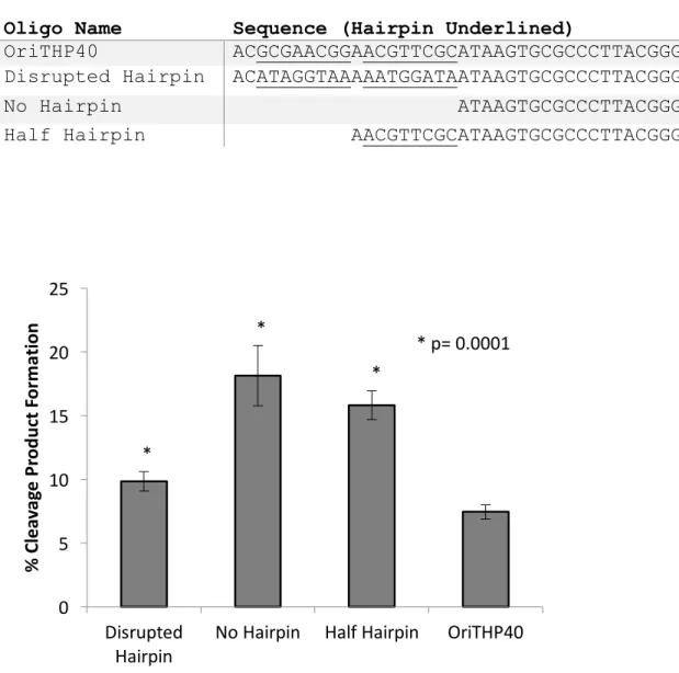
Table 4.1. Altered Hairpin Oligonucelotides ... 90
Table 5.1. Catalytic Activity of GUS ... 105
Table 5.2. In vitro GUS Inhibition, Ki (μM) ... 105
Table 6.1. Key Active Site Residues Used for β-glucuronidase Identification. ... 138
Table 6.2. Criteria Used for β-glucuronidase Loop Classification ... 138
LIST OF FIGURES
Figure 1.1. Mechanism of Conjugative Plasmid Transfer ... 10
Figure 1.2. Classification of Plasmids and MOB Genes ... 11
Figure 1.3. Proposed Mechanism of DNA Cleavage by NES ... 12
Figure 1.4. NES Structure from S. aureus pSK41 ... 13
Figure 1.5. Effect of NES Mutants on Conjugation in S aureus ... 14
Figure 2.1. Structure of NES Relaxase Domain and pSK41 oriT ... 42
Figure 2.3. Functional Analysis of NES Loop Deletion Mutants ... 44
Figure 2.4. Modeled Structure of the pSK41, pSK156, and pCA347 oriTs ... 47
Figure 2.5. NES Processing of pSK41, pSK156, and pCA347 oriT Oligonucleotides ... 49
Figure 2.6. Nucleotide Sequences for in vivo Transfer Assays ... 50
Figure 3.1. Polyamide Structure ... 70
Figure 3.2 Polyamide Impact on NES Activity ... 71
Figure 3.3. Chelator Fragment Library- 1.1 (CFL-1.1) ... 72
Figure 3.4. 100µM Screen of CFL-1.1 ... 75
Figure 3.5. Chelator Impact on NES Cleavage Product Formation ... 77
Figure 3.6. HPT1 E7 Derivative Library ... 79
Figure 4.1. NES cleavage of altered hairpin oligonucleotides ... 90
Figure 4.2. Gua-26 Binding Pocket ... 91
Figure 4.3. M3W Mutation Effects ... 92
Figure 5.1. Enzymatic Mechanism of E. coli GUS ... 107
Figure 5.2. Structure of Microbial GUS Enzymes ... 108
Figure 5.3. N-K and Y Motif ... 110
Figure 5.4. Presence of the Bacterial Loop in Representative GUS Enzymes ... 113
Figure 5.5. Metabolic Pathway of CPT-11 ... 115
Figure 5.6. Microbial GUS Inhibitors in Mice Treasted with CPT-11 ... 117
Figure 6.1. Workflow for Identification and Classification of Novel GUS Sequences ... 139
Figure 6.2. Identification of Novel GUS Sequences from HMGC- HMP Clustered Gene Indices Database ... 140
Figure 6.3. Identification of Novel GUS Sequences from HMGI- HMP Gene Indices Database ... 142
Figure 6.4. Classification of Novel GUS Sequences by Loop ... 144
Figure 6.5. Number of GUS Sequences of Each Loop Class Found per Individual ... 148
Figure 6.6. Predicted Bacteria Containing GUS Sequence of Each Loop Class ... 151
Figure 7.2. β-glucuronidase Activity Against SN-38G via Fluorescence ... 175
Figure 7.3. Catalytic Activity of β-glucuronidase Enzymes at Varying pH ... 176
Figure 7.4. Catalytic Activity of L. rhamnosus β-glucuronidase at Varying pH ... 177
LIST OF ABBREVIATIONS AND SYMBOLS
% percent
°C degrees Celsius
± plus or minus
< less than
> greater than
≥ greater than or equal to 6FAM/6-FAM/FAM fluorescein
A adenine
Å angstrom
ACIDO Acidobacteria Terriglobus roseus β-glucuronidase
ACTIN Actinobacteria Corynebacterium massiliense β-glucuronidase AI arabinose inducible
Ala alanine
Ap/ApR ampicillin/ampicillin resistance APS ammonium persulfate
ArchIa Archaea Ignisphaera aggregans β-glucuronidase B_dorei Bacteroides dorei β-glucuronidase
B. fragilis Bacteroides fragilis B. ovatus Bacteroides ovatus B. uniformis Bacteroides uniformis
BactNk Bacteroides Niastella koreensis β-glucuronidase BdGUS Bacteroides dorei β-glucuronidase
BfGUS Bacteroides fragilis β-glucuronidase BHI Brain Heart Infusion Broth
BLASTN Basic Local Alignment Search Tool for Nucleotide BLASTp Basic Local Alignment Search Tool for Protein BoGUS Bacteroides ovatus β-glucuronidase
BovineGUS Bos taurus β-glucuronidase BSA bovine serum albumin BtGUS Bos taurus β-glucuronidase
BuGUS Bacteroides uniformis β-glucuronidase C_perfringens Clostridium perfringens β-glucuronidase C-terminal, C-term carboxyl terminal
C, Cyt cytosine
C. perfringens Clostridium perfringens CaCl2 calcium chloride
CDC Center for Disease Control CFL chelator fragment library
Cl chloride
Cm/CmR chloramphenicol/chloramphenicol resistance CPD cysteine protease domain
CpGUS Clostridium perfringens β-glucuronidase
CPT conjugative plasmid transfer
CPT-11 irinotecan, 7-ethyl-10-[4-(1-piperidino)-1-piperidino] CRISPR clustered regularly interspaced short palindromic repeats
CV column volume
DICTY Dictyoglomi Dictyoglomus turgidum β-glucuronidase DMSO dimethyl sulfoxide
DNA deoxyribonucleic acid
dsDNA double stranded deoxyribonucleic acid DTT D,L-dithiothreitol
E glutamic acid
E_coli Escherichia coli β-glucuronidase E_eligens Eubacterium eligens β-glucuronidase E. coli Escherichia coli
E. eligens Eubacterium eligens E. lenta Eggerthella lenta
EcGUS Escherichia coli β-glucuronidase EDTA Ethylenediaminetetraacetic acid EeGUS Eubacterium eligens β-glucuronidase
Em emission
Ex excitation
f average fluorescence anisotropy signal F_prausnitzii Faecalibacterium prausnitzii β-glucuronidase F. prausnitzii Faecalibacterium prausnitzii
FBDD fragment-based drug design
FDA United States Food and Drug Administration FOLFIRI folinic acid, fluorouracil, irinotecan
FOLFIRINOX folinic acid, fluorouracil, irinotecan, oxaliplatin FpGUS Faecalibacterium prausnitzii β-glucuronidase FsR fusidic acid resistance
Fu fluorescence unit(s)
FUNGI Eukaryota Aspergillus niger β-glucuronidase g g-force, gravitational acceleration
G, Gly glycine G, Gua guanine
GH2 glycoside hydrolase family 2 GI gastrointestinal
Gm/GmR gentamycin/gentamycin resistance GUS β-glucuronidase
h hour(s)
H_sapiens Homo sapiens β-glucuronidase H, His histidine
H+ hydrogen ion
H2O water
HCl hydrochloric acid
HEPES 4-(2-hydroxyethyl)-1-piperazineethanesulfonic acid HGT horizontal gene transfer
His6 6 x histidine affinity tag
HMGC HMP Clustered Gene Indices HMGI HMP Gene Indices
HMP Human Microbiome Project
HPLC high-performance liquid chromatography HsGUS Homo sapiens β-glucuronidase
HTS high-throughput screen
HUH histidine residue-hydrophobic residue-histidine residue
I.V. intravenous
Im imidazole
Inh 1 Inhibitor 1
InsP6 inositol hexakisphosphate
IPTG isopropyl-β-D-thiogalactopyranoside
IR2 inverted repeats of the pWBG749 family oriT
IRI irinotecan
K KD
K, Lys lysine
kcat/KM catalytic efficiency KCl potassium chloride KD dissociation constant KH2PO4 potassium phosphate
Ki dissociation constant for the inhibitor KM Michaelis-Menten constant
KmR kanamycin resistance
KO knock out
L liter
L-DOPA levodopa, L-3,4-dihydroxyphenylalanine L, Leu leucine
L. rhamnosus Lactobacillus rhamnosus
L1 Loop 1
L2 Loop 2
LB lysogeny broth
LIC ligation independent cloning
LrGUS Lactobacillus rhamnosus β-glucuronidase
M methionine
MALDI-TOF matrix-assisted laser desorption/ionization-time of flight
max average fluorescence anisotropy signal of sample at saturating concentration of protein
MBP maltose binding protein
mg milligram
MIC minimum inhibitory concentration
min minute
min average fluorescence anisotropy signal of no protein control
mL milliliter
mL1 Mini Loop 1
mL1mL2 Mini Loop 1 Mini Loop 2 mL2 Mini Loop 2
mM millimolar
MOB mobility enzymes mob mobility genes
MRSA methicillin resistant Staphylococcus aureus MSA multiple sequence alignment
MSP multiple species
n the total number of reads in that HMP sequencing sample N the total number of HMP sequencing samples
N total number of GUS sequences in each individual N-terminal amino terminal
N, Asn asparagine NaCl sodium chloride
NAD+ nicotinamide adenine dinucleotide- oxidized form NADH nicotinamide adenine dinucleotide- reduced form Nb/NbR novobiocin/novobiocin resistance
NES nicking enzyme of Staphylococcus nes nicking enzyme of Staphylococcus gene
Ni nickel
ni no inhibition Ni2+ nickel cation
NIH National Institutes of Health
nL No Loop
nM nanomolar
nm nanometer
NmR neomycin resistance
NSAID nonsteroidal anti-inflammatory drug
NxKG asparagine residue, any residue, lysine residue, glycine residue
OD600 optical density at 600 nm wavelength
oligos oligonucleotides
Orf gene open reading frame
oriT origin of transfer
p p-value
P_merdae Parabacteroides merdae β-glucuronidase P, Pro proline
P. merdae Parabacteroides merdae PAGE polyacrylamide gel
PDB ID PDB identification code PEG polyethylene glycol
pH negative log (base 10) of the molar concentration of hydronium ions Phe phenylalanine
PmGUS Parabacteroides merdae β-glucuronidase
PNP p-nitrophenyl
PNPG p-nitrophenyl glucuronide
PROT Proteobacteria Vibrio harveyi β-glucuronidase
Py pyrrole
R arginine
RC read count for a gene in a particular HMP sequencing sample RCSB Research Collaboratory for Structural Bioinformatics
RfR rifampin resistance RNA ribonucleic acid
rRNA ribosomal ribonucleic acid
S_agalactiae Streptococcus agalactiae β-glucuronidase
s-1 per second
s-1mM-1 per second per millimolar S. agalactiae Streptococcus agalactiae S. aureus Staphylococcus aureus
SaGUS Streptococcus agalactiae β-glucuronidase SDS sodium dodecyl sulfate
Ser serine
∑x the total number of reads in all HMP sequencing samples Sm/SmR streptomycin/streptomycin resistance
SN-38 7-ethyl-10-hydroxyl-camptothecin
SN-38-glucuronide 7-ethyl-10-hydroxyl-camptothecin glucuronide SN-38G 7-ethyl-10-hydroxyl-camptothecin glucuronide SN2 bimolecular nucleophilic substitution
sp. species
T Thymine
T total DNA concentration T-strand transfer strand
T4CP type 4 coupling protein T4SS type four secretion system TAE tris base, acetic acid, EDTA TBE tris, borate, EDTA
TbR tobramycin resistance
TCEP tris(2-carboxyethyl)phosphine
TEMED tetramethylethylenediamine
TherTn Thermatogae Thermotoga naphthophila β-glucuronidase Tm melting temperature
tra conjugative transfer gene
UDP uridine 5'-diphosphate
UGT UDP-glucuronosyltransferase
UNC University of North Carolina at Chapel Hill UNC-CH University of North Carolina at Chapel Hill
UV ultraviolet
vol/vol, v/v volume per volume
VRSA vancomycin resistant S. aureus
vs versus
W tryptophan
WT wild type
x total protein concentration Y, Tyr tyrosine
α alpha
β beta
Δ deletion
ΔL1 Hairpin Loop 1 deletion
ΔL1ΔL2 Hairpin Loop 1 Loop 2 deletion ΔL2 Hairpin Loop 2 deletion
λ wavelength
μg microgram
μL microliter
μM micromolar
CHAPTER 1: INTRODUCTION TO CONJUGATIVE PLASMID TRANSFER OF pSKS41 IN STAPHYLOCOCCUS AUREUS
Staphylococcus aureus and Antibiotic Resistance
Staphylococcus aureus is a gram-positive bacterium that is found as a commensal in the nose, respiratory tract, gastrointestinal tract, and on the skin of humans (1). Despite this potentially beneficial role in the human microbiota, S. aureus is best known as a pathogen that causes skin and respiratory infections. While these infections are normally treatable, infections resistant to antibiotic treatment can enter the bloodstream and cause sepsis and death. Since the introduction of penicillin and other antibiotic compounds, resistant strains of S. aureus have been appearing in human infections. It took just two years after the introduction of methicillin in the clinic for methicillin-resistant Staphylococcus to begin to present a barrier to treatment (2). In 2011, methicillin-resistant S. aureus (MRSA) caused an estimated 80,461 infections in the United States leading to 11,285 deaths (2). Staphylococcal bacteria are a leading cause of healthcare-associated infections leading the Center for Disease Control (CDC) to classify MRSA as a serious threat (2).
resistances found in S. aureus can include oxacillin, erythromycin, levofloxacin, clindamycin, mupirocin, doxycycline, gentamicin, trimethoprim-sulfamethoxazole, and tetracycline, which when found in the same strain can leave only harsh compounds available for treatment (3). It has also been shown that MRSA can obtain resistance to vancomycin, the current front-line treatment, leading to
infections that are resistant to almost all approved antibiotics (4-7). Despite the rise of antibiotic resistance in S. aureus and other bacteria, the number of new antibiotics approved by the FDA has consistently decreased over the past two decades.
Even as new antibiotics are introduced, it is expected that resistance will arise quickly as seen in the past. Most antibiotics to date have been discovered as or derived from small molecules produced by bacteria or plants. In these natural environments, these small molecules function as bacteriostatic or bacteriotoxic agents in order to regulate the surrounding bacterial community, much like how they function as antibiotics relevant to human health. This means the bacteria producing the compound or any bacteria that persist in this natural environment must maintain a resistance element to avoid toxicity from the compound. In fact, environmental bacteria including those preserved for hundreds of years in permafrost, isolated caves, and ancient human specimens contain resistance genes (8). Because resistance to a compound similar to any new antibiotics almost certainly already exists in nature, it is likely only a matter of time until that resistance element moves into pathogenic bacteria.
mechanisms by which antibiotic resistance is spread between bacteria. We hope that characterization of these processes will lead to drugs that specifically target antibiotic resistant bacterial communities or prevent the spread of antibiotic resistance within bacterial communities.
Conjugative Plasmid Transfer
Bacteria become resistant to the effects of antibiotics and other small molecules by obtaining genetic elements that encode for proteins that allow the bacteria to breakdown or transport the lethal substance out of their cell or encode modified targets with reduced antibiotic drug-reactivity. These resistance genetic elements can be incorporated into the chromosome of a bacterium or may reside on extra-chromosomal elements such as gene cassettes, transposons, bacteriophages, and single- or double-stranded DNA plasmids. Bacteria can transfer their genetic material, including these resistance elements, through two routes: vertical or horizontal gene transfer. Vertical gene transfer takes place between a mother and her daughter cells via duplication of the chromosomal and extra-chromosomal
genetic material and distribution of a complete copy to each daughter cell. Horizontal gene transfer (HGT) is the transmission between mature, parent bacteria and
involves only extra-chromosomal elements. HGT takes many forms including scavenging of DNA by naturally competent bacterial cells, bacteriophage facilitated transfer, and regulated transfer for single- and double-stranded plasmids.
movement through a dsDNA plasmid; therefore, this work will focus on the transfer mechanism of these plasmids between bacterium. Double-stranded DNA plasmids can be beneficial to the larger bacterial community as many of these plasmids are readily involved in HGT, allowing an entire population of bacteria to quickly acquire antibiotic resistance (9). HGT of dsDNA plasmids is called conjugative plasmid transfer (CPT). During CPT, a donor bacterium transfers one strand of a double-stranded DNA plasmid to a recipient bacterium (Figure 1.1).
There are several sets of machinery required for this process that may be expressed on the plasmid being transferred or on a co-resident plasmid in the donor bacterium. This machinery includes the mobility (MOB) set of enzymes that is
responsible for the replication of the plasmid as well as genes for a type 4 secretion system (T4SS) and type 4 coupling protein (T4CP) that facilitate transfer of the plasmid (10). The MOB set of proteins forms a large complex called the relaxosome. The relaxosome complex includes the highly conserved relaxase (NES in Figure 1.1) as well as the replication machinery of the plasmid. The T4SS and T4CP form a secretion system through which DNA travels to the recipient cell (blue shaded box in Figure 1.1).
some data indicate that the covalently bound relaxase also travels into the recipient bacterium where the relaxase can then ligate the T-strand back together (12). A DNA polymerase within the donor bacterium replaces the T-strand as it is
transferred and a DNA polymerase within the recipient bacterium synthesizes a copy of the T-strand so that the process culminates with two bacteria each containing a full, double-stranded plasmid that can confer antibiotic resistance.
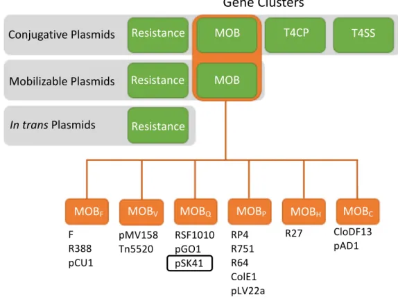
Types ofPlasmids
Plasmids that undergo CPT are divided into three classes based on the presence or absence of the MOB, T4CP, and T4SS gene clusters (Figure 1.2). The best studied type of plasmid is the conjugative plasmid which encodes all of the machinery needed to replicate and transmit itself including the MOB, T4CP, and T4SS clusters (Figure 1.2, 10). Mobilizable plasmids do not encode all of the machinery needed for their transmission; they express only the MOB proteins required for replication but do not encode their own T4SS and therefore must use the conjugation machinery expressed by a conjugative plasmid co-resident in the same bacterium (10). A small number of mobilizable plasmids encode their own T4CP but most utilize the T4CP from the co-resident plasmid expressing the T4SS.
There is also a third type of plasmid that does not encode its own replication or conjugative machinery. Until recently it was assumed these plasmids were transferred via mechanisms besides conjugative plasmid transfer such as
advantage of both the replication (MOB) and conjugation (T4CP/T4SS) machinery of a co-resident plasmid (Figure 1.2,13, 14). For this relaxase-in trans transfer, it is important that the MOB proteins of a conjugative or mobilizable plasmid co-resident in the bacterium match an oriT found on the in trans plasmid. Therefore,
understanding of MOB proteins is important for characterization of CPT of all three plasmid classes.
The relaxase is the only highly conserved member of the MOB family of proteins and is used to identify and classify conjugative and mobilizable plasmids (Figure 1.2). A relaxase enzyme is defined by the presence of two motifs (15). The first motif is the Y motif which consists of one or more tyrosines that are responsible for plasmid nicking and ligation. This conserved tyrosine (Y25 in Figure 1.3) initiates a nucleophilic substitution-type (SN2) attack on the scissile phosphate linking two nucleotides at the nic site (16). This generates a free 3’ hydroxyl and a 5’ covalent phosphotyrosine bond. The reaction is reversible with the 3’ hydroxyl acting as a nucleophile to ligate the strand together in the recipient cell.
Based on the sequence of their respective relaxase and in particular, the sequence in and around the Y and HUH motifs, conjugative and mobilizable plasmids have been classified into six families. These families are MOBF, MOBH, MOBC, MOBQ, MOBP, and MOBV (Figure 1.2, 20, 21). The relaxases of the MOBH and MOBC families have a different architecture and may not contain regions that resemble the Y and HUH motifs (15, 22). The MOBF family is the best characterized of the six families and contains relaxases with two to three tyrosines in the Y motif (15). The MOBQ, MOBP, and MOBV plasmid families encode relaxases containing only one tyrosine in the Y motif (15). Because the relaxase is responsible for the initiation and termination of CPT, understanding of these proteins could lead to development of novel antibiotics targeting resistance-spreading bacterial infections. Indeed, it has already been shown that inhibiting the relaxase can disrupt
propagation of the host plasmid (23).
Introduction to S. aureus plasmid pSK41
pSK41 is a 46,445 nucleotide, conjugative, multiresistance plasmid from S. aureus (24). Related plasmids were first detected in the mid-1970s; the transfer region of pSK41 was analyzed by Firth and colleagues in 1993 and the complete sequence of pSK41 was published in 1998 (24, 25). pSK41 is now considered the prototype plasmid for this family.
compounds, trimethoprim, mupirocin, macrolides, lincosamides, streptogramins, bleomycin, and β-lactams (3, 24-26). Of special note is the incorporation of vancomycin resistance into pSK41 to form the new plasmid pLW1043 (4). pSK41 has also been shown to contribute to the spread of antibiotic resistance through facilitating the transfer of mobilizable plasmids such as pC221 and pSK639 (27, 28). Because pSK41 and related plasmids confer such a wide range of resistances, it is an important target for understanding and modulating the spread of antibiotic resistance in S. aureus.
Introduction to Nicking Enzyme of Staphylococcus (NES)
The relaxase of pSK41 is named nicking enzyme of staphylococci (NES). This gene and its oriT target were first identified on the pGO1 plasmid by Climo et al. in 1996 (29). The nes gene was confirmed in pSK41 by Berg and colleagues (24). NES is a single-tyrosine relaxase in the MOBQ family and is considered the
prototype relaxase for this family.
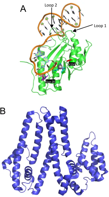
instead has two unique features to properly position the oriT DNA (17). The first feature is two protein loops (loop 1 and loop 2 in Figure 1.4A) that extend into the major and minor groove of the DNA hairpin formed by the oriT DNA. The second feature is nucleotide guanine-26 (magenta in Figure 1.4A) of the oriT that is flipped in the opposite direction from the surrounding nucleotides and makes significant contacts with amino acid side chains of NES.
Relaxases from the MOBQ, MOBP, and MOBV families including NES are two domain proteins with the N-terminal domain containing the relaxase motif. The C-terminal domain is variable and often functions as a helicase, primase, or DNA-binding domain that contributes to the DNA replication process (15). Interestingly, the NES terminal domain shows no similarity to previously characterized
C-terminal domains of relaxases or other relaxosome proteins. The C-C-terminal domain of NES was also crystallized and its structure solved; however, both this C-terminal domain structure and the SAXS envelope of the full-length protein did not give any suggestion of the potential function of this domain (Figure 1.4B,17).
Figure 1.1. Mechanism of Conjugative Plasmid Transfer
Figure 1.2. Classification of Plasmids and MOB Genes
Bacterial plasmids are classified according to the genes encoded on the plasmid. Conjugative plasmids encode resistance elements, mobilization (MOB) genes, a type 4 coupling protein (T4CP), and a type 4 secretion system (T4SS). Mobilizable plasmids encode only resistance elements and MOB genes while in trans plasmids encode on resistance elements. The MOB genes on conjugative and mobilizable plasmids are in turn classified as MOBF, MOBH, MOBC, MOBQ, MOBP, AND MOBV according to the motifs found in their relaxase gene.
Gene Clusters
Conjugative Plasmids
Mobilizable Plasmids
In trans Plasmids
Resistance
Resistance
Resistance
MOB
MOB
T4CP T4SS
MOBF MOBV MOBQ MOBP MOBH MOBC
F R388 pCU1
pMV158
Tn5520 RSF1010pGO1 pSK41
RP4 R751 R64 ColE1 pLV22a
Figure 1.3. Proposed Mechanism of DNA Cleavage by NES
The tyrosine residue of the relaxase (Y25 in NES) initiates a nucleophilic
Figure 1.4. NES Structure from S. aureus pSK41
A. 2.9 Å crystal structure of the NES relaxase domain (residues 1-195) in complex with a 30-nucleotide DNA sequence. PDB ID: 4HT4
B. 3.0 Å crystal structure of the NES C-terminal domain (residues 254-593). PDB
A
B
Loop 1 Loop 2
Ni Y25F
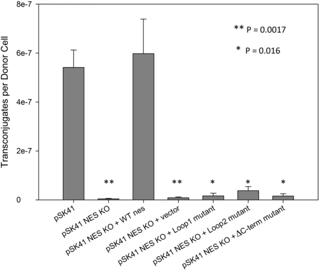
Figure 1.5. Effect of NES Mutants on Conjugation in S aureus
Conjugation of pSK41 in S. aureus and the effect of nes deletion (KO) or
REFERENCES
1. Brown AF, Leech JM, Rogers TR, McLoughlin RM. 2014. Staphylococcus aureus Colonization: Modulation of Host Immune Response and Impact on Human Vaccine Design. Front Immunol 4:507.
2. Centers for Disease Control and Prevention (CDC). 2013. Antibiotic Resistance Threats in the United States, 2013. cdcgov.
3. McDougal LK, Fosheim GE, Nicholson A, Bulens SN, Limbago BM, Shearer JES, Summers AO, Patel JB. 2010. Emergence of resistance among USA300 methicillin-resistant Staphylococcus aureus isolates causing invasive disease in the United States. Antimicrobial Agents and Chemotherapy 54:3804–3811.
4. Chang S, Sievert DM, Hageman JC, Boulton ML, Tenover FC, Downes FP, Shah S, Rudrik JT, Pupp GR, Brown WJ, Cardo D, Fridkin SK,
Vancomycin-Resistant Staphylococcus aureus Investigative Team. 2003. Infection with vancomycin-resistant Staphylococcus aureus containing the vanA resistance gene. N Engl J Med 348:1342–1347.
5. Sievert DM, Rudrik JT, Patel JB, McDonald LC, Wilkins MJ, Hageman JC. 2008. Vancomycin-resistant Staphylococcus aureus in the United States, 2002-2006. Clin Infect Dis 46:668–674.
6. Weigel LM, Clewell DB, Gill SR, Clark NC, McDougal LK, Flannagan SE, Kolonay JF, Shetty J, Killgore GE, Tenover FC. 2003. Genetic analysis of a high-level vancomycin-resistant isolate of Staphylococcus aureus. Science 302:1569–1571.
7. Zhu W, Clark N, Patel JB. 2013. pSK41-Like Plasmid Is Necessary for Inc18-Like vanA Plasmid Transfer from Enterococcus faecalis to Staphylococcus aureus In Vitro. Antimicrobial Agents and Chemotherapy 57:212.
8. Perry J, Waglechner N, Wright G. 2016. The Prehistory of Antibiotic Resistance. Cold Spring Harb Perspect Med 6.
9. Chen I, Christie PJ, Dubnau D. 2005. The ins and outs of DNA transfer in bacteria. Science 310:1456–1460.
10. Smillie C, Garcillán-Barcia MP, Francia MV, Rocha EPC, la Cruz de F. 2010. Mobility of plasmids. Microbiol Mol Biol Rev 74:434–452.
12. Dostál L, Shao S, Schildbach JF. 2011. Tracking F plasmid TraI relaxase processing reactions provides insight into F plasmid transfer. Nucleic Acids Research 39:2658–2670.
13. O'Brien FG, Ramsay JP, Monecke S, Coombs GW, Robinson OJ, Htet Z, Alshaikh FAM, Grubb WB. 2014. Staphylococcus aureus plasmids without mobilization genes are mobilized by a novel conjugative plasmid from community isolates. Journal of Antimicrobial Chemotherapy 70:649–652. 14. O'Brien FG, Eto KY, Murphy RJT, Fairhurst HM, Coombs GW, Grubb WB,
Ramsay JP. 2015. Origin-of-transfer sequences facilitate mobilisation of non-conjugative antimicrobial-resistance plasmids in Staphylococcus aureus. Nucleic Acids Research 43:7971–7983.
15. Chandler M, la Cruz de F, Dyda F, Hickman AB, Moncalián G, Ton-Hoang B. 2013. Breaking and joining single-stranded DNA: the HUH endonuclease superfamily. Nat Rev Microbiol 11:525–538.
16. Boer R, Russi S, Guasch A, Lucas M, Blanco AG, Pérez-Luque R, Coll M, la Cruz de F. 2006. Unveiling the molecular mechanism of a conjugative relaxase: The structure of TrwC complexed with a 27-mer DNA comprising the recognition hairpin and the cleavage site. Journal of Molecular Biology
358:857–869.
17. Edwards JS, Betts L, Frazier ML, Pollet RM, Kwong SM, Walton WG, Ballentine WK, Huang JJ, Habibi S, Del Campo M, Meier JL, Dervan PB, Firth N, Redinbo MR. 2013. Molecular basis of antibiotic multiresistance transfer in Staphylococcus aureus. Proceedings of the National Academy of Sciences of the United States of America 110:2804–2809.
18. Xia S, Robertus JD. 2009. Effect of divalent ions on the minimal relaxase domain of MobA. Arch Biochem Biophys 488:42–47.
19. Larkin C, Haft RJF, Harley MJ, Traxler B, Schildbach JF. 2007. Roles of active site residues and the HUH motif of the F plasmid TraI relaxase. J Biol Chem 282:33707–33713.
20. Francia MV, Varsaki A, Garcillán-Barcia MP, Latorre A, Drainas C, la Cruz de F. 2004. A classification scheme for mobilization regions of bacterial
plasmids. FEMS Microbiol Rev 28:79–100.
22. Francia MV, Clewell DB, la Cruz de F, Moncalián G. 2013. Catalytic domain of plasmid pAD1 relaxase TraX defines a group of relaxases related to
restriction endonucleases. Proceedings of the National Academy of Sciences of the United States of America 110:13606–13611.
23. Lujan SA, Guogas LM, Ragonese H, Matson SW, Redinbo MR. 2007. Disrupting antibiotic resistance propagation by inhibiting the conjugative DNA relaxase. Proceedings of the National Academy of Sciences of the United States of America 104:12282–12287.
24. Berg T, Firth N, Apisiridej S, Hettiaratchi A, Leelaporn A, Skurray RA. 1998. Complete nucleotide sequence of pSK41: evolution of staphylococcal conjugative multiresistance plasmids. J Bacteriol 180:4350–4359.
25. Firth N, Ridgway KP, Byrne ME, Fink PD, Johnson L, Paulsen IT, Skurray RA. 1993. Analysis of a transfer region from the staphylococcal conjugative plasmid pSK41. Structure 136:13–25.
26. Liu MA, Kwong SM, Jensen SO, Brzoska AJ, Firth N. 2013. Biology of the staphylococcal conjugative multiresistance plasmid pSK41. Plasmid 70:42–51. 27. Caryl JA, Thomas CD. 2006. Investigating the basis of substrate recognition
in the pC221 relaxosome. Mol Microbiol 60:1302–1318.
28. Apisiridej S, Leelaporn A, Scaramuzzi CD, Skurray RA, Firth N. 1997. Molecular analysis of a mobilizable theta-mode trimethoprim resistance plasmid from coagulase-negative staphylococci. Plasmid 38:13–24.
29. Climo MW, Sharma VK, Archer GL. 1996. Identification and characterization of the origin of conjugative transfer (oriT) and a gene (nes) encoding a single-stranded endonuclease on the staphylococcal plasmid pGO1. J Bacteriol 178:4975–4983.
30. Larkin C, Datta S, Harley MJ, Anderson BJ, Ebie A, Hargreaves V,
Schildbach JF. 2005. Inter- and intramolecular determinants of the specificity of single-stranded DNA binding and cleavage by the F factor relaxase.
CHAPTER 2: PROCESSING OF NONCONJUGATIVE RESISTANCE PLASMIDS BY CONJUGATIVE NICKING ENZYME OF STAPHYLOCOCCI1
Introduction
Antimicrobial resistant strains of Staphylococcus aureus are a growing concern for hospital- and community-acquired infections. Most S. aureus bacteria examined clinically harbor at least one plasmid that encodes for antimicrobial
resistance, and many plasmids carry multiple antimicrobial resistance determinants. The pSK41 family of plasmids is made up of large, low-copy-number, conjugative plasmids for which pSK41 is used as a prototype for characterization (1-4). These plasmids carry a variety of antimicrobial resistance determinants, including those against aminoglycosides, penicillins, tetracycline, bleomycin, trimethoprim, macrolides, lincosamides, mupirocin, antiseptics, and disinfectants (2, 4-9). This family of plasmids also played a key role in the rise of vancomycin resistant S. aureus (VRSA) (7, 8, 10). In addition to the antimicrobial resistance, they also carry transfer (tra) genes encoding the proteins necessary to conduct conjugative plasmid transfer that spread these plasmids among S. aureus and other gram-positive
bacteria (6, 7, 10, 11).
1 This chapter adapted from the previously published work Pollet RM, Ingle JD, Hymes JP, Eakes TC,
One of the proteins essential for conjugative plasmid transfer is the relaxase enzyme. A relaxase is responsible for initiation and completion of the transfer process as it cleaves one strand of the double-stranded plasmid to begin transfer, and then ligates that strand back together to complete transfer (8, 12-15). There are two classes of relaxases: multi-tyrosine relaxases that use a “thumb” motif to
position the plasmid DNA for processing, and single-tyrosine relaxases which lack this thumb motif (9, 16). The relaxase of pSK41 is termed NES, nicking enzyme in S. aureus, and is a single-tyrosine relaxase (1-6, 8, 9). NES contains a relaxase N-terminal 220 residues and a C-N-terminal 350 residues necessary for in vivo function but via an uncertain mechanism (5, 7, 8, 10). Here we determine in which steps of conjugation the C-terminal domain plays an important role.
The crystal structure of the relaxase domain of NES was the first of a single-tyrosine relaxase bound to its target DNA, allowing for more detailed
characterization of the protein-DNA interactions than previously possible (6-8, 11). This structure revealed two sets of important protein-DNA interactions. The first is that the “thumb” used by multi-tyrosine relaxases to position the DNA appears to be replaced by 12 protein-DNA contacts including a buried nucleotide 3 bases
phosphate backbone. NES Hairpin Loop 2, green in Figure 2.1A and B, contacts the DNA more extensively, with six base specific interactions and four phosphate
contacts in the major groove of the DNA hairpin. Edwards et al. previously showed in vitro that these loops disrupt DNA cleavage by the relaxase domain alone and in vivo that full-length NES protein lacking these loops was not able to facilitate plasmid transfer (7, 8, 10, 14). However, this important protein-DNA interaction had not been characterized in vitro in the context of the full-length 665-residue NES protein and we set out to determine in which steps of conjugation this interaction plays a role.
pSK41-like conjugative plasmids have been shown to mobilize several smaller co-resident plasmids such as pC221 and pSK639, which encode their own mob genes (9, 12, 13, 15, 16). Recently, O’Brien et al. showed that another
staphylococcal conjugative plasmid pWBG749, which is unrelated to pSK41, can facilitate the mobilization of other plasmids that lack mob genes (17). They
demonstrated that this transfer is facilitated by origin-of-transfer sequences on the non-conjugative plasmids that mimic the pWBG749 origin-of-transfer sequence, suggesting a conjugative relaxase-in trans mechanism (18). We have identified sequences similar to the pSK41 origin of transfer on numerous non-conjugative staphylococcal resistance plasmids (Appendices 1 and 2), raising the possibility that pSK41 family plasmids might likewise facilitate mobilization of other plasmids by an analogous relaxase-in trans mechanism mediated by NES. To investigate this possibility, we have characterized the pSK41 oriT mimic sequences from two
(19). The second plasmid, pCA347, was first sequenced in 2013 after isolation from a USA600 methicillin-resistant strain of S. aureus and encodes resistance to
penicillin and heavy metals (20). Importantly, the variation in the origin-of-transfer sequence of pSK41 and the mimics of pSK156 and pCA347 is in the hairpin region of the DNA (Figure 2.4A). Based on these observations we sought to explore the ability of NES to bind to and process putative oriT regions from pSK156 and pCA347 and the ability of pSK41 to facilitate transfer of plasmids containing these putative oriT regions in order to examine the potential for mobilization of plasmids containing pSK41 oriT mimics in staphylococci.
Materials and Methods
Cloning, Expression and Purification of the Relaxase Domain of NES The relaxase domain of NES was previously cloned into the cysteine
pH 7.4, and 0.02% (vol/vol) sodium azide] along with protease inhibitor tablets (Roche), DNase, and lysozyme. The slurry was lysed using a Fisher Scientific Sonic Dismembrator. The lysed cells were then spun at 18,500 × g for 1 h. The
supernatant was filtered and flowed over a 5 mL HisTrap column (GE Healthcare). The column and bound protein was then washed with Buffer A followed by
incubation with 1 CV of 2 mM inositol hexakisphosphate (InsP6) for 3 hours at 4 °C. The cleaved protein was washed off the column with Buffer A and flowed directly onto a Superdex 200 column (GE Healthcare) preequilibrated in sizing buffer [300 mM NaCl, 50 mM Tris buffer, pH 7.4, and 0.02% (vol/vol) sodium azide]. Fractions containing protein as assessed by UV absorbance were analyzed via SDS-PAGE and fractions containing >95% pure protein were combined and stored at -80 °C. Cloning, Expression and Purification of Full-Length NES
minutes, protein expression was induced with 100 μM
isopropyl-β-D-thiogalactopyranoside (IPTG) and cells were allowed to grow for 16 hours. The cells were pelleted and stored at -80°C. Individual cell pellets were resuspended in Buffer A [500 mM NaCl, 20 mM KH2PO4 pH 7.4, 25 mM Imidazole, 0.02% (v/v) sodium azide] along with protease inhibitor tablets (Roche), DNase, and lysozyme. The mixture was sonicated and then clarified via centrifugation. The supernatant was filtered and loaded onto a 5 mL HisTrap column (GE Healthcare). The CPD expression system contains a His6 tag in addition to the CPD tag, which has self-cleavage abilities in the presence of inositol hexakisphosphate (InsP6). Therefore, after the His-CPD-NES fusion protein was bound to the column via the His6 tag, the column was washed with 2 column volumes (CV) Buffer A and then incubated with 2 mM InsP6 for 3 hours at 4°C. The NES protein was then eluted off the column in Buffer A while the His6 and CPD tags remained bound to the column. The NES protein was then passed over a Superdex 200 column (GE Healthcare) pre-equilibrated in sizing buffer [25 mM HEPES pH 7.4, 300 mM NaCl, 0.02% (v/v) sodium azide]. Purity of each fraction was assessed by SDS-PAGE gel and fractions containing >95% pure protein were combined and concentrated to approximately 1.2 mg/ml.
DNA Binding Studies
binding was calculated using fluorescence anisotropy as described in Edwards et al. (7). Briefly, protein was serially diluted into a buffer of 100 mM NaCl, 0.1 mg/ml BSA, 5 mM Magnesium Acetate, 25 mM Tris Acetate, pH 7.5 to give 40 μL at final protein concentrations ranging from 0 to 0.5 μM. Assays were conducted in a 384-well black assay plate (Costar) allowing for 16 concentrations of protein. 10 μL of the DNA probe was added to the 40 μL protein solution resulting in a final concentration of DNA of 50 nM in a total volume of 50 μL in each well. Fluorescence anisotropy of the fluorescein-labeled DNA was observed via excitation at 485 nm and emission at 520 nm using a PHERAstar plate reader (BMG Labtech). Measurements were made in triplicate and reported values are the average of three separate triplicate runs. Data were plotted as average fluorescence anisotropy as a function of protein concentration using Graphpad PRISM v6.05 (Graphpad, 2014). The following equation was employed to fit the data and to calculate the KD for the substrate:
where is average fluorescence anisotropy signal; T, total DNA concentration (set to
50 nM); x, total protein concentration; K, KD; min, average fluorescence anisotropy signal of no protein control; and max, average fluorescence anisotropy signal of sample at saturating concentration of protein. A single binding site was assumed and standard error is reported for each measurement. The reported values are an average of at least 5 independent experiments.
€
f =min+(max−min)
T+x+K
(
)
− −[
(
T−x−K)
2−4Tx]
DNA Cleavage Assays
The same 5’-end 6-FAM labeled DNA oligos used for the DNA binding studies were used to measure equilibrium DNA cleavage via polyacrylamide gel
electrophoresis (PAGE) gels. Each 10 μL reaction contained 1.52 μM NES protein (relaxase or full-length constructs), 1 μM DNA substrate, and EMSA buffer (50 mM NaCl, 20 mM Tris, pH 7.4, 0.02% (vol/vol) sodium azide). The reaction was
incubated at 37°C for 1 hour and quenched by the addition of 2X running buffer (0.01% xylene cyanol, 0.01% Bromophenol Blue, 85% formamide, 20 mM EDTA, 2X TAE, 0.2% SDS). The resulting 20 μL reactions were run through a denaturing 16% polyacrylamide gel [35 mL 16% acrylamide gel stock (8 M urea, 16%
polyacrylamide/bisacrylamide, 1X TBE), 300 μL 10% ammonium persulfate (APS), 33 μL tetramethylethylenediamine (TEMED)] in 1X TBE running buffer to separate cleaved product DNA from the substrate. Using the fluorescein tag, oligos were visualized using a VersaDoc Imaging System, 4400 MP (BioRad) and the
QuantityOne software (BioRad). ImageJ 1.45s software was used to quantify band intensities and the percent cleavage product formation was calculated as a
percentage of the product band intensity divided by the product plus the substrate band intensities. The average of at least six individual cleavage experiments are presented.
DNA Strand Transfer Assays
DNA strand transfer assays were performed similarly to DNA cleavage assays except two pieces of DNA were used. The first piece of DNA was an
binding and cleavage studies (red DNA in Figure 2.3B). The second piece of DNA was a 5’-end 6-FAM labeled DNA oligo of the same sequence as the unlabeled substrate, but ending at the NES cleavage site (black DNA in Figure 2.3B). Each 10 μL reaction contained 1.52 μM NES protein (relaxase or full-length constructs), 1 μM unlabeled DNA substrate, 1 μM labeled DNA substrate, and EMSA buffer. The reaction was incubated, run, and analyzed as in the DNA cleavage assays. Percent strand transfer was calculated as a percentage of the product band intensity divided by the product plus the labeled substrate band intensities. The unlabeled DNA substrate was not visualized or quantified. The average of at least six individual cleavage experiments are presented.
Structure Modeling
The NES relaxase domain-DNA complex structure reported previously
(Edwards et al.; RCSB accession code 4HT4) was employed for Figures 2.1A-B and 2.4B-C. (7). For Figures 2.4D and E in which pSK156 and pCA347 were modeled in place of the original pSK41 DNA, Coot was used to mutate each DNA residue, and the final figures were rendered in PyMol (22, 23).
Plasmid Sequence Analysis
in the DNA hairpin. Plasmids determined to carry potential pSK41 oriT-mimics were then searched for the NES relaxase gene (Accession Code: NC_005024.1,
nucleotides 8115 to 10112) to determine if the plasmid is a conjugative plasmid. Bacterial Strains, Plasmids, Growth and Assay Conditions
Strains and plasmids used are listed in Table 2.1. E. coli and S. aureus were cultured at 37°C on LB agar or in liquid LB medium with aeration (200 rpm). When
required, growth medium was supplemented with antibiotics at the following concentrations: ampicillin (Ap) 100 μg/mL; chloramphenicol (Cm) 10 μg/mL;
gentamicin (Gm) 20 μg/mL; novobiocin (Nb) 5 μg/mL; streptomycin (Sm) 50 μg/mL. DNA fragments encompassing oriT regions were synthesised as GeneArt Strings (Figure 2.6; Life Technologies) and cloned into HindIII and/or BamHI sites of the pSK1-based S. aureus/E. coli shuttle vector pSK5632. The insert integrity was verified by sequencing. pSK5632 constructs were introduced into the restriction-deficient S. aureus strain RN4220 by electroporation. pSK41 was introduced into each resulting strain by conjugation with strain SK5428 and resulting CmR/GmR -transconjugants were used as donors in mobilization experiments. Mobilization assays were conducted in BHI liquid medium (Sigma Aldrich) containing 40% (final) polyethylene glycol (PEG) as described previously (O’Brien et al., 2015). The
Results Role of the C-terminal Domain of NES
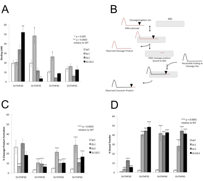
As discussed in Chapter 1, the C-terminal domain of NES is essential for function in vivo; Edwards et al. showed that a NES mutant lacking its C-terminal domain was not able to successfully facilitate conjugation of pSK41 (25). We set out to characterize the role of the C-terminal domain in vitro to determine in which step of conjugation it plays a significant role. To do so we compared the activity of the relaxase domain (1-220) of NES and the full-length (1-665) NES in both cleavage and strand transfer assays. In each case we also tested a range of oligonucleotide lengths as this has been previously shown to effect NES activity but it is unclear which lengths are biologically relevant (25). The oligonucleotides used are shown in Figure 2.1C.
In the cleavage assay, NES 1-220 and NES 1-665 showed differential activity (p<0.005) against OriTHP30 and OriTHP35 (Figure 2.2A). NES 1-665 was able to process OriTHP30 at a higher rate than NES 1-200; however, NES 1-220 was able to process OriTHP35 at a higher rate. In contrast, NES 1-220 and NES 1-665 did not show differential activity against OriTHP40 and OriTHP45 leading us to conclude that the C-terminal domain does not play a major role in the cleavage activity of NES.
We conclude that the C-terminal domain is important for proper regulation of the strand transfer activity of NES.
Characterization of NES Hairpin Loop 1 and 2
The crystal structure of the relaxase domain of NES in complex with pSK41 oriT DNA hairpin, reported previously by Edwards et al., revealed two features unique to this class of relaxase: two protein loops, termed Hairpin Loop 1 and 2 (Figure 2.1A and B), that clamp around the hairpin duplex of the oriT DNA (7). These contacts are unique to this class of relaxase compared to those observed with the longer, multi-tyrosine relaxases like F TraI, and they have yet to be characterized in the context of the full-length protein. Hence, we sought to determine the impact deleting these unique loops would have on NES functions in vitro. Hairpin Loop 1 deletion (ΔL1), Hairpin Loop 2 deletion (ΔL2), or double-deletion (ΔL1ΔL2) forms of full-length NES protein were created using site-directed mutagenesis in which the loops were replaced with Gly-Ser linkers. The proteins were expressed
recombinantly in E. coli and purified to homogeneity. DNA binding, cleavage, and strand transfer assays were conducted using DNA oligonucleotides similar to that employed in the complex presented in the crystal structure and possessing the same sequence as the origin of transfer (oriT) of NES conjugated plasmid pSK41 (Figure 2.1C).
mimic in vivo plasmid transfer. As shown in Figure 2.3A, the ΔL1 form of full-length NES exhibited increased DNA binding (p<0.005) compared to wild-type NES on the OriTHP35 and OriTHP40 oligonucleotides. ΔL2 NES did not demonstrate
significantly different DNA binding on any oligo. In contrast, ΔL1ΔL2 NES showed significantly increased DNA binding (p=0.0002) on the shortest oligo tested,
OriTHP30, but decreased binding (p<0.005) on OriTHP35. For the longest oligonucleotide tested, OriTHP45, no difference in binding was observed for any variant proteins compared to wild-type NES. Thus, we conclude that eliminating Hairpin Loops 1 or 2 from full-length NES can alter DNA binding in vitro in an oligonucleotide length-dependent manner.
DNA cleavage and strand transfer assays were conducted as described in Figure 2.3B. The cleavage assay mimics the cleavage of the plasmid oriT to produce the single strand transferred during conjugation. On OriTHP30, only ΔL1 NES
exhibited a significant (p<0.0001) difference in DNA cleavage, in this case a reduction, relative to wild-type NES (Figure 2.3C). On the longer OriTHP35, OriTHP40 and OriTHP45 oligos, all three variant proteins (ΔL1, ΔL2, ΔL1ΔL2)
demonstrated statistically significant (p<0.0001) increases in levels of DNA cleavage relative to wild-type NES. For these longer oligos, wild-type cleavage was observed at ~4%, while the variant proteins exhibited 2- to 7-fold increases in cleavage. We conclude that eliminating the DNA hairpin-associating loops from NES increases DNA cleavage by the enzyme.
ligation of a portion of DNA covalently linked to NES following cleavage to a new piece of DNA containing the hairpin characteristic of the oriT (Figure 2.3B). This mimics the ligation step of conjugation that ends plasmid transfer. For OriTHP30, all NES variants (ΔL1, ΔL2, and ΔL1ΔL2) showed 5- to 15-fold increases in DNA strand transfer relative to wild-type NES (Figure 2.3D). For OriTHPs 35, 40 and 45, the increases were even larger – 25% to nearly 50% of the substrate oligos provided to the NES variants were processed to strand transfer, while ~5% of the oligos formed strand transfer products with wild-type NES. Thus, eliminating the DNA hairpin contacting loops of NES produces significant and dramatic increases in the level of DNA strand transfer in vitro compared to wild-type NES. It can be concluded that the Hairpin Loop 1 and Loop 2 regions of NES play an important role, particularly on longer DNA substrates more relevant to transfer in vivo, in limiting the level of DNA religation during conjugation.
Modeling of NES Bound to pSK156 and pCA347
(Figure 2.4A and B). Interestingly, within the predicted DNA hairpins of pSK156 and pCA347, nucleotides at the base of the DNA hairpin (G3, C17 and G18) are conserved with the sequence of pSK41 (Figure 2.4A, C, D and E). Furthermore, we noted that the 8 base pair hairpins predicted for pSK156 and pCA347 are nearly identical in sequence to each other (Figure 2.4A, D and E).
We next modeled the pSK156 and pCA347 DNA sequences into the pSK41 NES relaxase domain-DNA hairpin complex crystal structure. For reference, Figure 2.4B shows the NES relaxase domain in complex with the pSK41 DNA hairpin, highlighting the interactions between the protein and DNA; the boxed region
nitrogen can form only one. Thus, in spite of sequence differences between pSK41 and these other two S. aureus plasmids, contacts between NES and the predicted oriTs of all three plasmids are largely maintained.
An additional contact is predicted to be lost between NES and pCA347. While a cytosine is conserved in the same positions in pSK41 (position -4) and pSK156 (position -2), it is a thymine in pCA347 (T2; Figure 2.4E). In pSK41 and pSK156 the amine group of C4 donates a hydrogen bond to the oxygen of N154; however, the para-oxygen of thymine cannot form the same interaction. It is possible that the asparagine side chain could rotate to allow the thymine oxygen to receive a
hydrogen bond from the N154 side chain amine. In doing so, though, this side chain would lose an interaction with C17. Despite this potential change, five base-specific contacts and six phosphate contacts are maintained in our models between NES and the sequences of plasmids pSK156 and pCA347 in this region. Thus, we hypothesize that NES is capable of binding to and utilizing these potential oriT regions of pSK156 and pCA347 as substrates.
Characterization of NES Processing of pSK156 and pCA347
We next analyzed the ability of pSK41 NES to process the potential oriT regions of pSK156 and pCA347 by measuring the protein’s ability to employ these DNAs for binding, cleavage and strand transfer. For DNA binding studies, wild-type full-length NES with an active site Y25F mutation was employed along with
2.2, 7) but significant differences between OriTHP40 and 45 were not seen in assays with the NES loop deletion protein mutants. NES bound the oriT mimic regions of pSK156 and pCA347 but less well compared to its binding of the pSK41 oriT (Figure 2.5A). The KD of NES binding to pSK41 is 19.3 ± 3 nM; in contrast, NES binds to pSK156 and pCA347 3- and 9-fold weaker, with KDs of 55.8 ± 9 nM and 175 ± 30 nM, respectively. While the loss of one or two hydrogen bonds is not sufficient to explain this decrease in binding affinity, it is interesting that the changes in binding affinity reflect the degree of change in sequence and interactions seen in our
models.
Relaxase-in trans Mobilizationby pSK41 In Vivo
To investigate the ability of pSK41 to facilitate relaxase-in trans mobilization, the oriT-like sites corresponding to pSK156 and pCA347, and the pSK41 oriT sequence itself were synthesized and cloned into the non-mobilizable shuttle vector pSK5632 (26) to generate the plasmids pSK6881, pSK6879 and pSK6877,
respectively; the DNA fragments cloned are shown in Figure 2.6. These new plasmid constructs and pSK5632 were electroporated into S. aureus strain RN4220 and pSK41 was subsequently introduced via conjugation. These strains were then used as donors in mobilization assays with the recipient strain S. aureus WBG4515. As shown in Table 2.2, pSK41 was found to mobilize pSK6877, containing its own cognate oriT sequence, at a frequency of 2.9 x 10⁻⁵, approximately five-fold lower than pSK41 itself transferred in the same assay (1.4 x 10⁻⁴). However, despite
repeated efforts, mobilization of the plasmids containing the pSK156 or pCA347 oriT mimics was never detected. These results demonstrate that pSK41-encoded NES can mediate in trans mobilization of a plasmid containing a copy of its own oriT site, but suggest its activity on the variant oriT-like sites from pSK156 and pCA347 is inadequate to facilitate plasmid transfer in vivo. As discussed below, an accessory protein may be required to complete relaxase-in trans transfer in vivo.
Discussion
focus on the mechanism of action of the NES relaxase enzyme encoded by pSK41 and related plasmids from staphylococci. NES is a two domain protein where the C-terminal domain is distinct from that which is normally paired with a relaxase domain. Rather than possessing a function important to the conjugation process such as a helicase, NES does not seem to possess any catalytic activity but was previously shown to be essential for successful conjugation (25). Here we show that the C-terminal domain is important for the strand-transfer (ligation) function but not cleavage activity of NES (Figure 2.2). It is likely that the increase in DNA strand transfer causes DNA to be ligated before transfer is complete. It has been suggested that the ligation action of single-tyrosine relaxases such as NES requires homo-dimerization, which could be mediated by the C-terminal domain. Alternatively, although we have not measured the DNA binding ability of the C-terminal domain, it could play a role in DNA sequence discrimination to ensure ligation only occurs to the 3’ end of the T-strand rather than any DNA blunt end it encounters.
complex with NES through interactions with the NES Hairpin Loop 1 and 2, amplifying the effect of loss of these protein features.
Because relaxases are essential for transfer, share many common features, and are unique to the conjugative plasmid system, they represent a novel
therapeutic target for decreasing the spread of antimicrobial resistance to allow current antimicrobial compounds to maintain efficacy. As explored previously and continued in Chapter 3, there are two potential sites of disruption common to relaxases: the metal binding site and specific protein-DNA interactions (7, 27). These results validate the NES Hairpin Loop 1 and 2 DNA interactions as a target site for such therapeutics. By disrupting the specific protein-DNA interactions in the NES Hairpin Loops, a molecule such as a sequence-specific polyamide could specifically disrupt cleavage and religation during pSK41 conjugation (7). As there seems to be some sequence conservation at the base of the DNA hairpin, this inhibitor molecule could target mobilizable plasmids in addition to the conjugative plasmid. Interestingly, there is a biological example of relaxase interference from Staphylococcus epidermidis strains carrying a CRISPR spacer that matches the nes gene of pSK41 and limits conjugative transfer (28). Targeted disruption of
conjugation after initiation of the process and formation of the mating pore could cause cell death specifically in conjugative plasmid containing bacteria. This targeted approach to bactericidal compounds is desirable as we learn more about the importance of the human skin microbiome (29).
sequences from pSK156 and pCA347 to be processed by NES. The origin-of-transfer-mimic sequences of pSK156 and pCA347 maintain all but one or two protein-DNA contacts, respectively, and are able to be bound, cleaved, and ligated by NES, although with altered efficiency. We were therefore somewhat surprised to find that plasmid constructs containing pCA347 or pSK156 oriT mimics could not be mobilized from cells harboring pSK41 co-resident, in contrast to a pSK41 oriT construct. However, the analogous relaxase-in trans mobilization phenomena recently described for the distinct pWBG749-like conjugative plasmids provide a precedent that likely explains this apparent paradox. Namely, pWBG749 oriT-like sequences exist as sub-types differentiated by sequence divergence in an inverted repeat (IR2) located adjacent to the nic site-containing core sequence (18). This results in specificity between various mobilizable plasmids and particular like conjugative plasmids. Thus, pWBG749 can mobilize plasmids with a pWBG749-like oriT of sub-type OT49 but not those carrying an OT45 sub-type, which instead can be mobilized by pWBG749-like conjugative plasmids that possess a cognate OT45 sub-type oriT (18). Despite this, pWBG749 was able to stimulate
recombination between OT49- and OT45-type oriT sequences carried on the same mobilizable plasmid, indicating that the pWBG749 relaxosome could recognize the OT45-type oriT even though it can’t mediate transfer of that sub-type (18). By analogy, it would seem plausible that the pSK156 and pCA347 oriT-mimics
examined here represent sub-types of pSK41-like oriTs that can be recognized by NES but cannot be mobilized by the pSK41 relaxosome. In the case of the