Microbiota regulate intestinal epithelial gene
expression by suppressing the transcription factor
Hepatocyte nuclear factor 4 alpha
James M. Davison,
1,2Colin R. Lickwar,
1Lingyun Song,
3Ghislain Breton,
4Gregory E. Crawford,
3and John F. Rawls
11
Department of Molecular Genetics and Microbiology, Center for the Genomics of Microbial Systems, Duke University, Durham,
North Carolina 27710, USA;
2Department of Cell Biology and Physiology, University of North Carolina, Chapel Hill, North Carolina
27599, USA;
3Department of Pediatrics, Division of Medical Genetics, Center for Genomic and Computational Biology, Duke
University, Durham, North Carolina 27708, USA;
4Department of Integrative Biology and Pharmacology, McGovern Medical School,
Houston, Texas 77030, USA
Microbiota influence diverse aspects of intestinal physiology and disease in part by controlling tissue-specific transcription
of host genes. However, host genomic mechanisms mediating microbial control of intestinal gene expression are poorly
un-derstood. Hepatocyte nuclear factor 4 (HNF4) is the most ancient family of nuclear receptor transcription factors with
im-portant roles in human metabolic and inflammatory bowel diseases, but a role in host response to microbes is unknown.
Using an unbiased screening strategy, we found that zebrafish Hnf4a specifically binds and activates a
microbiota-sup-pressed intestinal epithelial transcriptional enhancer. Genetic analysis revealed that zebrafish
hnf4a
activates nearly half
of the genes that are suppressed by microbiota, suggesting microbiota negatively regulate Hnf4a. In support, analysis of
genomic architecture in mouse intestinal epithelial cells disclosed that microbiota colonization leads to activation or
inacti-vation of hundreds of enhancers along with drastic genome-wide reduction of HNF4A and HNF4G occupancy. Interspecies
meta-analysis suggested interactions between HNF4A and microbiota promote gene expression patterns associated with
human inflammatory bowel diseases. These results indicate a critical and conserved role for HNF4A in maintaining
intes-tinal homeostasis in response to microbiota.
[Supplemental material is available for this article.]
All animals face the fundamental challenge of building and main-taining diverse tissues while remaining sensitive and responsive to their environment. This is most salient in the intestinal epithelium which performs important roles in nutrient absorption and barrier function while being constantly exposed to complex microbial communities (microbiota) and nutrients within the intestinal lu-men. The presence and composition of microbiota in the intesti-nal lumen influence diverse aspects of intestiintesti-nal development and physiology including dietary nutrient metabolism and ab-sorption, intestinal epithelial renewal, and edification of the host immune system. Abnormal host-microbiota interactions are strongly implicated in the pathogenesis of inflammatory bowel diseases (IBD), including Crohn’s disease (CD) and ulcerative coli-tis (UC) (Sartor and Wu 2016). Studies in mouse and zebrafish models of IBD have established that impaired intestinal epithelial cell (IEC) responses to microbiota are a key aspect of disease pro-gression (Bates et al. 2007; Kamada et al. 2013; Marjoram et al. 2015). Improved understanding of the molecular mechanisms by which microbiota evoke host responses in the intestinal epitheli-um can be expected to lead to new strategies for preventing or treating IBD and other microbiota-associated diseases.
The ability of IECs to maintain their physiologic functions and respond appropriately to microbial stimuli is facilitated
through regulation of gene transcription. Genome-wide compari-son of transcript levels in intestinal tissue or isolated IECs from mice reared in the absence of microbes (germ-free or GF) to those colonized with a microbiota (conventionalized or CV) have re-vealed hundreds of genes that have significantly increased or de-creased mRNA levels following microbiota colonization (Camp et al. 2014). Interestingly, many mouse genes that are transcrip-tionally regulated by microbiota have zebrafish homologs that are similarly responsive, suggesting the existence of evolutionarily conserved regulatory mechanisms (Rawls et al. 2004). For example, the protein hormone Angiopoetin-like 4 (ANGPTL4, also called FIAF) is encoded by a single ortholog in the mouse and zebrafish genomes, and microbiota colonization results in significant reduc-tions in transcript levels in the intestinal epithelium of both host species (Bäckhed et al. 2004; Camp et al. 2012). Whereas these impacts of microbiota on host IEC transcriptomes and their down-stream consequences have been extensively documented, the upstream transcriptional regulatory mechanisms remain poorly understood.
Specification and tuning of gene transcription proceeds, in part, through interactions between transcription factors (TFs) and their sequence-specific binding tocis-regulatory DNA.Cis -regula-tory regions (CRRs) harbor binding sites for multiple activating or repressing TFs and are generally associated with nucleosome
Corresponding author: [email protected]
Article published online before print. Article, supplemental material, and publi-cation date are at http://www.genome.org/cgi/doi/10.1101/gr.220111.116. Freely available online through theGenome ResearchOpen Access option.
© 2017 Davison et al. This article, published inGenome Research, is available under a Creative Commons License (Attribution 4.0 International), as described at http://creativecommons.org/licenses/by/4.0/.
depletion and specific post-translational modifications of histone proteins within adjacent nucleosomes when acting as poised (H3K4me1) or active (H3K27ac) enhancers (Creyghton et al. 2010). Antibiotic administration can impact transcript levels and histone modifications in IECs (Thaiss et al. 2016); however, it’s un-clear if these changes are indirect effects caused by alterations to microbiota composition, direct effects of the antibiotic on host cells, or the effects of remaining antibiotic-resistant microbiota (Morgun et al. 2015). Previous studies have shown that histone deacetylase 3 is required in IECs to maintain intestinal homeostasis in the presence of microbiota (Alenghat et al. 2013) and that overall histone acetylation and methylation in the intestine is altered by microbiota colonization (Krautkramer et al. 2016). However, the di-rect and specific effects of the microbiota on host CRRs and subse-quent transcriptional responses in IECs remain unknown.
Our previous studies predicted key roles for one or more nucle-ar receptor (NR) TFs in microbial down-regulation of IEC gene expression (Camp et al. 2014), but the specific TF(s) were not identified. Nuclear receptors are ideal candidate TFs for integrating microbe-derived signals since, for many, their transcriptional activity can be positively or negatively regulated by the binding of metabolic or hormonal ligands (Evans and Mangelsdorf 2014). However, the roles of nuclear receptors in host responses remain poorly understood, and no previous study has defined the impact of microbiota on nuclear receptor DNA binding. Nuclear receptors are a metazoan innovation. The earliest animals encoded a single nuclear receptor orthologous to Hepatocyte nuclear factor 4 (HNF4; nuclear receptor subfamily NR2A) (Bridgham et al. 2010). Despite subsequent duplication and diversification, distinct HNF4 TFs remain encoded in extant animals including mam-mals (HNF4A, HNF4G) and fishes (Hnf4a, Hnf4b, Hnf4g) (Supplemental Fig. S1G). HNF4A serves particularly important roles in IECs, where it binds CRRs and activates expression of genes involved in IEC maturation and function (Stegmann et al. 2006). The IEC-specific knockout of mouseHnf4aresults in spontaneous intestinal inflammation similar to human IBD (Darsigny et al. 2009). In accord, genetic variants at humanHNF4Aare associated with risk for both UC and CD as well as colon cancer (Barrett et al. 2009; Jostins et al. 2012; Marcil et al. 2012; Chellappa et al. 2016). HNF4A is predicted to bind a majority of IBD-linked CRRs and to regulate IBD-linked genes (Haberman et al. 2014; Meddens et al. 2016). Similarly, genetic variants near humanHNF4Ghave been associated with obesity and CD (Franke et al. 2007; Berndt et al. 2013). Importantly, these diverse roles for HNF4 TFs in host phys-iology have only been studied in animals colonized with micro-biota. Therefore, the role of HNF4 in host-microbiota interactions and the implications for human IBD remain unknown.
Results
Hnf4a is essential for transcriptional activity from a
microbiota-suppressed
cis
-regulatory DNA region
To identify transcriptional regulatory mechanisms underlying mi-crobial control of host gene expression, we took advantage of a pre-viously identified microbiota-responsive CRR termed in3.4 located within the third intron of zebrafishangptl4(Fig. 1A). A GFP report-er construct undreport-er control of in3.4 treport-ermedin3.4:cfos:gfpdrives IEC-specific expression of GFP in zebrafish IECs and is suppressed by microbiota colonization, recapitulating the microbial suppression of zebrafishangptl4(Camp et al. 2012). However, the factor(s) that mediate microbial suppression of in3.4 were unknown. Using a
yeast one-hybrid (Y1H) assay, we tested the capacity of 150 TFs expressed in the zebrafish digestive system to bind in3.4 (Supplemental Fig. S1A,B; Supplemental Table S1) and detected an interaction only with Hnf4a, Hnf4b, and Hnf4g (Fig. 1B). When either of two predicted HNF4A motifs in in3.4 are mutated, Hnf4-in3.4 interactions in the Y1H assay and intestinal GFP expression in in3.4:cfos:gfp zebrafish were strongly reduced (Supplemental Fig. S1C–F). Interestingly, while Gata4, Gata5, and Gata6 have predicted motifs in in3.4 (Camp et al. 2012), these TFs did not interact in the Y1H assay. This suggests that zebrafish Hnf4 TFs are capable of binding in3.4 directly and predicted HNF4A binding motifs are necessary for directing in3.4-based tran-scription in vitro and in the intestine.
We hypothesized that thehnf4transcription factor family is required to mediate microbial suppression of in3.4 activity. Although the Y1H assay demonstrated that all three zebrafish Hnf4 members are capable of binding in3.4, we concentrated our efforts on understanding the function of Hnf4a because it is the most highly conserved Hnf4 family member (Supplemental Fig. S1G;Supplemental Table S10) and has well-documented roles in intestinal physiology (San Roman et al. 2015). To that end, we generatedhnf4amutant zebrafish using the CRISPR/Cas9 system (Fig. 1C;Supplemental Fig. S2A–C,E). Whole-animalHnf4a knock-out mice die during early embryogenesis due to failure to develop a visceral endoderm (Duncan et al. 1997), but zebrafish and other fishes do not develop that extra-embryonic tissue. We found that zebrafish homozygous for a nonsense allele ofhnf4aare viable and survive to sexual maturity (Supplemental Fig. S2D), providing new opportunities to study the roles of HNF4A in host-microbiota interactions.
To determine if Hnf4a is essential for in3.4 activity, we crossed mutanthnf4aalleles to thein3.4:cfos:gfptransgenic report-er line. GFP expression was significantly reduced in the absence of Hnf4a, suggesting that Hnf4a activates in3.4 (Fig. 1D,E,G,H). This loss of GFP expression inhnf4a−/−mutants was not associated with overt defects in brush border development or epithelial polarity in larval stages (Fig. 1F) nor in the establishment of intestinal folds during adult stages (Fig. 1G). However, intestinal lumen of mutant larvae was reduced in size at 6 d post-fertilization (dpf) compared to WT siblings (Fig. 1F;Supplemental Fig. S2F). Together, these data indicate Hnf4a is essential for robust activity of a micro-biota-suppressed CRR. Unlikein3.4:cfos:gfp,angptl4 is expressed in multiple tissues and cell types (Camp et al. 2012). To determine if intestinalangptl4expression is dependent on Hnf4a function, we isolated RNA from IECs from hnf4a+/+ and hnf4−/− adult in3.4:cfos:gfp zebrafish and performed qRT-PCR. Adult IECs (AIECs) fromhnf4a−/−have significant reductions in mRNA for gfpandhnf4acompared tohnf4a+/+controls. However,angptl4 expression remained unchanged inhnf4−/−AIECs compared to WT, suggestingangptl4transcript levels in the adult intestine are regulated by additional mechanisms and not solely from in3.4 or Hnf4a (Fig. 1H). Transcript levels forhnf4gandhnf4bin hnf4a−/−AIEC were also unchanged. Together, these results estab-lish that Hnf4a is required for in3.4 activity in IECs and raises the possibility that Hnf4a may have broader roles in mediating host transcriptional and physiological responses to microbiota.
Hnf4a activates transcription of genes that are suppressed
upon microbiota colonization
mRNA levels from digestive tracts isolated from hnf4a+/+ and hnf4a−/− zebrafish larvae in the presence (CV) or absence of a microbiota (GF) (Fig. 2A). Consistent with our previous studies (Rawls et al. 2004; Kanther et al. 2011), comparison of wild-type zebrafish reared under CV vs. GF conditions revealed differential expression of 598 genes that were enriched for processes such as DNA replication, oxidation reduction, and response to bacterium (Fig. 2B,D;Supplemental Fig. S3; Supplemental Tables S2, S4). Strikingly, disruption of thehnf4agene caused gross dysregulation of the transcriptional response to microbiota colonization, with the
total number of microbiota responsive genes (CV vs. GF) increasing to 2217. Furthermore, comparison of the hnf4a mutant (Mut) vs. wild-type (WT) geno-types revealed differential expression of many genes in the CV condition (2741 genes) and GF condition (1441 genes) that inform a general role for Hnf4a in regulating genes in the intestinal tract (Fig. 2D,E). Principal components analy-sis (Supplemental Fig. S3A) and hierarchi-cal clustering (Fig. 2B) of FPKM values indicated that thehnf4agenotype had a complex contribution to regulating genes involved in both responses to the micro-biota and digestive physiology.
Because we found that Hnf4a acti-vates the microbiota-suppressed intesti-nal CRR, in3.4, we hypothesized that this may represent a general regulatory paradigm for other microbiota-influ-enced CRRs and genes across the genome. When we compared the 598 genes that were microbiota responsive in wild-type digestive tracts with the 2741 genes that Hnf4a regulates in CV digestive tracts, we found these lists shared 295 genes that included fads2 and saa, both of which have human orthologs that are ei-ther implicated (FADS1/2) or markers (SAA) of IBD (Fig. 2C–F; Plevy et al. 2013; Costea et al. 2014). While loss of Hnf4a could be pleiotropic, strikingly, the overlap between these subsets reveals that a disproportionate 88 of the 98 (∼90%) microbiota-suppressed genes are activated by Hnf4a (Fig. 2F; Supplemen-tal Table S2). These 88 genes represent al-most half of all 185 genes suppressed by the microbiota. These data suggest, like its role at in3.4, Hnf4a plays a critical role in directly activating a large percent-age of genes that are suppressed by micro-bial colonization. This set of Hnf4a-activated microbiota-suppressed genes is enriched for ontologies and pathways involved in lipid and carbohydrate me-tabolism, suggesting microbiota might regulate these processes through sup-pression of Hnf4a (Fig. 2G). Interestingly, the top two diseases associated with this gene set were obesity-related traits and IBD (Fig. 2G;Supplemental Table S11). Based on these results, we hypothesized that Hnf4a DNA binding is lost upon microbial colonization within CRRs associated with microbiota-suppressed genes.
HNF4A binding sites are enriched in promoters near genes
associated with microbiota-regulated H3K27ac marks
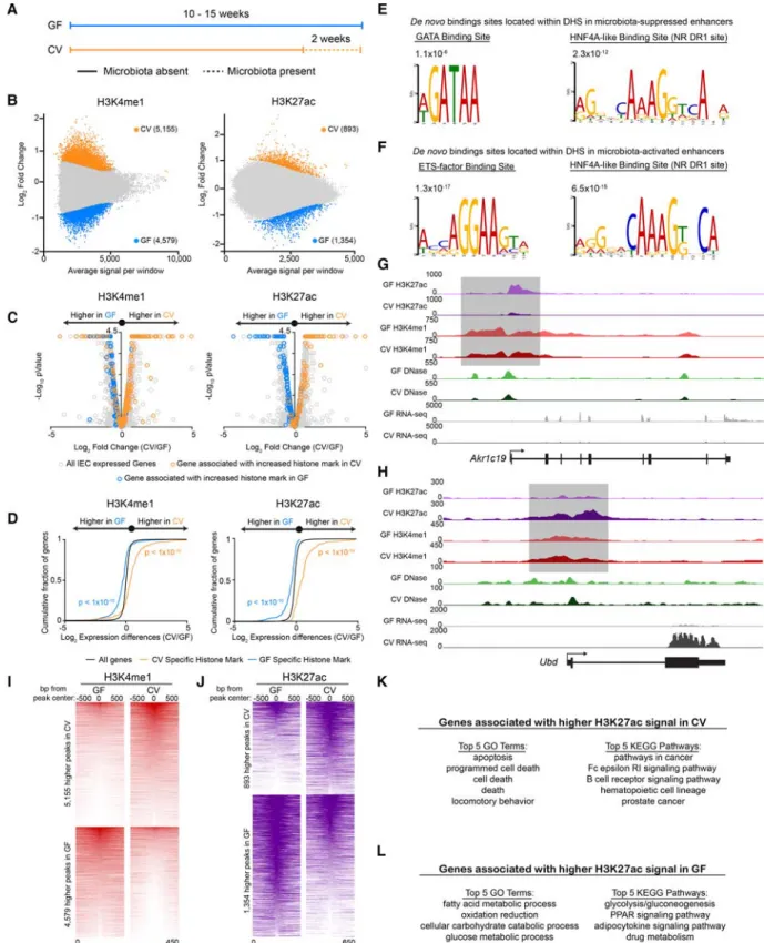
Previous attempts to identify microbial responsive enhancers ge-nome-wide were complicated by the lack of significant changes Figure 1. Zebrafishhnf4ais required for robustin3.4:cfos:gfpactivity. (A) Schematic of the
microbiota-suppressed zebrafish enhancer, in3.4, highlighting the regions required for intestinal activity (purple) which both contain putative HNF4 binding sites (Site 1 and Site 2) (Camp et al. 2012). (B) Image of four plates from the Y1H assay showing the Hnf4 family of transcription factors capable of binding in3.4 and driving expression of the antibiotic resistance reporter gene. (C) Hnf4a+/+and Hnf4a−/−protein cartoons showing the DNA binding domain (DBD) and hinge domain (HD). We characterized the two with the largest lesions, a−43 bp deletion in the hinge domain (allele designationrdu14) and a +25 bp insertion in the hinge domain (allele designationrdu15), which both result in frame-shift and early stop codons and significantly reduced transcript. (D) Stereofluorescence GFP and bright-field microscopy showing representativehnf4a+/+(top3) andhnf4a−/−(bottom3) 6dpfin3.4:cfos:gfpzebrafish. Genotype was blinded, and samples were arranged by intensity of GFP fluorescence. (E) GFP fluorescence (mean ± SEM) inhnf4a+/+(n= 8),hnf4a+/−(n= 8), andhnf4a−/−(n= 8) 6-dpfin3.4:cfos:gfpzebrafish (Two-tailedt -test:t= 17.84, 16.51, respectively, df = 14, and [∗∗∗∗]P< 0.0001). (F) Confocal microscopy showing rep-resentative axial cross sections in 6-dpfhnf4a+/+(n= 4) andhnf4a−43/−43(n= 4) larval zebrafish. 4E8 antibody (yellow) labels the intestinal brush border, DAPI (blue), and phalloidin (red), and nephros (n). (G) Bright-field microscopy (top) and stereofluorescence GFP (bottom) for representativehnf4a+/+ (n= 3) (left) andhnf4a−/−(n= 3) (right) dissected intestinal folds from adultin3.4:cfos:gfpzebrafish. (H) Relative mRNA levels (mean ± SEM) inhnf4a+/+(n= 3) andhnf4a−/−(n= 3) adult zebrafish intestinal epithelial cell as measured by qRT-PCR. Two-tailedt-test:t= 0.93, 5.22, 6.56, 10.65, 0.75, 0.94, respec-tively, df = 4, and (∗)P< 0.05, (∗∗∗)P< 0.001. See alsoSupplemental Figures S1 and S2.
in chromatin DNase accessibility between GF and CV IECs from mouse colon and ileum (Camp et al. 2014). These previous findings suggested other chromatin dynamics may be involved in regulat-ing the IEC response to microbiota. We therefore sought to provide a genomic context for understanding how the microbiota alter HNF4A activity and chromatin modifications in IECs by perform-ing RNA-seq, DNase-seq, and ChIP-seq for the enhancer histone modifications H3K4me1 and H3K27ac and the HNF4 TF family members HNF4G and HNF4A in CV and GF conditions, totaling 35 data sets. We conducted these experiments in jejunal IECs from gnotobiotic mice because (1) ChIP-grade antibodies for mouse HNF4A and HNF4G are available, (2) the relatively large or-gan size in mice provided sufficient numbers of IECs for ChIP-seq
experiments, and (3) we speculated that the roles of HNF4A in host response to microbiota may be conserved to mammals. We first performed DNase-seq in jejunal IECs from mice reared GF or colonized for 2 wk with a conventional mouse microbiota to deter-mine the impact of microbiota colonization on chromatin accessi-bility (Fig. 3A). In accord with previous studies that tested for chromatin accessibility in ileal or colonic IECs from GF or CV mice (Camp et al. 2014), we similarly found no differential DNase hypersensitivity sites (DHSs) in GF or CV jejunum (data not shown, but see Supplemental Fig. S4A–D; Supplemental Tables S6, S8). These data indicate that gross accessibility changes in chromatin do not underlie the transcription of microbiota-re-sponsive genes in IECs.
Figure 3. Microbiota colonization results in targeted alterations in enhancer activity near microbiota-responsive genes. (A) Schematic showing the gno-tobiotic experimental timeline for testing mRNA levels and chromatin architecture in GF and CV mice. (B) MA plots from DESeq2 analysis (FDR < 0.01) of H3K4me1 (n= 3 per condition) (left) and H3K27ac (n= 2 per condition) (right) ChIP-seq from GF and CV mouse jejunal IECs. Colored dots signify regions significantly enriched for a histone mark in GF (blue) or CV (orange). We found 4579 unique H3K4me1 and 1354 unique H3K27ac peaks in GF and 5155 unique H3K4me1 and 893 unique H3K27ac peaks in CV. (C) Volcano plots showing pairwise comparison of RNA expression between GF (n= 2) and CV (n
To test if other metrics of chromatin utilization were dynam-ically regulated by microbiota, we performed ChIP-seq from GF and CV mouse jejunal IECs for histone marks H3K4me1 and H3K27ac that are enriched at poised enhancers and active enhanc-ers, respectively (Fig. 3B). By determining the single-nearest gene TSS within 10 kb of the differential histone marks and overlaying these data with our new RNA-seq data sets, we found that regions that gain poised (H3K4me1) and activated (H3K27ac) enhancers upon colonization are associated with genes that have increased transcript levels upon colonization (Fig. 3C,H–K; Supplemental Fig. S4I;Supplemental Tables S3, S6,S8). Similarly, regions that lose poised and active enhancers upon colonization are associated with microbiota-suppressed genes (Fig. 3C,G,I,J,L;Supplemental Fig. S4J;Supplemental Tables S3, S6,S8). A two-sided Kolmogo-rov-Smirnov goodness-of-fit test shows a positive relationship be-tween differential H3K4me1/H3K27ac regions and increased transcript abundance of nearby genes in the same colonization state (Fig. 3D). Collectively, we identified for the first time a ge-nome-wide map of hundreds of newly identified microbial regulat-ed CRRs, suggesting that microbiota regulation of host genes in the intestinal epithelium is mechanistically linked to histone modifi-cation changes more than gross chromatin accessibility changes (Camp et al. 2014).
We leveraged this novel atlas of microbiota-regulated enhanc-ers and accessible chromatin to determine which TFs are predicted to bind to these regions. An unbiased analysis found that three HNF4A binding site motifs were significantly (P< 1 × 10−45,P<
1 × 10⁻²⁸, andP< 1 × 10⁻¹³) enriched in promoters of genes associ-ated with microbiota-suppressed enhancers (Supplemental Fig. S4E), and STAT1 binding site motifs were significantly (P< 1 × 10⁻¹⁶) enriched in promoters of genes associated with
micro-biota-activated enhancers (Supplemental Fig. S4F). Interestingly, DHSs associated with differentially active enhancers were enriched for two different sets of TF binding sites. DHSs flanked by micro-biota-inactivated enhancers were enriched for nuclear receptor DR1 sites, which can be recognized by HNF4A (Fang et al. 2012), and GATA binding sites (P= 2.3 × 10⁻¹²and 1.1 × 10⁻⁶, respective-ly) (Fig. 3E). DHSs associated with microbiota-activated enhancers were similarly enriched for the nuclear receptor DR1 binding sites but also for STAT/IRF-like and ETS binding sites (P= 6.5 × 10⁻¹⁵ and 1.3 × 10⁻¹⁷, respectively) (Fig. 3F). These data suggest that nu-clear receptors like HNF4A may play a central role in IEC responses to microbial colonization.
Microbiota colonization is associated with a reduction in HNF4A
and HNF4G cistrome occupancy
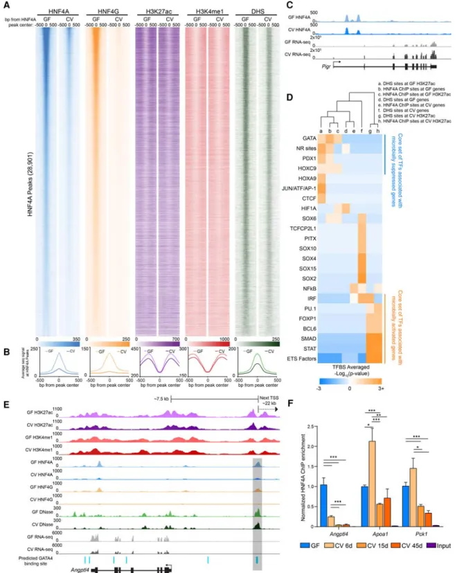
To directly evaluate the impact of microbiota on HNF4A activity, we tested the plasticity of the genome-wide distribution of HNF4 family members in response to microbial colonization. HNF4A bound 28,901 sites and HNF4G bound 21,875 sites across the ge-nome in GF conditions in jejunal IECs, with∼80% of these sites being bound by both TFs. In striking contrast, the number of sites bound by HNF4A and HNF4G in CV conditions was∼10-fold less (Fig. 4A,B;Supplemental Fig. S5A–D;Supplemental Tables S5, S8). Of the 3964 HNF4A binding sites detected in CV, there were only 267 HNF4A sites that were specific to the CV condition ( Supple-mental Fig. S6A,C;Supplemental Table S8). Yet, the genes associat-ed with these HNF4A sites that are retainassociat-ed in CV are enrichassociat-ed for ontologies and pathways fundamental to intestinal epithelial biol-ogy (Supplemental Fig. S6B). Surprisingly, we found that HNF4A sites are equally distributed between genes that are up-regulated
in both GF and CV conditions (Supplemental Fig. S6E). However, we did find that the average CV HNF4A ChIP-seq signal strength was significantly increased at HNF4A sites associated with micro-biota-induced genes relative to those HNF4A sites associated with microbiota-suppressed genes, suggesting HNF4A may play a limited role in genes up-regulated by colonization (Supplemental Fig. S6F). In contrast, GF HNF4A ChIP-seq signal was equivalent at HNF4A sites associated with microbiota-suppressed and induced genes (Supplemental Fig. S6F). Interestingly, we found that HNF4A sites correspond with increased H3K27ac, H3K4me1, and DHS sig-nal in GF compared to these same chromatin marks in CV ( Supple-mental Fig. S6G). We do not believe that the reduction of HNF4A binding is the result of chromatin quality in a particular condition since there are genomic locations where GF and CV HNF4A sites appeared to have equivalent signal (Fig. 4C). Furthermore, ChIP enrichment in these IEC preparations for another zinc finger TF, CTCF, was unaffected by microbiota colonization (Supplemental Fig. S6D). This indicates that the observed reduction of HNF4 ChIP-seq signal in CV IECs is a result of microbiota on HNF4 bind-ing and is not the result of altered ChIP efficiency or sample quality in the different conditions. To test if microbial suppression of HNF4A occupancy is persistent, we performed ChIP-PCR from ex-GF mice that were colonized with microbiota for 6, 15, or 45 d. We found that even after 45 d post-colonization, HNF4A occu-pancy at binding sites was significantly reduced compared to GF (Fig. 4F). The dramatic loss of HNF4A and HNF4G DNA binding upon colonization is consistent with HNF4A acting as a potent ac-tivator of microbiota-suppressed genes.
We further speculated that certain coregulatory sequence-specific transcription factors may also contribute to regulating transcription with HNF4 at these sites. To explore this possibility, we searched for TF motifs associated with HNF4A ChIP sites and found an enrichment of putative binding sites for TFs known to be involved in small intestinal physiology (GATA and HOXC9) as well as nutrient metabolism (PDX1) at both HNF4A-bound re-gions associated with genes and enhancers suppressed by mi-crobes (Fig. 4D). We similarly found GATA sites located within an HNF4A-bound CRR near murineAngptl4(Fig. 4E), similar to the coincident HNF4 and GATA motifs in zebrafish in3.4 (Camp et al. 2012). Furthermore, binding sites for TFs known to be in-volved in cell proliferation and cell death (ETS transcription factor family) are enriched near HNF4A bound regions that intersect microbiota-induced enhancers (Fig. 4D). Collectively, our integra-tive analyses of these novel ChIP-seq, DNase-seq, and RNA-seq data sets identify a core set of putative microbiota-responsive TFs that may interact with HNF4A to mediate microbial control of IEC gene expression. These results suggest HNF4A plays a major role in integrating microbial signals to regulate gene expression and raise the possibility that this novel microbiota-HNF4A axis might contribute to human disease.
Microbiota-mediated suppression of HNF4A may contribute
to gene expression profiles associated with human IBD
Figure 4. Microbiota colonization results in extensive loss of HNF4A and HNF4G DNA binding in IECs. (A) Heat maps showing the average GF and CV ChIP-seq or DNase-seq signal at the 1000 bp flanking HNF4A sites found in GF. (B) Line plots showing the average GF (light-colored line) and CV (dark-colored line) ChIP-seq and DNase-seq RPKM-normalized signal for the indicated TF, histone mark, or DHS at the 1000 bp flanking HNF4A sites found in GF (HNF4A:n= 3 per condition; HNF4G:n= 4 per condition; H3K27ac:n= 2 per condition; H3K4me1:n= 3 per condition; DNase:n= 3 for CV,n= 2 for GF). (C) Representative signal tracks highlighting a microbiota-induced gene (Pigr, polymeric immunoglobulin receptor) that is associated with an HNF4A peak with similar signal in both GF and CV jejunal IECs. (D) Heat map showing the enrichment of TFBS motifs within 50 bp of the DHS or HNF4A peak maxima. (E) Representative signal track atAngptl4highlighting two GATA4 sites within an HNF4A-bound region. (F) Bar graph showing HNF4A ChIP-PCR results at
Angptl4,Apoa1, andPck1loci from jejunal IECs from mice colonized for 0 (n= 2), 6 (n= 3), 15 (n= 2), and 45 (n= 3) d. Data are relative to the GF condition and normalized against a negative control locus (Neurog1). (∗)P< 0.5, (∗∗)P< 0.005, (∗∗∗)P< 0.0005. See alsoSupplemental Figures S5 and S6.
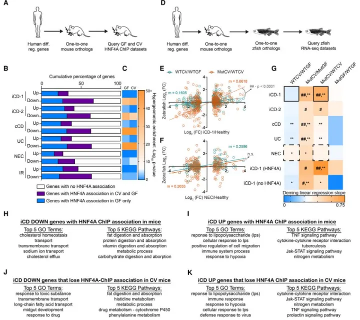
gene lists to identify one-to-one orthologs in mice and referenced them against our new gnotobiotic mouse jejunal HNF4A ChIP-seq data (Fig. 5A). Strikingly, the majority of human genes down-reg-ulated in each of these IBD data sets have mouse orthologs that are associated with an HNF4A-bound region (Fig. 5B,C;
Supplemental Table S7). Focusing on the iCD data set from the largest of these previous studies (Haberman et al. 2014), we found that differential iCD genes associated with HNF4A sites are en-riched for distinct ontologies and pathways that are dysregulated in IBD (Fig. 5H–K). In contrast to IBD, analysis of intestinal
transcriptomic data sets from human subjects with necrotizing en-terocolitis (NEC) (Tremblay et al. 2016) or insulin-resistance (IR) (Veilleux et al. 2015) did not reveal strong enrichment of HNF4A-bound regions near down-regulated genes (Fig. 5C). Notably, in each of these CD, UC, NEC, and IR data sets, a greater percentage of down-regulated genes were linked to HNFA-bound regions compared to up-regulated genes (Fig. 5B). These data sug-gest that microbiota-dependent and microbiota-independent suppression of HNF4A activity in the intestine might play an im-portant role in IBD pathologies. To assess if microbiota suppression of HNF4A activity regulates genes differentially expressed in IBD, we queried the published human IBD and NEC gene expression data sets to identify human-mouse-zebrafish one-to-one-to-one orthologs that were differentially expressed in our RNA-seq analy-sis of gnotobiotic zebrafishhnf4amutants (Fig. 5D). We found that ortholog expression fold changes in human IBD/healthy compar-isons most closely resembled the expression fold changes of MutCV/MutGF and MutCV/WTCV (Fig. 5E–G). Neither the WTCV/WTGF nor the MutGF/WTGF comparisons faithfully reca-pitulate the expression profiles of IBD/healthy comparisons. These data indicate that microbiota colonization in the absence ofhnf4a function in zebrafish is sufficient to induce a gene expression pro-file that resembles human IBD. Strikingly, the positive correlation and significant resemblance to the iCD-like gene signatures in the colonizedhnf4a−/−compared to colonizedhnf4a+/+zebrafish di-gestive tracts become even stronger when we limited our analysis to one-to-one orthologs that have an association with an HNF4A-bound region in mouse IECs (Fig. 5G). Together, these re-sults indicate that intestinal suppression of HNF4A target genes is a prevalent feature of human CD and UC and suggest a model wherein HNF4A maintains transcriptional homeostasis in the presence of a microbiota and protects against an evolutionarily conserved IBD-like gene expression signature.
Discussion
Over the course of animal evolution, the intestinal epithelium has served as the primary barrier between animal hosts and the com-plex microbial communities they harbor. IECs maintain this barri-er and pbarri-erform their physiological roles in nutrient transport and metabolism through dynamic transcriptional programs. The regu-latory mechanisms that orchestrate these transcriptional programs represent potential therapeutic targets for a variety of human in-testinal diseases, including IBD. Here, we discovered that HNF4A activity and its transcriptional network are suppressed by micro-biota. HNF4 is the oldest member of the nuclear receptor TF family (Bridgham et al. 2010), and our findings in fish and mammals sug-gest that microbial suppression of HNF4A may be a conserved fea-ture of IEC transcriptional programs present in the common ancestor.
We discovered HNF4A as a microbiota-suppressed transcrip-tion factor by demonstrating that it specifically binds to a micro-biota-suppressedcis-regulatory element, in3.4, which is located at the zebrafish geneangptl4. This finding, combined with our zebra-fish RNA-seq data (Fig. 2F,G), revealed a broad role for HNF4A in ac-tivation of microbially suppressed transcripts. Though hnf4a mutant zebrafish have reduced in3.4 activity in the intestinal epi-thelium based on transgenic reporter assays, the transcript levels of the endogenous zebrafishangptl4gene appears unaffected in both larval digestive tracts and adult IECs. The zebrafish genome encodes two additional HNF4 family members (hnf4b, hnf4g), and previous studies in mammals have shownAngptl4can be
regu-lated by other metabolically activated nuclear receptors (Staiger et al. 2009; Korecka et al. 2013). We hypothesize that loss of HNF4A function may lead to a metabolic imbalance leading to atypical or compensatory activation of othertrans- andcis-factors that control expression ofangptl4and other genes in the intestine. Our results suggest new links between HNF4A and microbiota in the context of human IBD. IBD patients, particularly those suf-fering from Crohn’s disease, often present with decreased serum low-density lipoprotein levels and reduced total cholesterol levels compared to healthy individuals (Hrabovsky et al. 2009; Agouridis et al. 2011). These serum levels are consistent with re-duced transcript levels for genes involved in intestinal absorption and transport of lipid and cholesterol in ileal and colonic biopsies from UC and CD patients (Arijs et al. 2009; Haberman et al. 2014). Transcription factors, including nuclear receptors like HNF4A and FXR, are known to regulate bile acid production and lipid and cholesterol absorption and have already been implicated in IBD (Ahn et al. 2008; Nijmeijer et al. 2011). Previous studies have shown that some IBD-associated H3K27ac-activated regions that also overlap with IBD-associated SNPs contain HNF4A binding sites (Mokry et al. 2014). This is consistent with our findings and supports a role for HNF4A in regulating gene expression and in-flammation in the context of IBD. However, our work is the first to demonstrate a role for microbiota in suppressing HNF4A and to implicate microbiota-HNF4A interactions in driving an IBD-like gene expression signature (Fig. 5). In addition to IBD, human HNF4A variants are associated with metabolic syndrome (Weissglas-Volkov et al. 2006) and type 2 diabetes (Ma et al. 2016). Interestingly, microbiota have also been implicated in both of these diseases (Qin et al. 2012; Vrieze et al. 2012), raising the possibility that microbiota suppression of HNF4Atransactivity could play a role in these diseases as well. Indeed, we find that genes down-regulated in intestinal tissue from IR-obese patients have in-creased HNF4A binding associations compared to up-regulated genes (Veilleux et al. 2015), similar to the enrichment of HNF4A as-sociations at down-regulated genes in IBD (Fig. 5B,C). Interestingly, up-regulated genes in these IR-obese patients were enriched for pro-inflammatory markers. This underscores the relationship between metabolic impairments and inflammation in the intestine and prompts further investigation of how HNF4A might contribute to this relationship. HNF4A has been shown to play key roles in anti-oxidative and anti-inflammatory defense mechanisms (Marcil et al. 2010), so aberrant microbial suppression could pro-mote an inflammatory state. HNF4A target genes are down-regulat-ed in human IBD (Arijs et al. 2009; Haberman et al. 2014) and mouse experimental colitis (Chahar et al. 2014), and the HNF4A targetAPOA1has been shown to be protective against intestinal in-flammation in mice (Gkouskou et al. 2016). We speculate that the genes governed by this novel microbiota–HNF4A axis may include additional anti- and pro-inflammatory factors that could provide new targets for IBD therapy.
is achieved within 15 d and persists through at least 45 d after col-onization. These data collectively suggest that microbiota suppress HNF4A activity in the jejunum through mechanisms distinct from those utilized in DSS-induced colitis.
HNF4A has been characterized as a master metabolic regula-tor for its conserved roles in gluconeogenesis, glucose homeostasis, and fatty acid metabolism (Palanker et al. 2009; Frochot et al. 2012; Barry and Thummel 2016). Despite its clear importance in meta-bolic health, relatively little insight into its regulation in a biolog-ical context has been reported. In vitro and cell culture studies have identified possible suppressors and activators of HNF4A, including acetylation by CREB-binding protein (CREBBP, also known as CBP), which has been shown to induce HNF4A activity (Soutoglou et al. 2000; Hong et al. 2003). The nuclear receptor cofactor and master regulator of mitochondrial biogenesis PPARGC1A binds HNF4A and promotes activation of HNF4A tar-get genes (Rha et al. 2009). Colonization of GF animals with microbiota leads to increased energy harvest (Rabot et al. 2010; Semova et al. 2012) and changes in metabolic homeostasis includ-ing decreased AMPK complex activity in skeletal muscle and liver (Backhed et al. 2007). Previous studies have also shown that the ac-tivated AMPK complex phosphorylates and activates PPARGC1A (Jager et al. 2007); therefore, microbiota might suppress HNF4A ac-tivity indirectly through induced alterations in metabolic homeo-stasis. Other studies have shown that HNF4A activity is controlled through use of alternative promoters which generate different iso-forms (Huang et al. 2009). However, we did not detect differential Hnf4aexon usage by DEXseq (Li et al. 2015) in our RNA-seq data from GF and CV IECs (data not shown). Another facet of HNF4A biology that remains unresolved is the identity of its endogenous ligand(s). Although historically considered an orphan nuclear re-ceptor, several fatty acids (FA), including linoleic acid, have been identified as ligands for HNF4A (Hertz et al. 1998; Palanker et al. 2009; Yuan et al. 2009). Fatty acids are an attractive class of puta-tive regulators of HNF4A, since the microbiota are known to regu-late FA absorption in zebrafish IECs (Semova et al. 2012). Further, specific bacterial taxa are known to modify the structure of poly-unsaturated FAs (PUFAs), and these native and modified PUFAs have distinct impacts on animal health (O’Shea et al. 2012) and may serve as therapeutics for IBD (Mbodji et al. 2013).
In our attempt to understand how the microbiota regulate HNF4A activity and host gene transcription, we were motivated to investigate if microbiota impact histone modification and chro-matin accessibility in the mouse jejunum. Our findings support the model that microbiota alter IEC gene expression by affecting TF binding and histone modification at tissue-defined open chro-matin sites (Camp et al. 2014). We provide the genomic addresses of hundreds of microbiota-regulated enhancers as well as the genes associated with these enhancers and HNF4A binding sites. Similar to other findings in intra-epithelial lymphocytes (Semenkovich et al. 2016), our work demonstrates a clear microbial contribution to the modification of the histone landscape in IECs and provides another important layer of regulation that orchestrates microbiota regulation of host genes involved in intestinal physiology and hu-man disease. We were also able to establish a link between micro-biota-regulated genes and enhancers and NR binding sites. These NR binding sites are coincident with a core set of TFs that are enriched near microbiota-suppressed enhancers/genes (GATA) or induced enhancers/genes (ETS-factors and IRF) (Supplemental Fig. S7). GATA4 was previously shown to be a positive regulator of genes suppressed by microbiota in the mouse jejunum (Shulzhenko et al. 2011), supporting potential coregulatory
inter-actions with HNF4A. Coregulation by other TFs represents one possible mode of HNF4A regulation by which the microbiota could suppress HNF4A activity without impacting the gene transcription of all HNF4A-associated genes.
Methods
Yeast one-hybrid ORFeome screen
The yeast one-hybrid ORFeome screen was performed using the Clontech Matchmaker Gold Yeast One-hybrid Library Screening System (cat. #630491) protocol with the following exceptions: The Y1HGold yeast strain was transformed using standard yeast transformation procedures with BstBI-digested pBait-AbAi containing either the WT or a SDM in3.4 or the TP53 binding site sequence, and positive transformants were selected on SD/−URA media. In addition, a ORFeome library consisting of 148 zebrafish transcription factors cloned from adult zebrafish liv-er (Supplemental Table S1) plus hnf4a and hnf4g cDNAs in pDEST22 prey vectors containing an N-terminal GAL4-activation domain were utilized (Boyle et al. 2017). For additional informa-tion, seeSupplemental Methods.
Mouse IEC isolation for DNase-, ChIP-, and RNA-seq
The small intestine was removed from the mouse, and the jejunum was excised from the duodenum and ileum. Duodenum was de-fined as the anterior 5 cm of the midgut, and ileum was dede-fined as posterior 6 cm of midgut as described (Camp et al. 2014). Adipose and vasculature were removed from the tissue. The jeju-num was opened longitudinally along the length of the tissue, ex-posing the lumen and epithelial cell layer. Luminal debris was washed away from the epithelia with ice-cold sterile PBS. The tissue was temporarily stored in 10 mL of ice-cold sterile PBS with 1× pro-tease inhibitors (cOmplete EDTA-Free, Roche, #11873580001) and 10 µM Y-27632 (ROCK I inhibitor, Selleck Chemicals, #S1049) to inhibit spontaneous apoptosis. The jejunum was moved into a 15-mL conical tube containing 3 mM EDTA in PBS with 1× prote-ase inhibitors and 10 µM Y-27632. The tissue was placed on a nuta-tor in a cold room for 15 min. The jejunum was removed from the 3 mM EDTA and placed on an ice-cold glass Petri dish with PBS containing 1 mM MgCl2and 2 mM CaCl2with protease inhibitors
and 10 µM Y-27632. Villi were scraped off of the tissue using the side of a sterile plastic micropipette and transferred into a new 15-mL conical tube. The isolated IECs were then crosslinked for ChIP-seq, ChIP-PCR or used for DNase-seq or RNA-seq. For addi-tional information, seeSupplemental Methods.
Bioinformatic and statistical analysis
Sample sizes for zebrafish experiments (noted in figure legends) were selected based on genotype availability and transgenesis effi-ciency. All sample collection was performed two or more times on independent days. For sequencing experiments, statistical calls for differential gene expression were made by Cuffdiff2 (Trapnell et al. 2013). For the zebrafish RNA-seq experiment, next-generation se-quencing was performed once and at the same time to avoid batch effects: WTGF and WTCV (n= 3); MutGF and MutCV (n= 2). We originally collectedn= 3 MutGF and MutCV biological replicates; however, using pre-established criteria and to avoid RNA contam-ination, we excluded one biological replicate from all analysis from these groups because of sequencing reads that mapped within the deletedhnf4aexon in thehnf4a−/−genotype.
condition on independent days. GF and CV mouse samples were collected on different days. For sequencing experiments, statistical calls for differential gene expression and differential peak calls were made by Cuffdiff2, MACS2, and DESeq2 (Zhang et al. 2008; Anders and Huber 2010; Trapnell et al. 2013; Love et al. 2014). For the mouse RNA-seq experiment, next-generation sequencing was performed once and at the same time to avoid batch effects: GF (n= 2) and CV (n= 2). Paired GF and CV ChIP and library am-plification was performed simultaneously. Typically, biological ChIP replicates were sequenced on different days and were always paired with the other condition (i.e., CV and GF were always se-quenced together). The number of biological ChIP replicates (not-ed in figure legends) was dependent on reproducibility between ChIP samples and/or our ability to determine statistical differential sites using DESeq2 (for H3K4me1 and H3K27ac).
All statistical metrics (except where otherwise noted) were performed in Graphpad Prism 7.01. Deming linear regression was used for Figure 5 because it is a stronger and more accurate as-sessment of correlation when both thexandyvariables have ex-perimental error. Details regarding the other statistical tests used in this study can be found in the figure legends or above.
For detailed methods on animal husbandry, zebrafish trans-genesis, zebrafish mutatrans-genesis, imaging, immunostaining, site-di-rected mutagenesis, and ChIP-, RNA-, and DNase-seq preparation and analysis, please seeSupplemental Methods.
Data access
Transcription factor ChIP-seq, Histone ChIP-seq, DNase-seq, and RNA-seq data sets from this study have been submitted to the NCBI Gene Expression Omnibus (GEO; http://www.ncbi.nlm. nih.gov/geo/) under accession number GSE90462.
Acknowledgments
We thank Balfour Sartor, Scott Magness, Maureen Bower, Jeremy Herzog, and Scott T. Espenschied for assistance with gnotobiotic mice, and Wenbiao Chen and Stacy Horner for sharing reagents.
We also thank the Genomic Sequencing Laboratory at
HudsonAlpha Institute for Biotechnology and the Duke Sequenc-ing and Genomic Technologies Facility. This work was supported by grants from the National Institutes of Health (National Institute of Diabetes and Digestive and Kidney Diseases) (R01-DK081426, U24-DK097748, P01-DK094779, and P30-DK34987) and NIH Of-fice of the Director (R24-OD016761).
References
Agouridis AP, Elisaf M, Milionis HJ. 2011. An overview of lipid abnormali-ties in patients with inflammatory bowel disease.Ann Gastroenterol
24:181–187.
Ahn SH, Shah YM, Inoue J, Morimura K, Kim I, Yim S, Lambert G, Kurotani R, Nagashima K, Gonzalez FJ, et al. 2008. Hepatocyte nuclear factor 4α in the intestinal epithelial cells protects against inflammatory bowel dis-ease.Inflamm Bowel Dis14:908–920.
Alenghat T, Osborne LC, Saenz SA, Kobuley D, Ziegler CG, Mullican SE, Choi I, Grunberg S, Sinha R, Wynosky-Dolfi M, et al. 2013. Histone deacetylase 3 coordinates commensal-bacteria-dependent intestinal ho-meostasis.Nature504:153–157.
Anders S, Huber W. 2010. Differential expression analysis for sequence count data.Genome Biol11:R106.
Arijs I, De Hertogh G, Lemaire K, Quintens R, Van Lommel L, Van Steen K, Leemans P, Cleynen I, Van Assche G, Vermeire S, et al. 2009. Mucosal gene expression of antimicrobial peptides in inflammatory bowel dis-ease before and after first infliximab treatment.PLoS One4:e7984. Bäckhed F, Ding H, Wang T, Hooper LV, Koh GY, Nagy A, Semenkovich CF,
Gordon JI. 2004. The gut microbiota as an environmental factor that regulates fat storage.Proc Natl Acad Sci101:15718–15723.
Backhed F, Manchester JK, Semenkovich CF, Gordon JI. 2007. Mechanisms underlying the resistance to diet-induced obesity in germ-free mice.Proc Natl Acad Sci104:979–984.
Barrett JC, Lee JC, Lees CW, Prescott NJ, Anderson CA, Phillips A, Wesley E, Parnell K, Zhang H, Drummond H, et al. 2009. Genome-wide associa-tion study of ulcerative colitis identifies three new susceptibility loci, in-cluding the HNF4A region.Nat Genet41:1330–1334.
Barry WE, Thummel CS. 2016. TheDrosophilaHNF4 nuclear receptor pro-motes glucose-stimulated insulin secretion and mitochondrial function in adults.eLife5:e11183.
Bates JM, Akerlund J, Mittge E, Guillemin K. 2007. Intestinal alkaline phospha-tase detoxifies lipopolysaccharide and prevents inflammation in zebra-fish in response to the gut microbiota.Cell Host Microbe2:371–382. Berndt SI, Gustafsson S, Magi R, Ganna A, Wheeler E, Feitosa MF, Justice AE,
Monda KL, Croteau-Chonka DC, Day FR, et al. 2013. Genome-wide meta-analysis identifies 11 new loci for anthropometric traits and pro-vides insights into genetic architecture.Nat Genet45:501–512. Boyle G, Richter K, Priest HD, Traver D, Mockler TC, Chang JT, Kay SA,
Breton G. 2017. Comparative analysis of vertebrate diurnal/circadian transcriptomes.PLoS One12:e0169923.
Bridgham JT, Eick GN, Larroux C, Deshpande K, Harms MJ, Gauthier ME, Ortlund EA, Degnan BM, Thornton JW. 2010. Protein evolution by mo-lecular tinkering: diversification of the nuclear receptor superfamily from a ligand-dependent ancestor.PLoS Biol8:e1000497.
Camp JG, Jazwa AL, Trent CM, Rawls JF. 2012. Introniccis-regulatory mod-ules mediate tissue-specific and microbial control of angptl4/fiaf tran-scription.PLoS Genet8:e1002585.
Camp JG, Frank CL, Lickwar CR, Guturu H, Rube T, Wenger AM, Chen J, Bejerano G, Crawford GE, Rawls JF. 2014. Microbiota modulate tran-scription in the intestinal epithelium without remodeling the accessible chromatin landscape.Genome Res24:1504–1516.
Chahar S, Gandhi V, Yu S, Desai K, Cowper-Sal-lari R, Kim Y, Perekatt AO, Kumar N, Thackray JK, Musolf A, et al. 2014. Chromatin profiling re-veals regulatory network shifts and a protective role for hepatocyte nu-clear factor 4αduring colitis.Mol Cell Biol34:3291–3304.
Chellappa K, Deol P, Evans JR, Vuong LM, Chen G, Briancon N, Bolotin E, Lytle C, Nair MG, Sladek FM. 2016. Opposing roles of nuclear receptor HNF4α isoforms in colitis and colitis-associated colon cancer.eLife5:e10903. Costea I, Mack DR, Lemaitre RN, Israel D, Marcil V, Ahmad A, Amre DK.
2014. Interactions between the dietary polyunsaturated fatty acid ratio and genetic factors determine susceptibility to pediatric Crohn’s dis-ease.Gastroenterology146:929–931.
Creyghton MP, Cheng AW, Welstead GG, Kooistra T, Carey BW, Steine EJ, Hanna J, Lodato MA, Frampton GM, Sharp PA, et al. 2010. Histone H3K27ac separates active from poised enhancers and predicts develop-mental state.Proc Natl Acad Sci107:21931–21936.
Darsigny M, Babeu JP, Dupuis AA, Furth EE, Seidman EG, Levy E, Verdu EF, Gendron FP, Boudreau F. 2009. Loss of hepatocyte-nuclear-factor-4α af-fects colonic ion transport and causes chronic inflammation resembling inflammatory bowel disease in mice.PLoS One4:e7609.
Duncan SA, Nagy A, Chan W. 1997. Murine gastrulation requires HNF-4 reg-ulated gene expression in the visceral endoderm: tetraploid rescue of Hnf-4(−/−) embryos.Development124:279–287.
El Aidy S, van Baarlen P, Derrien M, Lindenbergh-Kortleve DJ, Hooiveld G, Levenez F, Dore J, Dekker J, Samsom JN, Nieuwenhuis EE, et al. 2012. Temporal and spatial interplay of microbiota and intestinal mucosa drive establishment of immune homeostasis in conventionalized mice.Mucosal Immunol5:567–579.
Evans RM, Mangelsdorf DJ. 2014. Nuclear receptors, RXR, and the Big Bang. Cell157:255–266.
Fang B, Mane-Padros D, Bolotin E, Jiang T, Sladek FM. 2012. Identification of a binding motif specific to HNF4 by comparative analysis of multiple nuclear receptors.Nucleic Acids Res40:5343–5356.
Franke A, Hampe J, Rosenstiel P, Becker C, Wagner F, Hasler R, Little RD, Huse K, Ruether A, Balschun T, et al. 2007. Systematic association map-ping identifies NELL1 as a novel IBD disease gene.PLoS One2:e691. Frochot V, Alqub M, Cattin AL, Carriere V, Houllier A, Baraille F, Barbot L,
Saint-Just S, Ribeiro A, Lacasa M, et al. 2012. The transcription factor HNF-4α: a key factor of the intestinal uptake of fatty acids in mouse. Am J Physiol Gastrointest Liver Physiol302:G1253–G1263.
Gkouskou KK, Ioannou M, Pavlopoulos GA, Georgila K, Siganou A, Nikolaidis G, Kanellis DC, Moore S, Papadakis KA, Kardassis D, et al. 2016. Apolipoprotein A-I inhibits experimental colitis and colitis-pro-pelled carcinogenesis.Oncogene35:2496–2505.
Haberman Y, Tickle TL, Dexheimer PJ, Kim MO, Tang D, Karns R, Baldassano RN, Noe JD, Rosh J, Markowitz J, et al. 2014. Pediatric Crohn disease patients exhibit specific ileal transcriptome and micro-biome signature.J Clin Invest124:3617–3633.
Hertz R, Magenheim J, Berman I, Bar-Tana J. 1998. Fatty acyl-CoA thioesters are ligands of hepatic nuclear factor-4α.Nature392:512–516.
Hong YH, Varanasi US, Yang W, Leff T. 2003. AMP-activated protein kinase regulates HNF4αtranscriptional activity by inhibiting dimer formation and decreasing protein stability.J Biol Chem278:27495–27501. Hrabovsky V, Zadak Z, Blaha V, Hyspler R, Karlik T, Martinek A, Mendlova A.
2009. Cholesterol metabolism in active Crohn’s disease.Wien Klin Wochenschr121:270–275.
Huang J, Levitsky LL, Rhoads DB. 2009. Novel P2 promoter-derived HNF4α isoforms with different N-terminus generated by alternate exon inser-tion.Exp Cell Res315:1200–1211.
Jager S, Handschin C, St-Pierre J, Spiegelman BM. 2007. AMP-activated pro-tein kinase (AMPK) action in skeletal muscle via direct phosphorylation of PGC-1α.Proc Natl Acad Sci104:12017–12022.
Jostins L, Ripke S, Weersma RK, Duerr RH, McGovern DP, Hui KY, Lee JC, Schumm LP, Sharma Y, Anderson CA, et al. 2012. Host-microbe interac-tions have shaped the genetic architecture of inflammatory bowel dis-ease.Nature491:119–124.
Kamada N, Seo SU, Chen GY, Nunez G. 2013. Role of the gut microbiota in immunity and inflammatory disease.Nat Rev Immunol13:321–335. Kanther M, Sun X, Muhlbauer M, Mackey LC, Flynn EJ III, Bagnat M, Jobin
C, Rawls JF. 2011. Microbial colonization induces dynamic temporal and spatial patterns of NF-κB activation in the zebrafish digestive tract. Gastroenterology141:197–207.
Korecka A, de Wouters T, Cultrone A, Lapaque N, Pettersson S, Dore J, Blottiere HM, Arulampalam V. 2013. ANGPTL4 expression induced by butyrate and rosiglitazone in human intestinal epithelial cells utilizes independent pathways. Am J Physiol Gastrointest Liver Physiol304:
G1025–G1037.
Krautkramer KA, Kreznar JH, Romano KA, Vivas EI, Barrett-Wilt GA, Rabaglia ME, Keller MP, Attie AD, Rey FE, Denu JM. 2016. Diet-micro-biota interactions mediate global epigenetic programming in multiple host tissues.Mol Cell64:982–992.
Li Y, Rao X, Mattox WW, Amos CI, Liu B. 2015. RNA-seq analysis of differ-ential splice junction usage and intron retentions by DEXSeq.PLoS One
10:e0136653.
Love MI, Huber W, Anders S. 2014. Moderated estimation of fold change and dispersion for RNA-seq data with DESeq2.Genome Biol15:550. Ma R, Yang H, Li J, Yang X, Chen X, Hu Y, Wang Z, Xue L, Zhou W. 2016.
Association of HNF4αgene polymorphisms with susceptibility to type 2 diabetes.Mol Med Rep13:2241–2246.
Marcil V, Seidman E, Sinnett D, Boudreau F, Gendron FP, Beaulieu JF, Menard D, Precourt LP, Amre D, Levy E. 2010. Modification in oxidative stress, inflammation, and lipoprotein assembly in response to hepato-cyte nuclear factor 4αknockdown in intestinal epithelial cells.J Biol Chem285:40448–40460.
Marcil V, Sinnett D, Seidman E, Boudreau F, Gendron FP, Beaulieu JF, Menard D, Lambert M, Bitton A, Sanchez R, et al. 2012. Association be-tween genetic variants in the HNF4A gene and childhood-onset Crohn’s disease.Genes Immun13:556–565.
Marjoram L, Alvers A, Deerhake ME, Bagwell J, Mankiewicz J, Cocchiaro JL, Beerman RW, Willer J, Sumigray KD, Katsanis N, et al. 2015. Epigenetic control of intestinal barrier function and inflammation in zebrafish. Proc Natl Acad Sci112:2770–2775.
Mbodji K, Charpentier C, Guerin C, Querec C, Bole-Feysot C, Aziz M, Savoye G, Dechelotte P, Marion-Letellier R. 2013. Adjunct therapy of n-3 fatty acids to 5-ASA ameliorates inflammatory score and decreases NF-κB in rats with TNBS-induced colitis.J Nutr Biochem24:700–705.
Meddens CA, Harakalova M, van den Dungen NA, Foroughi Asl H, Hijma HJ, Cuppen EP, Bjorkegren JL, Asselbergs FW, Nieuwenhuis EE, Mokry M. 2016. Systematic analysis of chromatin interactions at disease associ-ated loci links novel candidate genes to inflammatory bowel disease. Genome Biol17:247.
Mokry M, Middendorp S, Wiegerinck CL, Witte M, Teunissen H, Meddens CA, Cuppen E, Clevers H, Nieuwenhuis EE. 2014. Many inflammatory bowel disease risk loci include regions that regulate gene expression in immune cells and the intestinal epithelium. Gastroenterology 146:
1040–1047.
Morgun A, Dzutsev A, Dong X, Greer RL, Sexton DJ, Ravel J, Schuster M, Hsiao W, Matzinger P, Shulzhenko N. 2015. Uncovering effects of anti-biotics on the host and microbiota using transkingdom gene networks. Gut64:1732–1743.
Nijmeijer RM, Gadaleta RM, van Mil SW, van Bodegraven AA, Crusius JB, Dijkstra G, Hommes DW, de Jong DJ, Stokkers PC, Verspaget HW, et al. 2011. Farnesoid X receptor (FXR) activation and FXR genetic var-iation in inflammatory bowel disease.PLoS One6:e23745.
O’Shea EF, Cotter PD, Stanton C, Ross RP, Hill C. 2012. Production of bio-active substances by intestinal bacteria as a basis for explaining probiot-ic mechanisms: bacteriocins and conjugated linoleprobiot-ic acid.Int J Food Microbiol152:189–205.
Palanker L, Tennessen JM, Lam G, Thummel CS. 2009.DrosophilaHNF4 reg-ulates lipid mobilization andβ-oxidation.Cell Metab9:228–239.
Plevy S, Silverberg MS, Lockton S, Stockfisch T, Croner L, Stachelski J, Brown M, Triggs C, Chuang E, Princen F, et al. 2013. Combined serological, ge-netic, and inflammatory markers differentiate non-IBD, Crohn’s dis-ease, and ulcerative colitis patients.Inflamm Bowel Dis19:1139–1148. Qin J, Li Y, Cai Z, Li S, Zhu J, Zhang F, Liang S, Zhang W, Guan Y, Shen D, et al. 2012. A metagenome-wide association study of gut microbiota in type 2 diabetes.Nature490:55–60.
Rabot S, Membrez M, Bruneau A, Gerard P, Harach T, Moser M, Raymond F, Mansourian R, Chou CJ. 2010. Germ-free C57BL/6J mice are resistant to high-fat-diet-induced insulin resistance and have altered cholesterol metabolism.FASEB J24:4948–4959.
Rawls JF, Samuel BS, Gordon JI. 2004. Gnotobiotic zebrafish reveal evolu-tionarily conserved responses to the gut microbiota.Proc Natl Acad Sci
101:4596–4601.
Rha GB, Wu G, Shoelson SE, Chi YI. 2009. Multiple binding modes between HNF4αand the LXXLL motifs of PGC-1αlead to full activation.J Biol Chem284:35165–35176.
San Roman AK, Aronson BE, Krasinski SD, Shivdasani RA, Verzi MP. 2015. Transcription factors GATA4 and HNF4A control distinct aspects of in-testinal homeostasis in conjunction with transcription factor CDX2.J Biol Chem290:1850–1860.
Sartor RB, Wu GD. 2016. Roles for intestinal bacteria, viruses, and fungi in pathogenesis of inflammatory bowel diseases and therapeutic ap-proaches.Gastroenterology152:327–339.e4.
Semenkovich NP, Planer JD, Ahern PP, Griffin NW, Lin CY, Gordon JI. 2016. Impact of the gut microbiota on enhancer accessibility in gut intraepi-thelial lymphocytes.Proc Natl Acad Sci113:14805–14810.
Semova I, Carten JD, Stombaugh J, Mackey LC, Knight R, Farber SA, Rawls JF. 2012. Microbiota regulate intestinal absorption and metabolism of fatty acids in the zebrafish.Cell Host Microbe12:277–288.
Shulzhenko N, Morgun A, Hsiao W, Battle M, Yao M, Gavrilova O, Orandle M, Mayer L, Macpherson AJ, McCoy KD, et al. 2011. Crosstalk between B lymphocytes, microbiota and the intestinal epithelium governs immu-nity versus metabolism in the gut.Nat Med17:1585–1593.
Soutoglou E, Katrakili N, Talianidis I. 2000. Acetylation regulates transcrip-tion factor activity at multiple levels.Mol Cell5:745–751.
Staiger H, Haas C, Machann J, Werner R, Weisser M, Schick F, Machicao F, Stefan N, Fritsche A, Haring HU. 2009. Muscle-derived angiopoietin-like protein 4 is induced by fatty acids via peroxisome proliferator-acti-vated receptor (PPAR)-δ and is of metabolic relevance in humans. Diabetes58:579–589.
Stegmann A, Hansen M, Wang Y, Larsen JB, Lund LR, Ritie L, Nicholson JK, Quistorff B, Simon-Assmann P, Troelsen JT, et al. 2006. Metabolome, transcriptome, and bioinformaticcis-element analyses point to HNF-4 as a central regulator of gene expression during enterocyte differentia-tion.Physiol Genomics27:141–155.
Thaiss Christoph A, Levy M, Korem T, Dohnalová L, Shapiro H, Jaitin Diego A, David E, Winter Deborah R, Gury-BenAri M, Tatirovsky E, et al. 2016. Microbiota diurnal rhythmicity programs host transcriptome oscilla-tions.Cell167:1495–1510.e12.
Trapnell C, Hendrickson DG, Sauvageau M, Goff L, Rinn JL, Pachter L. 2013. Differential analysis of gene regulation at transcript resolution with RNA-seq.Nat Biotech31:46–53.
Tremblay E, Thibault MP, Ferretti E, Babakissa C, Bertelle V, Bettolli M, Burghardt KM, Colombani JF, Grynspan D, Levy E, et al. 2016. Gene ex-pression profiling in necrotizing enterocolitis reveals pathways com-mon to those reported in Crohn’s disease.BMC Med Genomics9:6. Veilleux A, Mayeur S, Berube JC, Beaulieu JF, Tremblay E, Hould FS, Bosse Y,
Richard D, Levy E. 2015. Altered intestinal functions and increased local inflammation in insulin-resistant obese subjects: a gene-expression pro-file analysis.BMC Gastroenterol15:119.
Vrieze A, Van Nood E, Holleman F, Salojarvi J, Kootte RS, Bartelsman JF, Dallinga-Thie GM, Ackermans MT, Serlie MJ, Oozeer R, et al. 2012. Transfer of intestinal microbiota from lean donors increases insulin sen-sitivity in individuals with metabolic syndrome.Gastroenterology143:
913–916.e7.
Weissglas-Volkov D, Huertas-Vazquez A, Suviolahti E, Lee J, Plaisier C, Canizales-Quinteros S, Tusie-Luna T, Aguilar-Salinas C, Taskinen MR, Pajukanta P. 2006. Common hepatic nuclear factor-4αvariants are asso-ciated with high serum lipid levels and the metabolic syndrome. Diabetes55:1970–1977.
Yuan X, Ta TC, Lin M, Evans JR, Dong Y, Bolotin E, Sherman MA, Forman BM, Sladek FM. 2009. Identification of an endogenous ligand bound to a native orphan nuclear receptor.PLoS One4:e5609.
Zhang Y, Liu T, Meyer CA, Eeckhoute J, Johnson DS, Bernstein BE, Nusbaum C, Myers RM, Brown M, Li W, et al. 2008. Model-based analysis of ChIP-Seq (MACS).Genome Biol9:R137.