COMMON GENETIC VARIATION IN CELL CYCLE REGULATORY GENES AND ETIOLOGY OF INTRINSIC BREAST CANCER SUBTYPE: A CANDIDATE GENE
APPROACH
Nicholas J. Taylor
A dissertation submitted to the faculty of the University of North Carolina at Chapel Hill in partial fulfillment of the requirements for the degree of Doctor of Philosophy in the Department
of Epidemiology in the Gillings School of Global Public Health.
Chapel Hill 2013
2
ABSTRACT
Nicholas J. Taylor: Common Genetic Variation in Cell Cycle Regulatory Genes and Etiology of Intrinsic Breast Cancer Subtype: A Candidate Gene Approach
(Under the direction of Andrew F. Olshan)
A large proportion of unexplained risk for breast cancer remains to be accounted for. Contributing factors may be environmental, genetic, or a combination of both and there is considerable debate about which factors are most important. However, the scope and magnitude of the genetic contribution to the causation of breast cancer remains unclear. Genetic risk factors for breast cancer remain to be discovered, and with heterogeneity of breast cancer being
characterized into intrinsic molecular subtypes, the difficulty in identifying these risk factors is diminishing.
2
associations were also estimated by intrinsic subtype of breast cancer in a similarly adjusted combined race group.
The intronic SNP rs6092309 on AURKA showed an inverse association with rate of breast cancer among African Americans (OR=0.69, 95%CI=0.53-0.90), with inverse associations also noted across all strata of intrinsic subtype. Exploratory race-stratified, subtype-specific analyses for some AURKA SNPs suggested race-specific effects. Three SNPs in high LD on
BRCA1 (rs16941, rs16942, and rs1799966) had positive associations with overall rate of breast
cancer among Caucasians. One SNP on BARD1 (rs28997576: OR=1.42, 95%CI: 1.00-2.03) showed a positive association with rate of breast cancer among Caucasians.
iv
ACKNOWLEDGEMENTS
I would like to think the members of my dissertation committee for their guidance and patience through this process. I would like to thank David Richardson for his support and advice. I would also like to thank Dr. Danyu Lin for his consultation and direction regarding HAPSTAT. Many thanks to Dr. Patricia Basta and the UNC Biospecimen Processing Facility for their work handling CBCS biological specimens. I would also like to acknowledge the late Dr. Robert C. Millikan, my academic advisor upon my entrance in the PhD program at UNC, without whom I would not have gained the rich appreciation I have for cancer epidemiology. Finally, I would like to thank the Department of Epidemiology and the Lineberger
v
TABLE OF CONTENTS
LIST OF TABLES x
LIST OF FIGURES ... xiii
LIST OF ABBREVIATIONS ...xv
CHAPTER 1: BACKGROUND AND SIGNIFICANCE...1
Section 1.1 The Public Health Burden of Breast Cancer ...1
Section 1.2 Genetic Risk Factors for Breast Cancer ...2
Section 1.3 Other Risk Factors for Breast Cancer ...4
Subsection 1.3.1 Age ...4
Subsection 1.3.2 BMI ...5
Subsection 1.3.3 Physical Activity ...6
Subsection 1.3.4 Menarche ...6
Subsection 1.3.5 Breast Density ...7
Subsection 1.3.6 Breast Feeding ...7
Subsection 1.3.7 Exogenous Hormone Use ...8
Subsection 1.3.8 Other Reproductive Factors ...9
Subsection 1.3.9 Height ...10
Subsection 1.3.10 Ionizing Radiation ...10
Section 1.4 Intrinsic Breast Cancer Subtypes ...11
Subsection 1.4.1 Gene Expression Patterns & Hormonal Receptor Status ...11
vi
Subsection 1.4.3 Etiology ...14
Section 1.5 Cell Cycle Regulation and Cancer ...15
Section 1.6 The Centrosome and Centrosome Cycle ...16
Section 1.7 Centrosomal Amplification and Breast Cancer ...19
Section 1.8 AURKA ...20
Section 1.9 BRCA1 and Interacting Genes: BARD1, BRIP1, and ZNF350 ...22
Section 1.10 Summary—Background and Significance ...24
References ...26
CHAPTER 2: STUDY DESIGN AND METHODS ...41
Section 2.1 Specific Aims ...41
Section 2.2 Purpose ...45
Section 2.3 The Carolina Breast Cancer Study (CBCS) ...47
Section 2.4 Immunohistochemistry ...49
Subsection 2.4.1 Receptor Status ...49
Subsection 2.4.2 Intrinsic Breast Cancer Subtypes ...50
Section 2.5 CBCS Participation ...50
Section 2.6 Characteristics of CBCS Case Participants ...52
Section 2.7 CBCS Genotyping ...52
Section 2.8 Population Stratification and Ancestry ...53
Section 2.9 Modeling Genotype Effects ...56
Section 2.10 Gene-gene Interaction ...58
Section 2.11 Methodological Considerations ...59
vii
Section 2.13 Public Health Impact and Scientific Significance ...63
Section 2.14 Strengths and Limitations ...64
Section 2.15 Summary—Study Design and Methods ...66
Section 2.16 Tables ...68
Section 2.17 Figures ...103
References ...117
CHAPTER 3: RESULTS MANUSCRIPT 1: GENETIC VARIATION IN CELL CYCLE REGULATORY GENE AURKA AND ASSOCIATION WITH INTRINSIC BREAST CANCER SUBTYPE ...125
Section 3.1 Background ...125
Section 3.2 Methods ...126
Section 3.3 Results ...131
Section 3.4 Discussion ...132
Section 3.5 Tables ...137
References ...142
CHAPTER 4: RESULTS MANUSCRIPT 2: GENETIC VARIATION IN BRCA1 AND BRCA1-INTERACTING GENES AND ASSOCIATION WITH INTRINSIC BREAST CANCER SUBTYPE ...148
Section 4.1 Background ...148
Section 4.2 Methods ...150
Section 4.3 Results ...155
Section 4.4 Discussion ...158
Section 4.5 Tables ...162
viii
CHAPTER 5: SUMMARY AND CONCLUSIONS ...195
Section 5.1 Main Findings ...195
Section 5.2 Future Directions ...198
ix
LIST OF TABLES
Table 2.1 – Breast cancer intrinsic subtype classification by
immunohistochemistry (IHC) ...68 Table 2.2 – Response/participation rates of women selected as
potential participants for the CBCS by case status, race, and age ...69 Table 2.3 – Attributes of CBCS case participants with
immunohistochemical (IHC) subtype data ...70 Table 2.4 – Single nucleotide polymorphisms (SNPs) genotyped in
CBCS participants ...71 Table 2.5 – Previous study results of the association between
polymorphisms on AURKA and odds/hazard of breast cancer ...72 Table 2.6 – Previous study results of the association between
polymorphisms on BRCA1 and odds of breast cancer ...73 Table 2.7 – Candidate gene single nucleotide polymorphisms (SNPs)
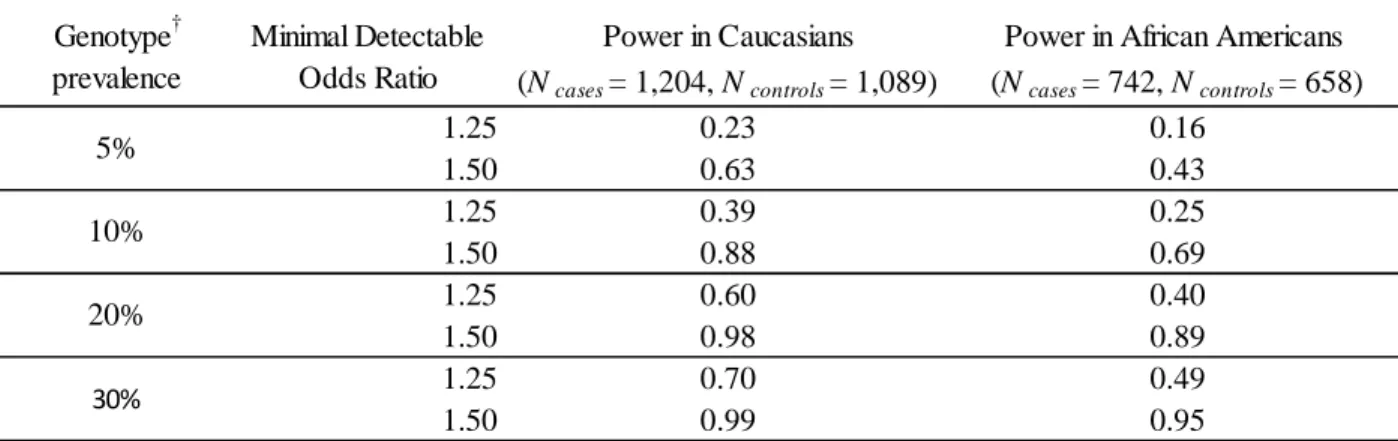
with extreme Hardy-Weinberg equilibrium (HWE) p-values ...74 Table 2.8 – Study power for main effects of genotype on all breast
cancer in CBCS participants by race (α=0.05) ...75 Table 2.9 – Study power for main effects of genotype on intrinsic
subtype of breast cancer in CBCS participants (α=0.05) ...76 Table 2.10 – Single nucleotide polymorphisms (SNPs) included
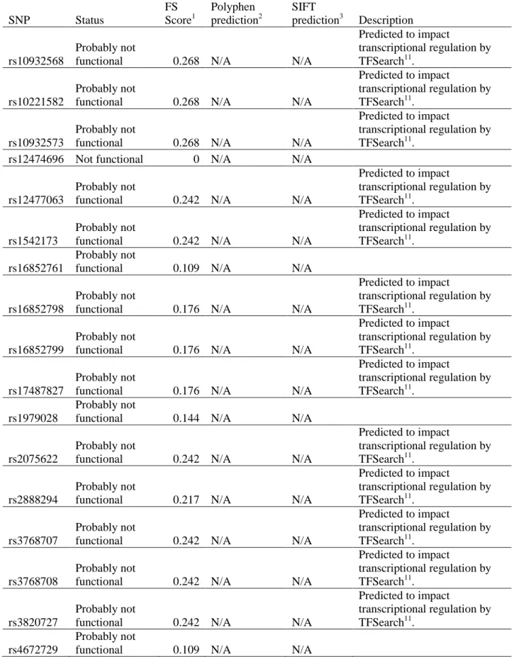
in additive interaction analysis ...77 Table 2.11 – Assessment of potential functionality of single nucleotide
polymorphisms (SNPs) on candidate gene ZNF350 genotyped in
the CBCS ...78 Table 2.12 – Assessment of potential functionality of single nucleotide
polymorphisms (SNPs) on candidate gene BARD1 genotyped in
the CBCS ...81 Table 2.13 – Assessment of potential functionality of single nucleotide
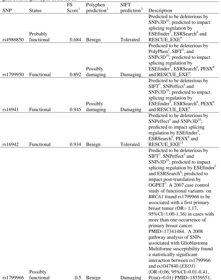
polymorphisms (SNPs) on candidate gene BRCA1 genotyped in
x
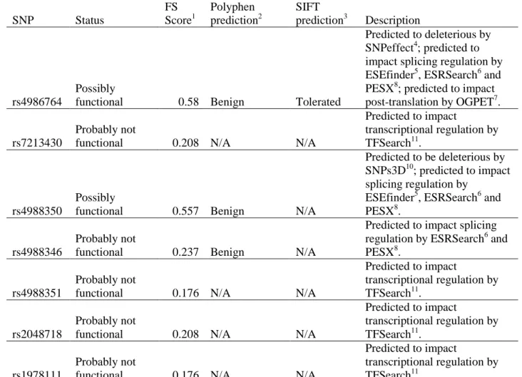
Table 2.14 – Assessment of potential functionality of single nucleotide polymorphisms (SNPs) on candidate gene BRIP1 genotyped in
the CBCS ...88 Table 2.15 – Assessment of potential functionality of single nucleotide
polymorphisms (SNPs) on candidate gene AURKA genotyped in
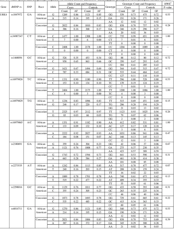
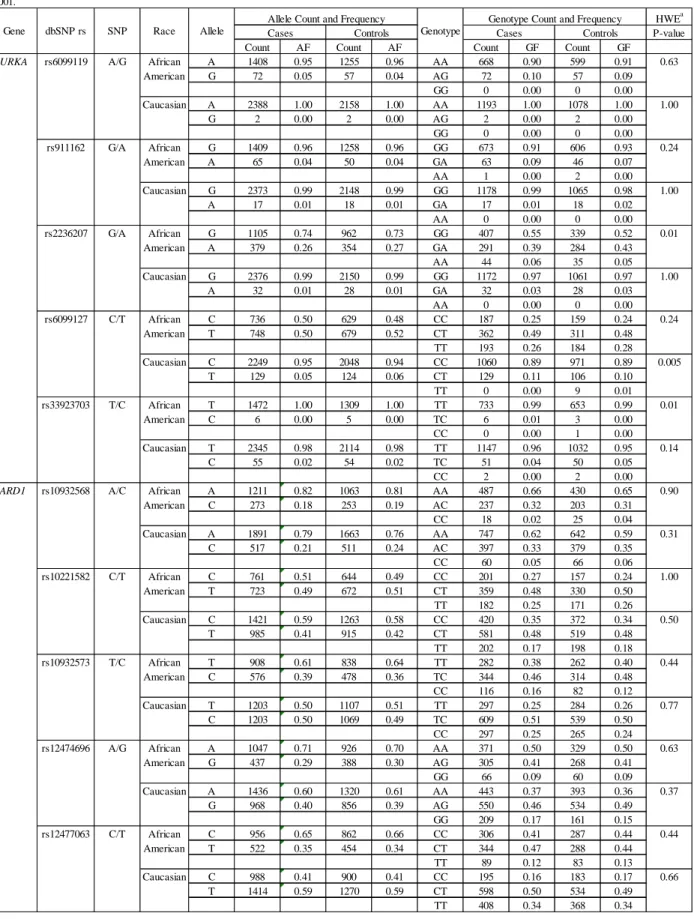
the CBCS ...90 Table 2.16 – Race-specific allele and genotype frequencies for AURKA,
BRCA1, and BRCA1-interacting genes genotyped in CBCS
participants enrolled 1993-2000 ...93 Table 2.17 – Characteristics of CBCS case participants with genotype
data (N=1,946), case participants missing data (N=331), controls with genotype data (N=1,747) and controls missing genotype
data (N=238) ...102 Table 3.1 – Characteristics of CBCS participants with genotype data ...137 Table 3.2 – Odds ratios (ORs) and 95% confidence intervals (CIs)
for the association between single nucleotide polymorphisms (SNPs) on AURKA and all incident cases of breast cancer by
race ...138 Table 3.3 – Odds ratios (ORs) and 95% confidence intervals (CIs)
for the association between single nucleotide polymorphisms
(SNPs) on AURKA and intrinsic subtype of breast cancer ...140 Table 4.1 – Odds ratios (ORs) and 95% confidence intervals (CIs)
for the association between single nucleotide polymorphisms (SNPs) on BRCA1 and BRCA1-interacting genes and all
incident cases of breast cancer by race ...162 Table 4.2 – Odds ratios (ORs) and 95% confidence intervals (CIs)
for the association between single nucleotide polymorphisms (SNPs) on BRCA1 and BRCA1-interacting genes and breast cancer
subtype ...167 Table 4.3 – Additive interaction analysis between select SNPs on AURKA
and BRCA1 ...172 Table 4.4 – Additive interaction analysis between select SNPs on AURKA
xi
Table 4.5 – Additive interaction analysis between select SNPs on AURKA
and BRIP1 ...183 Table 4.6 – Additive interaction analysis between select SNPs on AURKA
xii
LIST OF FIGURES
Figure 2.1 – Age-specific (crude) SEER incidence rates by race
and sex, female breast cancer, all ages, 2000-2007 ...103 Figure 2.2 – Age-adjusted SEER incidence rates by race and sex,
female breast cancer, all ages, 2000-2007 (SEER17) ...104 Figure 2.3 – Age-adjusted U.S. Mortality rates by race and sex,
female breast cancer, all ages, 2000-2007 ...105 Figure 2.4 – Age-specific (crude) U.S. Mortality rates by race
and sex, female breast cancer, all ages, 2000-2007 ...106 Figure 2.5 – Immunohistochemical identification of breast tumor
intrinsic subtypes ...107 Figure 2.6 – Carolina Breast Cancer Study geographic study area ...108 Figure 2.7 – Decision tree for inclusion of single nucleotide
polymorphisms (SNPs) into interaction study ...109 Figure 2.8 – Power to detect an association between genotype and
overall rate of breast cancer in Caucasian participants (cases=1,204, controls=1,089) given a genotype prevalence
of 5% ...110 Figure 2.9 – Power to detect an association between genotype and
overall rate of breast cancer in Caucasian participants (cases=1,204, controls=1,089) given a genotype prevalence
of 10% ...111 Figure 2.10 – Power to detect an association between genotype and
overall rate of breast cancer in Caucasian participants (cases=1,204, controls=1,089) given a genotype prevalence
of 20% ...112 Figure 2.11 – Power to detect an association between genotype and
overall rate of breast cancer in African American participants
(cases=742, controls=658) given a genotype prevalence of 5%...113
Figure 2.12 – Power to detect an association between genotype and overall rate of breast cancer in African American participants
xiii
Figure 2.13 – Power to detect an association between genotype and overall rate of breast cancer in African American participants
(cases=742, controls=658) given a genotype prevalence of 20%...115 Figure 2.14 – Forest plot of the association between AURKA
functional polymorphism rs2273535 and breast cancer risk
xiv
LIST OF ABBREVIATIONS
AA – African American(s)
AIM – ancestry informative marker BMI – body mass index
Cau – Caucasian(s)
CBCS – Carolina Breast Cancer Study
CEU – Utah residents with ancestry from northern and western Europe genotyped by the International HapMap Project
CI – confidence interval CIS – carcinoma in situ CK – cytokeratin
CLR – confidence limit ratio DCIS – ductal carcinoma in situ EM – expectation maximization ER – estrogen receptor
GWAS – genome-wide association study
HER1 – human epidermal growth factor receptor 1 HER2 – human epidermal growth factor receptor 2 HRT – hormone replacement therapy
HWE – Hardy-Weinberg equilibrium IHC – immunohistochemistry
xv OR – odds ratio
PCM – pericentriolar material PJS – Peutz-Jeghers Syndrome PR – progesterone receptor
RERI – relative excess risk due to interaction SNP – single nucleotide polymorphism
r2 – pairwise correlation coefficient
UNC – University of North Carolina at Chapel Hill
1
Chapter 1. Background and Significance
1.1 The Public Health Burden of Breast Cancer
Breast cancer continues to represent a tremendous health burden in the United States. The American Cancer Society estimates that 30% of all cancers diagnosed among American women in 2010 will be breast cancers, making them the most commonly diagnosed cancers among women in the U.S. [1]. After cancers of the lung and bronchus, breast cancer is the leading cause of cancer death in American women [1]. Although recent data indicate a decline in incidence and mortality, a consistent disparity between African American and Caucasian women persists [1, 2].
Incident cases of breast cancer have been and continue to be more frequent in Caucasian women (126.5 per 100,000) compared to African American women (118.3 per 100,000) [2]. However, age-adjusted trends have been consistent, if not convergent since 1975 [1] (Figure 2.2). The racial disparity in incidence is highlighted in women aged 40 and above (Figure 2.1). Notably however, this trend is reversed in women under 35, with African American women displaying a higher incidence rate.
2
American women is distinguished by larger, higher-grade tumors that are diagnosed at later stages[3-6]. Even after controlling for stage at diagnosis, African American women still exhibit poorer survival when compared to Caucasian women [3, 6-8]. It has been suggested that differences in survival may be attributed to socioeconomic factors [5, 9-12] or
differences in access to care [8-13]. However, recent studies have reported that trends in screening by mammography among African Americans and Caucasians are similar [14-16]. In fact, controlling for socioeconomic factors, access to healthcare and co-morbidities does not diminish the racial disparity in mortality [11, 17-21]. This may suggest potential differences in tumor biology among African American and Caucasian women.
1.2 Genetic Risk Factors for Breast Cancer
A family history of breast cancer is a strong risk factor; women having a single first-degree relative with breast cancer are nearly twice as likely to develop the disease, while having two first-degree relatives with breast cancer approximately triples a woman’s risk [1, 22, 23]. Still, the vast majority of women who develop breast cancer (~85%) have no family history of the disease [22].
Hereditary breast cancers constitute between 5 and 10% of all cases [24]. The most common predisposing factors contributing to these cases are highly penetrant mutations in
BRCA1 and BRCA2. However, population-based epidemiologic studies have demonstrated
3
the causation of breast cancer remains unclear [26, 27]. Twin-studies and studies of familial inheritance have suggested that common, low penetrance genetic factors may account for the observed residual familial risk [26, 28]. This so-called polygenic model proposes that genetic susceptibility to breast cancer is not entirely predicted by rare, highly penetrant genes but more often stems from several common loci that each confer smaller independent increases in risk [28-31]. Acting multiplicatively, this aggregate of common risk variants may contribute a significant proportion of familial risk. Under this model it would be rare to observe multiple-case families (as is the case for those demonstrating mutations in highly penetrant genes such as BRCA1) since an individual would have to inherit each of several different variants.
Results from genome-wide association studies (GWAS) seem to support the polygenic model with respect to breast cancer. GWAS take advantage of technological advances allowing for hundreds of thousands to millions of single nucleotide polymorphisms (SNPs) to be analyzed as potential risk modifying loci without information regarding function. A large GWAS conducted by Easton et al. identified significant associations between SNPs on
FGFR2, TNRC9, MAP3K1, LSP1, H19 and breast cancer in European women from the
4
Additionally, there are several rare conditions that substantially increase the risk of breast cancer in a small proportion of the population. Li-Fraumeni Syndrome (LFS) is caused by a mutation in TP53 and is thought to account for approximately 1% of hereditary breast cancers [34]. LFS is characterized by early-onset cancers, including: breast cancer, soft-tissue sarcoma and leukemia [35]. LFS families experience an increased risk of cancer up to 90% by age 60 [36]. Cowden Syndrome is also associated with increased risk for breast cancer [37]. Cowden Syndrome is generally defined by germline mutations in PTEN, a putative tumor suppressor gene [38, 39]. Women with Cowden Syndrome have a lifetime risk of breast cancer between 25-50% [40]. Peutz-Jeghers Syndrome (PJS) has also been associated with an increased risk of cancer. PJS is characterized by germline mutations in the tumor suppressor gene STK11 [41]. Women with PJS have demonstrated increased risk for breast cancer of up to 30% by age 60 [42]. Ataxia telangiectasia, a rare childhood condition characterized by neurological deterioration and hypersensitivity to ionizing radiation, has also been associated with an increased risk for breast cancer [37].
1.3 Other Risk Factors for Breast Cancer
1.3.1 Age
5
1.3.2 BMI
Studies investigating the relationship between BMI and risk of breast cancer have been inconsistent. Reports have suggested that increased BMI is associated with an increased risk of breast cancer; in a pooled multivariate analysis of prospective cohorts, van den Brandt et
al. reported increased risk of breast cancer with increasing BMI and weight only in
postmenopausal women, but the trend was not linear. Women between 75-80kg showed a higher relative risk than women ≥80kg. Likewise, BMI results demonstrated the same trend, with postmenopausal women having a BMI of 31-33 demonstrating a higher relative risk than women of BMI ≥33 [46]. In premenopausal women, an inverse trend in risk was noted in both weight and BMI. Possible explanations for this inverse trend include more frequent anovulatory menstrual cycles resultant from decreased concentrations of estrogen and progesterone exhibited in obese women [46-49]. In contrast, several case-control studies have found both inverse and direct associations between BMI and odds of breast cancer among premenopausal women [50-52].
As a result of inconsistent findings for associations between BMI and risk of breast cancer, it has been suggested that distribution of adiposity may be an important factor in explaining the relationship between BMI, weight, and risk of breast cancer in
6
found no association between WHR and risk of breast cancer in premenopausal women [52]. In contrast, a meta-analysis of case-control and cohort studies performed by Connolly et al. reported significant associations between WHR and risk of breast cancer, regardless of menopausal status, after controlling for BMI [56].
1.3.3 Physical Activity
Studies of potential associations between physical activity and risk of breast cancer have been equivocal, probably due in part to the lack of any clear standardized instrument for measuring exposure and a failure to thoroughly evaluate confounding and effect measure modification [57]. Nevertheless, Monninkhoff’s systematic review of 29 case-control and 19 cohort studies found strong evidence for risk reductions with increased physical activity among postmenopausal women; evidence for risk reduction among physically active premenopausal women was weaker [58].
1.3.4 Menarche
Reproductive factors such as age at menarche and regularity of menstrual cycles have also been associated with breast cancer risk. Early age at menarche (12 years or earlier) has been associated with an increased risk of breast cancer, with modest declines in risk
7
adolescent years [59, 62]. Estrogen is known to influence normal breast epithelial cell growth by promoting cellular proliferation [59]. The increased exposure to estrogen during adolescence provides an environment of rapid cell proliferation that is thought to increase the risk of random mutations in the genome [59, 60, 63]. Supporting the role of estrogen in tumorigenesis, early menopause has been shown to decrease a woman’s risk for breast cancer [60].
1.3.5 Breast Density
Breast density based on parenchymal patterns has also been strongly and consistently associated with breast cancer risk. Mammographic studies have demonstrated increased risks among women with large nodular densities and/or extensive areas of homogenous density (i.e. high proportions of connective and epithelial tissues) compared to women whose breasts were largely composed of less dense fat tissue [64-67].
1.3.6 Breast Feeding
Bernstein et al. reported that breast feeding decreased risk of breast cancer in
8
than a dozen known and purported risk factors. This association was monotonically
strengthened with increasing number of children breastfed [70]. In a 2000 meta-analysis of the effects of breast feeding on risk for breast cancer, Bernier et al. reported a slight
protective effect in women who ever breast fed (Pooled OR=0.88, 95%CI: 0.84-0.92) [71].
1.3.7 Exogenous Hormone Use
9
1.3.8 Other Reproductive Factors
Other reproductive factors that influence endogenous estrogen exposure, such as parity and early age at first birth, have shown inverse associations with risk of breast cancer. Parity and early age at first full-term pregnancy are associated with an overall decreased risk of breast cancer [59, 80]. Lifetime risk decreases with increasing number of full-term pregnancies, but only among those women who experienced their first full-term pregnancy before the age of 20 [59, 80]. This reduction in risk observed among younger women at first birth is an overall reduction. In actuality, the short term effects of term pregnancies on breast cancer risk appear to increase risk [59, 81]. Bruzzi et al. found that full term
pregnancy at any age is followed by a short increase in risk of breast cancer, irrespective of the increase associated with aging alone, that distorts the long term inverse association between parity and risk of breast cancer [81]. One explanation for this short term increase in risk is the increased level of bioavailable estradiol during the first trimester of pregnancy. Exposure to high levels of estradiol is suspected to increase risk for breast cancer [47, 50, 59, 82].
10
1.3.9 Height
Associations between height and breast cancer risk have also been investigated, yielding conflicting results with respect to menopausal status. Several studies have found an
association between height and risk of breast cancer only among postmenopausal women [84-86]. However, Ahlgren et al. reported a significant increase in risk among women who were in the highest quintile of height at age 14 [87]. A pooled analysis conducted by van den Brandt et al. also found a significant association between height and risk for breast cancer, irrespective of menopausal status [46].
1.3.10 Ionizing Radiation
11
1.4 Intrinsic Breast Cancer Subtypes
1.4.1 Gene Expression Patterns & Hormonal Receptor Status
In an effort to improve molecular taxonomy and targeted therapies for breast cancers, Perou et al. identified four distinct subtypes of breast cancer based on differences in gene expression patterns using cDNA microarrays and hierarchical clustering [90]. Each subtype can also be described by immunohistochemical staining profiles based on hormonal receptor and cellular cytokeratin status, which are surrogates for the gene expression profiles [3, 90, 91]. Of these, estrogen receptor positive (ER+) tumors are characterized by high expression of genes expressed by luminal breast cells. ER+ tumors were also distinguished
immunohistochemically by staining with antibodies against luminal cytokeratins 8 and 18 [90]. Recent studies showed that ER+/luminal tumors can be further classified into luminal-A and luminal-B subtypes based on expression of human epidermal growth factor receptor-2 (HER2) (Figure 2.5) [3, 92, 93]. In comparison, luminal-A tumors are more common, express higher levels of estrogen receptor and little to no expression of HER2, and generally render a better prognosis [3, 92]. A second subtype was characterized by high expression of genes expressed by breast basal epithelial cells [90]. Support for this finding was evidenced by immunohistochemical staining of basal cell keratins 5/6 and 17 [90]. Basal-like tumors are also distinguished by the absence or low expression of estrogen receptor (ER-) and human epidermal growth factor receptor-2 (HER2-) [3, 90]. Basal-like tumors, often referred to as “triple-negative” breast cancers, are among the least responsive to hormonal and targeted therapies, and usually result in poorer prognoses [3]. A third subtype,
12
nearly all genes associated with ER expression [90]. HER2+/ER- tumors exhibit gene expression patterns similar to those of basal-like cancers [3]. However, the availability of Herceptin treatment renders a more favorable prognosis in women with HER2+ tumors. The final subtype includes those remaining tumors whose gene expression profiles are characteristic of basal epithelial and adipose cells. Tumors of this subtype are denoted “normal-like” due to their low expression of genes typified by ER+/luminal tumors and cannot be identified via immunohistochemistry [90].
In early 2006, Carey et al. used immunohistochemical surrogates for expression
profiling to identify subtypes, including ER and progesterone receptor (PR) status, and also to further distinguish between those tumors expressing HER2 [3]. PR was included in the definitions because it is a commonly used breast tumor marker that is regulated by ER and is associated with response to hormonal therapy [3]. The HER2+ tumors were further
categorized by ER expression due to the propensity for HER2+/ER- tumors to express genes that cluster closer to those of basal-like tumors, while HER2+/ER+ more closely resembled the clustering pattern of luminal cancers(Figure 2.5) [3]. Luminal-A tumors were defined as (ER+ and/or PR+, HER2-); luminal-B tumors were (ER+ and/or PR+, HER2+); HER2+/ER- tumors were further defined by PR status as (ER-, HER2+, PR-); basal-like tumors were defined as (ER-, PR-, HER2-, cytokeratin 5/6+, and/or HER1+) [3].
1.4.2 Epidemiologic Findings
It is well established that breast cancer subtypes differ in their responsiveness to
13
luminal-A subtype, while women exhibiting HER2+/ER- tumors and basal-like tumors demonstrated the worst survival [3, 95, 96]. In a population-based study of African
American and Caucasian women, Carey et al. reported women with HER2+/ER- tumors and luminal-B tumors were more likely to have lymph node metastases, while those women with basal-like tumors were not [3]. Several studies have reported a tendency for younger, premenopausal women to develop basal-like tumors when compared to older,
postmenopausal women [3, 97-102]. Basal-like tumors are associated with poor prognosis, often characterized by higher grade, higher mitotic index, and significant DNA mutations [3, 95, 100, 103-106]. Basal-like tumors are also characterized by aneuploidy [107, 108]. Other research has reported that basal-like tumors are more likely to be larger and exhibit a greater tendency to metastasize [95, 106, 109-112]. Basal-like breast cancers are also more likely to be associated with BRCA1 mutations compared to other subtypes, suggesting a distinct biological mechanism [92, 104, 113]. Since BRCA1 mutation carriers tend to develop basal-like breast tumors, there may be other inherited genetic variants that predispose to developing specific subtypes of breast cancer [3, 92, 113]. In addition to relatively worse prognoses and fewer treatment options, basal-like breast cancers tend to develop in younger African American women disproportionately [3, 97, 98, 114]. Carey et
al. found a high prevalence of basal-like tumors in African American women, all of whom
14
1.4.3 Etiology
As more epidemiologic evidence supports the biological heterogeneity of breast cancer, assessing risk factors by distinct breast cancer subtypes may reveal more accurate
associations. Lacroix et al. suggest that molecular tumor characteristics do not change appreciably over the progression from in situ carcinoma to invasive carcinoma [117]. As such, exposures that are associated with breast cancer etiology may show different associations according to molecular subtype. Several studies have found varying
associations between common risk factors for breast cancer (age, parity, age at first birth, age at menarche, race) and hormone receptor status [114, 118].
The Carolina Breast Cancer Study (CBCS), a population-based case-control study of African American and Caucasian women in North Carolina, reported increased odds of basal-like breast cancer as opposed to luminal breast cancer among women who were younger at first pregnancy [97]. On the other hand, a reduced odds of basal-like breast cancer was noted among women who breastfed more children for a longer duration, but not among luminal cases [97]. This finding is in contrast to other study findings indicating a reduced risk of breast cancer among Chinese women who breast fed, however those studies did not stratify by intrinsic subtype and were based on study populations that are not
comparable to CBCS [119, 120]. In addition to finding the highest prevalence of basal-like tumors in younger African American women, Millikan et al. also reported increased odds of basal-like breast cancer associated with higher waist-hip ratio in both pre- and
post-menopausal women [97].
15
among premenopausal women (OR=0.71, 95% CI: 0.57-0.88 per five-unit increase), while a slightly increased odds for basal-like breast cancer was noted among women with higher BMI (OR=1.18, 95% CI: 0.86-1.64) [114]. The same study also noted a significantly reduced risk of basal-like breast cancer with increasing age at menarche (OR=0.78, 95% CI: 0.68-0.89 per 2-year increase) [114].
Contrasting data from two centers participating in the Cancer and Steroid Hormone Study (CASH) suggests clear differences in risk associated with late age at first birth between African American and Caucasian women. A stronger association between ER negative tumors and late age at first birth was noted among African American women, whereas a strong association was noted with ER positive tumors in Caucasian women [118, 121, 122]. Significant heterogeneity of associations by subtype was also reported in a case only study of 2,544 breast cancer cases classified by ER, PR, and HER2 status [123]. Notable risk factors that may be related to the development of particular molecular subtypes of breast cancer included: BMI, alcohol consumption, and history of breastfeeding [123].
1.5 Cell Cycle Regulation and Cancer
Cancers are characterized by aberrations in cell cycle regulation, leading to inappropriate cell replication. This unchecked cell proliferation is associated with reduction in or loss of sensitivity to normal signals to either differentiate or initiate apoptosis. Many genes are responsible for adherence to proper cell cycle function, and interpreting the changes that can disrupt this process is integral for understanding the etiology of cancer [124].
16
mutations. Gain-of-function mutations are characterized by the transformation of proto-oncogenes into proto-oncogenes (mutated genes that once performed normal cellular functions as proto-oncogenes, and now contribute to aberrant cell proliferation) [125]. Proto-oncogenes perform important functions within the cell, from signal transduction to programmed cell death [125]. Conversion of proto-oncogenes into oncogenes can result in unregulated cell growth [125]. Studies have shown that individual oncogenes can have identical effects leading to gain-of-function or can be cell-type specific, suggesting different genetic
pathways resulting in cancer [124]. Gain-of-function mutations only require one copy of the mutant allele for transformation to the oncogene [124].
Loss-of-function mutations occur in tumor suppressor genes and are far more common. Only individuals who are homozygous for the mutant allele will exhibit loss-of-function, with heterozygotes demonstrating the normal wild-type phenotype. However, heterozygotes will bear an increased risk for developing cancer due to the fact that a subsequent deleterious mutation will prevent normal gene function [124]. Loss-of-function mutations in tumor suppressor genes have been shown to result in circumvention of normal negative regulation that controls entry into the cell cycle [126]. One such example is a loss-of-function
mutation in the tumor suppressor gene p53; p53 normally functions to arrest cell cycle progression in response to DNA damage [127]. Loss of normal p53 function allows for unchecked cellular proliferation of mutant DNA.
1.6 The Centrosome and Centrosome Cycle
17
an amorphous pericentriolar material (PCM) [129, 130]. Often referred to as the
microtubule organizing center of the cell, centrosomes determine the number, length and distribution of microtubules. Animal cells normally contain one centrosome which is duplicated once and only once per cell cycle. Centrosomal duplication involves centriolar duplication in G1 of interphase and culminates in dual centrosomes by G2/mitosis. As this is a semi-conservative process, one of the centrioles present in the centrosome will be more “mature” than the other and is denoted the mother centriole since it has experienced more cell cycles. Likewise a centriole that has not yet completed a full cell cycle is referred to as a daughter centriole. The distinction is in the number of microtubules each centriole can nucleate; the more mature centriole can be identified by appendages protruding from its distal end and is capable of nucleating more microtubules [129, 131]. During mitosis, the centrosomes nucleate microtubules in a polarized array with their positive ends directed outward from the electron-dense PCM [129, 130]. This polymerization of microtubules toward either pole of the cell forms the spindle apparatus that will facilitate alignment of the chromosomes in preparation for cell division. The centrosomes are also important for cytokinesis and in establishing a midpoint at metaphase for the cleavage furrow to form; studies have shown that removal of the centrosome from cells resulted in failure to complete cytokinesis [129, 132]. Further studies of individual centriole removal provided evidence of the same; after removing one of two centrioles from a cell’s centrosome, Piel et al.
18
Cells completing cytokinesis and exiting mitosis are characterized by a single
centrosome, composed of two orthogonally positioned centrioles. During G1, the centrioles separate in preparation for duplication. Centriole duplication is distinguished by the
formation of procentrioles on either parental centriole, a process referred to as centriole engagement [134]. Formation and orientation of the procentrioles and the duplication
process are thought to be tightly regimented to prevent more than one replication in the same cell cycle. Tsou and Stearns hypothesize that the physical presence and positioning of the procentrioles blocks reduplication [134]. During S phase and throughout G2, the
procentrioles grow until they achieve their maximum length. Maturation of the previously immature centriole begins during G2 and culminates in the development of distal
appendages. The maturation process requires approximately 1.5 cell cycles to complete [129, 131]. As the cell transitions from G2 into mitosis, it contains two centrosomes which will separate and migrate to either pole of the dividing cell to establish the mitotic spindle. A concomitant centrosomal and cell cycle are integral to ensure two independent
19
identical daughter cells each containing a single centrosome. The centrosome cycle is then repeated.
1.7 Centrosomal Amplification and Breast Cancer
It is important that a cell undergoing mitosis contain two independent centrosomes, each located at either pole. Since the centrosome acts as a microtubule organizing center in the dividing cell, the presence of more than two could result in improper formation of the spindle apparatus, aberrant segregation of chromosomes, or failure of cytokinesis [138]. Pihan et al. found amplified centrosomes (more than two centrosomes or more than four centrioles) as commonly characteristic of solid malignant tumors [139, 140]. In a study of high grade human breast tumors, Lingle et al. had similar findings, reporting increased microtubule nucleation in addition to amplified centrosomes [138].
Seven hundred eighty-two SNPs from 101 centrosomal genes were analyzed in a
population-based study of 798 invasive breast cancer cases and 843 controls from the Mayo Clinic Breast Cancer Study. Findings indicated that genes involved in the centrosome regulatory pathway were highly enriched with SNPs associated with risk of breast cancer (p=4.6x10-50) [141]. Amplified centrosomes are suspected of contributing to aneuploidy by increasing the rate of aberrant mitoses resulting in chromosomal missegregation [135, 142]. Furthermore, Lingle et al. found evidence to support the hypothesis that centrosomal
20
demonstrated unchecked centrosome replication in cells arrested at the G1/S boundary, supporting the hypothesis that centrosome replication is driven by activation and
inactivation of centrosomal regulatory genes during the cell cycle [143]. An investigation by Zhou et al. showed that the AURKA locus encoding a serine-threonine kinase associated with centrosome regulation was implicated in causing centrosome duplication abnormalities and aneuploidy in mammalian cells; overexpression of AURKA was associated with
centrosome amplification and chromosomal instability [135, 144]. Chromosomal instability is the rate of gains or losses of chromosomes, whereas aneuploidy is the cross-sectional disposition of the cell with respect to chromosome number [129]. Although aneuploidy is a common characteristic of cancer cells [129, 145-148], it is unclear as to whether or not it causes or results from disease progression.
1.8 AURKA
The Aurora A gene, also known as AURKA, encodes a serine/threonine kinase and is located on the q arm of chromosome 20 at amplicon 13.2, a region commonly amplified in human breast cancers [149, 150]. Isola et al. reported poorer prognosis and survival among breast cancer cases exhibiting highly amplified 20q13 [151]. Likewise, Tanner et al. found high amplification of 20q13 in primary breast carcinomas to be significantly associated with high histological grade, aneuploidy, short disease-free survival, and poor clinical outcome suggesting this region contains a gene involved in breast cancer progression [152]. AURKA functions in centrosomal maturation and separation, mitotic spindle formation and
21
progression [154-157]. In a study of mammary tumorigenesis in mice, Wang et al.
demonstrated centrosome amplification and aneuploidy in transgenic mice overexpressing
AURKA in mammary epithelium [158]. Notably, severe chromosomal abnormalities failed
to trigger apoptosis in cells overexpressing AURKA, allowing for continued proliferation of abnormal karyotypes [158]. Tanaka et al. showed overexpression of AURKA in 94% of invasive ductal carcinomas of the breast in a cohort of Japanese women [159].
Few population-based studies of genetic variation in AURKA have been conducted, and those that have been conducted have focused on a few functional variants in European and Asian populations. The T/A coding region polymorphism (F31I) on AURKA that results in an amino acid substitution (Phe Ile) has been studied extensively in European and Asian populations. Functional evidence for a biochemical difference between the proteins encoded by the Ile31 variant and the more common Phe31 variant has been reported. The Ile allele is more efficient in inducing cell growth in vitro, which facilitates the oncogenic effect of
AURKA [160]. The Ile-Ile genotype has been associated with increased aneuploidy in
22
valine isoleucine substitution has been investigated for potential association with risk of breast cancer. Egan et al. reported no association with breast cancer risk among European women with the Ile/Ile genotype [164]. However, examining a relatively common genotype combining the two polymorphisms (31I-57V/31I-57V), Egan et al. reported a 2-fold
increase in risk of breast cancer (OR=1.96; 95% CI: 1.01-3.79) [164]. A study by Dai et al. investigating both (F31I) and (V57I) reported no association between the combined (31I-57V/31I-57V) genotype and risk of breast cancer in a population-based study of Chinese women [161]. Evidence for purported breast risk loci on AURKA are summarized in Table 5.
Because AURKA is strongly involved in centrosomal regulation, and aberrations in the centrosomal cycle lead to aneuploidy, polymorphisms on AURKA may also be more strongly associated with subtypes of breast cancer that are known to demonstrate relatively high levels of aneuploidy; namely, triple-negative and basal like breast cancers [108, 113]. Relative to all other subtypes, basal like and triple-negative breast cancers are more likely to demonstrate higher levels of aneuploidy, with other subtypes demonstrating a more variable degree of aneuploidy [113]. This proposal will be the first to investigate AURKA’s
association with intrinsic subtype of breast cancer.
1.9 BRCA1 and Interacting Genes: BARD1, BRIP1, and ZNF350
AURKA is not the only centrosomal regulatory gene that has been associated with breast
23
[167]. Ruan et al. reported a significant interaction between haplotypes on AURKA and
BRCA1 in a Han Chinese population, warranting further investigation in different
populations [166]. Other purported risk loci on BRCA1 are summarized in Table 6.
Genetic variants conferring high risk for breast cancer are not sufficient to cause breast cancer. Even for carriers of mutations in the highly penetrant BRCA1 and BRCA2, the distribution of risk varies suggesting possible gene-gene or gene-environment interactions that affect overall risk [29]. Evidence supporting the multiplicative joint effects of low-penetrance genes on breast cancer risk has been reported by Antoniou et al. [28]. BARD1,
BRIP1 and ZNF350 are three putative low penetrance breast cancer susceptibility genes.
The BARD1 protein interacts with BRCA1 to form a heterodimer complex
BRCA1/BARD1 [168]. By itself, BRCA1 confers an ubiquitin ligase activity that is essential
for its normal tumor suppression action, namely in coordinating DNA repair [168].
However, when complexed with BARD1, the ubiquitin ligase activity is markedly enhanced [168]. This interaction with BRCA1 suggests a possible role for BARD1 in DNA repair processes. Mutations in BRCA1 are known to deactivate the ubiquitin ligase activity of the
BRCA1/BARD1 heterodimer complex [168, 169], which has been correlated with its tumor
suppression function [170].
Germline mutations in BARD1 were demonstrated in a cohort of 40 Italian families with a history of breast and breast/ovarian cancer whose probands were chosen due to their lack of BRCA1 mutations [171]. The Nordic collaborative study has reported that a specific
BARD1 variant (Cys557Ser) may represent a common breast cancer susceptibility allele
24
BRIP1 encodes a helicase that binds directly to the C-terminus of BRCA1 and directly
contributes to the double-strand break repair function of BRCA1 [175]. Cantor et al. found germline mutations in BRIP1 affecting the helicase domain among breast cancer patients with normal BRCA1 but not among controls, implicating BRIP1 as a potential
low-penetrance gene that contributes to familial breast cancer risk. Further evidence of BRIP1’s important association with BRCA1 was noted: missense and deletion mutations in the C-terminus of BRCA1, which inactivated its normal double-strand break repair function, also inhibited BRIP1 binding, suggesting a functional role in DNA repair [176].
ZNF350 is a corepressor of GADD45, which is involved in cell cycle arrest at the G2/M
checkpoint subsequent to DNA damage [177]. GADD45 is regulated by both ZNF350 and
BRCA1 [177, 178]. The interaction of ZNF350 with BRCA1 is necessary for the modulation
of GADD45 [179]. ZNF350 has been shown to negatively regulate overexpression of
BRCA1 [177], but its role in human carcinomas is largely unknown [180].
Due to their interactive roles with BRCA1, BARD1, BRIP1 and/or ZNF350 may show gene-gene interactions in association with odds of breast cancer with other BRCA1-interacting genes, such as AURKA.
1.10 Summary—Background and Significance
25
differences in breast cancer subtype using a candidate gene approach. This study
investigated important genes involved in cell cycle regulation that were carefully chosen for their suspected role in oncogenesis.
Due to its function as an important centrosomal and cell cycle regulator, AURKA may play a significant role in oncogenesis, especially with respect to the proliferation of aneuploid cells—a common feature of basal-like tumors. Although AURKA has been investigated in large population-based studies, these studies have been largely conducted among European and Chinese individuals. In light of a disproportionate number of basal-like cases among African American women, and due to previous investigations’ findings that basal-like and triple negative breast tumors demonstrate higher degrees of aneuploidy, the CBCS offers a novel opportunity to evaluate AURKA in African American women.
This study was the first to investigate statistical interactions between BRCA1 and
AURKA in African American women with breast cancer. Since BRCA1 is known to be an
important regulator of the cell cycle, and since BRCA1 proteins are known to interact with
AURKA proteins, it’s reasonable to suspect gene—gene interactions between them. In
26
REFERENCES
1. American Cancer Society. Cancer Facts and Figures 2011. In. Atlanta: American Cancer Society.
2. National Cancer Institute--Surveillance Epidemiology and End Results (SEER) Program 2010. In. Edited by Incidence - SEER 9 Regs Research Data NS-.
3. Carey LA, Perou CM, Livasy CA, Dressler LG, Cowan D, Conway K, Karaca G,
Troester MA, Tse CK, Edmiston S et al: Race, breast cancer subtypes, and survival in the Carolina Breast Cancer Study. JAMA 2006, 295(21):2492-2502.
4. Furberg H, Millikan R, Dressler L, Newman B, Geradts J: Tumor characteristics in African American and white women. Breast Cancer Res Treat 2001, 68(1):33-43. 5. Porter PL, Lund MJ, Lin MG, Yuan X, Liff JM, Flagg EW, Coates RJ, Eley JW: Racial
differences in the expression of cell cycle-regulatory proteins in breast carcinoma.
Cancer 2004, 100(12):2533-2542.
6. Eley JW, Hill HA, Chen VW, Austin DF, Wesley MN, Muss HB, Greenberg RS, Coates RJ, Correa P, Redmond CK et al: Racial differences in survival from breast cancer. Results of the National Cancer Institute Black/White Cancer Survival Study. JAMA 1994, 272(12):947-954.
7. Clegg LX, Li FP, Hankey BF, Chu K, Edwards BK: Cancer survival among US whites and minorities: a SEER (Surveillance, Epidemiology, and End Results) Program population-based study. Arch Intern Med 2002, 162(17):1985-1993.
8. Shavers VL, Brown ML: Racial and ethnic disparities in the receipt of cancer treatment. J
Natl Cancer Inst 2002, 94(5):334-357.
9. Li CI, Malone KE, Daling JR: Differences in breast cancer stage, treatment, and survival by race and ethnicity. Arch Intern Med 2003, 163(1):49-56.
10. O'Malley CD, Le GM, Glaser SL, Shema SJ, West DW: Socioeconomic status and breast carcinoma survival in four racial/ethnic groups: a population-based study. Cancer 2003, 97(5):1303-1311.
27
12. Cunningham JE, Butler WM: Racial disparities in female breast cancer in South Carolina: clinical evidence for a biological basis. Breast Cancer Res Treat 2004, 88(2):161-176.
13. del Carmen MG, Hughes KS, Halpern E, Rafferty E, Kopans D, Parisky YR, Sardi A, Esserman L, Rust S, Michaelson J: Racial differences in mammographic breast density.
Cancer 2003, 98(3):590-596.
14. Smigal C, Jemal A, Ward E, Cokkinides V, Smith R, Howe HL, Thun M: Trends in breast cancer by race and ethnicity: update 2006. CA Cancer J Clin 2006, 56(3):168-183. 15. Yankaskas BC, Gill KS: Diagnostic mammography performance and race: outcomes in
Black and White women. Cancer 2005, 104(12):2671-2681.
16. Gill KS, Yankaskas BC: Screening mammography performance and cancer detection among black women and white women in community practice. Cancer 2004, 100(1):139-148.
17. Amend K, Hicks D, Ambrosone CB: Breast cancer in African-American women: differences in tumor biology from European-American women. Cancer Res 2006, 66(17):8327-8330.
18. Miller BA, Hankey BF, Thomas TL: Impact of sociodemographic factors, hormone receptor status, and tumor grade on ethnic differences in tumor stage and size for breast cancer in US women. Am J Epidemiol 2002, 155(6):534-545.
19. Yood MU, Johnson CC, Blount A, Abrams J, Wolman E, McCarthy BD, Raju U,
Nathanson DS, Worsham M, Wolman SR: Race and differences in breast cancer survival in a managed care population. J Natl Cancer Inst 1999, 91(17):1487-1491.
20. Chlebowski RT, Chen Z, Anderson GL, Rohan T, Aragaki A, Lane D, Dolan NC, Paskett ED, McTiernan A, Hubbell FA et al: Ethnicity and breast cancer: factors influencing differences in incidence and outcome. J Natl Cancer Inst 2005, 97(6):439-448. 21. Smith-Bindman R, Miglioretti DL, Lurie N, Abraham L, Barbash RB, Strzelczyk J,
Dignan M, Barlow WE, Beasley CM, Kerlikowske K: Does utilization of screening mammography explain racial and ethnic differences in breast cancer? Ann Intern Med 2006, 144(8):541-553.
22. American Cancer Society--Breast Cancer Risk Factors (2010)
[http://www.cancer.org/cancer/breastcancer/detailedguide/breast-cancer-risk-factors]
23. Familial breast cancer: collaborative reanalysis of individual data from 52
28
24. Bradbury AR, Olopade OI: Genetic susceptibility to breast cancer. Rev Endocr Metab
Disord 2007, 8(3):255-267.
25. Balmain A, Gray J, Ponder B: The genetics and genomics of cancer. Nat Genet 2003, 33 Suppl:238-244.
26. Lichtenstein P, Holm NV, Verkasalo PK, Iliadou A, Kaprio J, Koskenvuo M, Pukkala E, Skytthe A, Hemminki K: Environmental and heritable factors in the causation of cancer--analyses of cohorts of twins from Sweden, Denmark, and Finland. N Engl J Med 2000, 343(2):78-85.
27. Peto J: Cancer epidemiology in the last century and the next decade. Nature 2001, 411(6835):390-395.
28. Antoniou AC, Pharoah PD, McMullan G, Day NE, Stratton MR, Peto J, Ponder BJ, Easton DF: A comprehensive model for familial breast cancer incorporating BRCA1, BRCA2 and other genes. Br J Cancer 2002, 86(1):76-83.
29. Pharoah PD, Antoniou A, Bobrow M, Zimmern RL, Easton DF, Ponder BA: Polygenic susceptibility to breast cancer and implications for prevention. Nat Genet 2002, 31(1):33-36.
30. Antoniou AC, Pharoah PD, McMullan G, Day NE, Ponder BA, Easton D: Evidence for further breast cancer susceptibility genes in addition to BRCA1 and BRCA2 in a population-based study. Genet Epidemiol 2001, 21(1):1-18.
31. Antoniou AC, Easton DF: Polygenic inheritance of breast cancer: Implications for design of association studies. Genet Epidemiol 2003, 25(3):190-202.
32. Easton DF, Pooley KA, Dunning AM, Pharoah PD, Thompson D, Ballinger DG, Struewing JP, Morrison J, Field H, Luben R et al: Genome-wide association study identifies novel breast cancer susceptibility loci. Nature 2007, 447(7148):1087-1093. 33. Zheng W, Long J, Gao YT, Li C, Zheng Y, Xiang YB, Wen W, Levy S, Deming SL,
Haines JL et al: Genome-wide association study identifies a new breast cancer susceptibility locus at 6q25.1. Nat Genet 2009, 41(3):324-328.
34. Hutter CM, Young AM, Ochs-Balcom HM, Carty CL, Wang T, Chen CT, Rohan TE, Kooperberg C, Peters U: Replication of breast cancer GWAS susceptibility loci in the Women's Health Initiative African American SHARe Study. Cancer Epidemiol
Biomarkers Prev 2011, 20(9):1950-1959.
29
36. Lustbader ED, Williams WR, Bondy ML, Strom S, Strong LC: Segregation analysis of cancer in families of childhood soft-tissue-sarcoma patients. Am J Hum Genet 1992, 51(2):344-356.
37. Hankinson S HD: Breast Cancer. In: Textbook of Cancer Epidemiology. Edited by Adami H HD, Trichopoulos D. New York: Oxford University Press, Inc.; 2002: 301-309.
38. Orloff MS, Eng C: Genetic and phenotypic heterogeneity in the PTEN hamartoma tumour syndrome. Oncogene 2008, 27(41):5387-5397.
39. Lynch ED, Ostermeyer EA, Lee MK, Arena JF, Ji H, Dann J, Swisshelm K, Suchard D, MacLeod PM, Kvinnsland S et al: Inherited mutations in PTEN that are associated with breast cancer, cowden disease, and juvenile polyposis. Am J Hum Genet 1997,
61(6):1254-1260.
40. Hampel H, Panescu J, Lockman J, Sotamaa K, Fix D, Comeras I, LaJeunesse J,
Nakagawa H, Westman JA, Prior TW et al: Comment on: Screening for Lynch Syndrome (Hereditary Nonpolyposis Colorectal Cancer) among Endometrial Cancer Patients.
Cancer Res 2007, 67(19):9603.
41. Hemminki A, Avizienyte E, Roth S, Loukola A, Aaltonen LA, Jarvinen H, de la Chapelle A: [A serine/threonine kinase gene defective in Peutz-Jeghers syndrome]. Duodecim 1998, 114(7):667-668.
42. Hearle N, Schumacher V, Menko FH, Olschwang S, Boardman LA, Gille JJ, Keller JJ, Westerman AM, Scott RJ, Lim W et al: Frequency and spectrum of cancers in the Peutz-Jeghers syndrome. Clin Cancer Res 2006, 12(10):3209-3215.
43. Altekruse SF KC, Krapcho M, Neyman N, Aminou R, Waldron W, Ruhl J, Howlader N, Tatalovich Z, Cho H, Mariotto A, Eisner MP, Lewis DR, Cronin K, Chen HS, Feuer EJ, Stinchcomb DG, Edwards BK (eds). : SEER Cancer Statistics Review, 1975-2007. In. Bethesda: National Cancer Institute; 2010.
44. SEER Stat Fact Sheets: Breast [http://seer.cancer.gov/statfacts/html/breast.html] 45. Breast Cancer Rates by Age [http://www.cdc.gov/cancer/breast/statistics/age.htm] 46. van den Brandt PA, Spiegelman D, Yaun SS, Adami HO, Beeson L, Folsom AR, Fraser
G, Goldbohm RA, Graham S, Kushi L et al: Pooled analysis of prospective cohort studies on height, weight, and breast cancer risk. Am J Epidemiol 2000, 152(6):514-527.
47. Sherman BM, Korenman SG: Measurement of serum LH, FSH, estradiol and
progesterone in disorders of the human menstrual cycle: the inadequate luteal phase. J
Clin Endocrinol Metab 1974, 39(1):145-149.
30
49. Key TJ, Pike MC: The role of oestrogens and progestagens in the epidemiology and prevention of breast cancer. Eur J Cancer Clin Oncol 1988, 24(1):29-43.
50. Ursin G, Longnecker MP, Haile RW, Greenland S: A meta-analysis of body mass index and risk of premenopausal breast cancer. Epidemiology 1995, 6(2):137-141.
51. Hunter DJ, Willett WC: Diet, body size, and breast cancer. Epidemiol Rev 1993, 15(1):110-132.
52. IARC: Weight Control and Physical Activity. In., vol. 6. Lyon; 2002.
53. Lahmann PH, Hoffmann K, Allen N, van Gils CH, Khaw KT, Tehard B, Berrino F, Tjonneland A, Bigaard J, Olsen A et al: Body size and breast cancer risk: findings from the European Prospective Investigation into Cancer And Nutrition (EPIC). Int J Cancer 2004, 111(5):762-771.
54. Cleary MP, Maihle NJ: The role of body mass index in the relative risk of developing premenopausal versus postmenopausal breast cancer. Proc Soc Exp Biol Med 1997, 216(1):28-43.
55. Kaaks R: Nutrition, hormones, and breast cancer: is insulin the missing link? Cancer
Causes Control 1996, 7(6):605-625.
56. Connolly BS, Barnett C, Vogt KN, Li T, Stone J, Boyd NF: A meta-analysis of published literature on waist-to-hip ratio and risk of breast cancer. Nutr Cancer 2002, 44(2):127-138.
57. Friedenreich CM, Orenstein MR: Physical activity and cancer prevention: etiologic evidence and biological mechanisms. J Nutr 2002, 132(11 Suppl):3456S-3464S. 58. Monninkhof EM, Elias SG, Vlems FA, van der Tweel I, Schuit AJ, Voskuil DW, van
Leeuwen FE: Physical activity and breast cancer: a systematic review. Epidemiology 2007, 18(1):137-157.
59. Bernstein L: Epidemiology of endocrine-related risk factors for breast cancer. J
Mammary Gland Biol Neoplasia 2002, 7(1):3-15.
60. Pike MC, Spicer DV, Dahmoush L, Press MF: Estrogens, progestogens, normal breast cell proliferation, and breast cancer risk. Epidemiol Rev 1993, 15(1):17-35.
61. Henderson BE, Pike MC, Casagrande JT: Breast cancer and the oestrogen window hypothesis. Lancet 1981, 2(8242):363-364.
31
63. Henderson BE, Feigelson HS: Hormonal carcinogenesis. Carcinogenesis 2000, 21(3):427-433.
64. Brisson J, Merletti F, Sadowsky NL, Twaddle JA, Morrison AS, Cole P: Mammographic features of the breast and breast cancer risk. Am J Epidemiol 1982, 115(3):428-437. 65. Boyd NF, Lockwood GA, Martin LJ, Knight JA, Byng JW, Yaffe MJ, Tritchler DL:
Mammographic densities and breast cancer risk. Breast Dis 1998, 10(3-4):113-126. 66. Threatt B, Norbeck JM, Ullman NS, Kummer R, Roselle P: Association between
mammographic parenchymal pattern classification and incidence of breast cancer.
Cancer 1980, 45(10):2550-2556.
67. McCormack VA, dos Santos Silva I: Breast density and parenchymal patterns as markers of breast cancer risk: a meta-analysis. Cancer Epidemiol Biomarkers Prev 2006,
15(6):1159-1169.
68. Enger SM, Ross RK, Henderson B, Bernstein L: Breastfeeding history, pregnancy experience and risk of breast cancer. Br J Cancer 1997, 76(1):118-123.
69. Newcomb PA, Storer BE, Longnecker MP, Mittendorf R, Greenberg ER, Clapp RW, Burke KP, Willett WC, MacMahon B: Lactation and a reduced risk of premenopausal breast cancer. N Engl J Med 1994, 330(2):81-87.
70. Enger SM, Ross RK, Paganini-Hill A, Bernstein L: Breastfeeding experience and breast cancer risk among postmenopausal women. Cancer Epidemiol Biomarkers Prev 1998, 7(5):365-369.
71. Bernier MO, Plu-Bureau G, Bossard N, Ayzac L, Thalabard JC: Breastfeeding and risk of breast cancer: a metaanalysis of published studies. Hum Reprod Update 2000, 6(4):374-386.
72. Breast cancer and hormonal contraceptives: collaborative reanalysis of individual data on 53 297 women with breast cancer and 100 239 women without breast cancer from 54 epidemiological studies. Collaborative Group on Hormonal Factors in Breast Cancer.
Lancet 1996, 347(9017):1713-1727.
73. Romieu I, Willett WC, Colditz GA, Stampfer MJ, Rosner B, Hennekens CH, Speizer FE: Prospective study of oral contraceptive use and risk of breast cancer in women. J Natl
Cancer Inst 1989, 81(17):1313-1321.
32
75. Brinton LA, Daling JR, Liff JM, Schoenberg JB, Malone KE, Stanford JL, Coates RJ, Gammon MD, Hanson L, Hoover RN: Oral contraceptives and breast cancer risk among younger women. J Natl Cancer Inst 1995, 87(11):827-835.
76. Davidson NE, Helzlsouer KJ: Good news about oral contraceptives. N Engl J Med 2002, 346(26):2078-2079.
77. Hunter DJ, Colditz GA, Hankinson SE, Malspeis S, Spiegelman D, Chen W, Stampfer MJ, Willett WC: Oral contraceptive use and breast cancer: a prospective study of young women. Cancer Epidemiol Biomarkers Prev 2010, 19(10):2496-2502.
78. Chlebowski RT, Hendrix SL, Langer RD, Stefanick ML, Gass M, Lane D, Rodabough RJ, Gilligan MA, Cyr MG, Thomson CA et al: Influence of estrogen plus progestin on breast cancer and mammography in healthy postmenopausal women: the Women's Health Initiative Randomized Trial. JAMA 2003, 289(24):3243-3253.
79. Martin LJ, Minkin S, Boyd NF: Hormone therapy, mammographic density, and breast cancer risk. Maturitas 2009, 64(1):20-26.
80. MacMahon B, Cole P, Lin TM, Lowe CR, Mirra AP, Ravnihar B, Salber EJ, Valaoras VG, Yuasa S: Age at first birth and breast cancer risk. Bull World Health Organ 1970, 43(2):209-221.
81. Bruzzi P, Negri E, La Vecchia C, Decarli A, Palli D, Parazzini F, Del Turco MR: Short term increase in risk of breast cancer after full term pregnancy. BMJ 1988,
297(6656):1096-1098.
82. Henderson BE, Ross RK, Judd HL, Krailo MD, Pike MC: Do regular ovulatory cycles increase breast cancer risk? Cancer 1985, 56(5):1206-1208.
83. Yuan JM, Yu MC, Ross RK, Gao YT, Henderson BE: Risk factors for breast cancer in Chinese women in Shanghai. Cancer Res 1988, 48(7):1949-1953.
84. Manjer J, Kaaks R, Riboli E, Berglund G: Risk of breast cancer in relation to
anthropometry, blood pressure, blood lipids and glucose metabolism: a prospective study within the Malmo Preventive Project. Eur J Cancer Prev 2001, 10(1):33-42.
85. Galanis DJ, Kolonel LN, Lee J, Le Marchand L: Anthropometric predictors of breast cancer incidence and survival in a multi-ethnic cohort of female residents of Hawaii, United States. Cancer Causes Control 1998, 9(2):217-224.
33
87. Ahlgren M, Melbye M, Wohlfahrt J, Sorensen TI: Growth patterns and the risk of breast cancer in women. N Engl J Med 2004, 351(16):1619-1626.
88. Ronckers CM, Erdmann CA, Land CE: Radiation and breast cancer: a review of current evidence. Breast Cancer Res 2005, 7(1):21-32.
89. Brooks JD, Teraoka SN, Reiner AS, Satagopan JM, Bernstein L, Thomas DC, Capanu M, Stovall M, Smith SA, Wei S et al: Variants in activators and downstream targets of ATM, radiation exposure, and contralateral breast cancer risk in the WECARE study. Hum
Mutat 2011.
90. Perou CM, Sorlie T, Eisen MB, van de Rijn M, Jeffrey SS, Rees CA, Pollack JR, Ross DT, Johnsen H, Akslen LA et al: Molecular portraits of human breast tumours. Nature 2000, 406(6797):747-752.
91. Nielsen TO, Hsu FD, Jensen K, Cheang M, Karaca G, Hu Z, Hernandez-Boussard T, Livasy C, Cowan D, Dressler L et al: Immunohistochemical and clinical characterization of the basal-like subtype of invasive breast carcinoma. Clin Cancer Res 2004,
10(16):5367-5374.
92. Sorlie T, Tibshirani R, Parker J, Hastie T, Marron JS, Nobel A, Deng S, Johnsen H, Pesich R, Geisler S et al: Repeated observation of breast tumor subtypes in independent gene expression data sets. Proc Natl Acad Sci U S A 2003, 100(14):8418-8423.
93. Sorlie T, Perou CM, Tibshirani R, Aas T, Geisler S, Johnsen H, Hastie T, Eisen MB, van de Rijn M, Jeffrey SS et al: Gene expression patterns of breast carcinomas distinguish tumor subclasses with clinical implications. Proc Natl Acad Sci U S A 2001,
98(19):10869-10874.
94. Sotiriou C, Neo SY, McShane LM, Korn EL, Long PM, Jazaeri A, Martiat P, Fox SB, Harris AL, Liu ET: Breast cancer classification and prognosis based on gene expression profiles from a population-based study. Proc Natl Acad Sci U S A 2003, 100(18):10393-10398.
95. Kim MJ, Ro JY, Ahn SH, Kim HH, Kim SB, Gong G: Clinicopathologic significance of the basal-like subtype of breast cancer: a comparison with hormone receptor and
Her2/neu-overexpressing phenotypes. Hum Pathol 2006, 37(9):1217-1226.
96. Kurebayashi J, Moriya T, Ishida T, Hirakawa H, Kurosumi M, Akiyama F, Kinoshita T, Takei H, Takahashi K, Ikeda M et al: The prevalence of intrinsic subtypes and prognosis in breast cancer patients of different races. Breast 2007, 16 Suppl 2:S72-77.
97. Millikan RC, Newman B, Tse CK, Moorman PG, Conway K, Dressler LG, Smith LV, Labbok MH, Geradts J, Bensen JT et al: Epidemiology of basal-like breast cancer. Breast
34
98. Olopade OI, Grushko TA, Nanda R, Huo D: Advances in breast cancer: pathways to personalized medicine. Clin Cancer Res 2008, 14(24):7988-7999.
99. Calza S, Hall P, Auer G, Bjohle J, Klaar S, Kronenwett U, Liu ET, Miller L, Ploner A, Smeds J et al: Intrinsic molecular signature of breast cancer in a population-based cohort of 412 patients. Breast Cancer Res 2006, 8(4):R34.
100. Ishihara A, Tsuda H, Kitagawa K, Yoneda M, Shiraishi T: Morphological characteristics of basal-like subtype of breast carcinoma with special reference to cytopathological features. Breast Cancer 2009, 16(3):179-185.
101. Huo D, Ikpatt F, Khramtsov A, Dangou JM, Nanda R, Dignam J, Zhang B, Grushko T, Zhang C, Oluwasola O et al: Population differences in breast cancer: survey in
indigenous African women reveals over-representation of triple-negative breast cancer. J
Clin Oncol 2009, 27(27):4515-4521.
102. Ihemelandu CU, Leffall LD, Jr., Dewitty RL, Naab TJ, Mezghebe HM, Makambi KH, Adams-Campbell L, Frederick WA: Molecular breast cancer subtypes in premenopausal and postmenopausal African-American women: age-specific prevalence and survival. J
Surg Res 2007, 143(1):109-118.
103. Fulford LG, Easton DF, Reis-Filho JS, Sofronis A, Gillett CE, Lakhani SR, Hanby A: Specific morphological features predictive for the basal phenotype in grade 3 invasive ductal carcinoma of breast. Histopathology 2006, 49(1):22-34.
104. Livasy CA, Karaca G, Nanda R, Tretiakova MS, Olopade OI, Moore DT, Perou CM: Phenotypic evaluation of the basal-like subtype of invasive breast carcinoma. Mod Pathol 2006, 19(2):264-271.
105. Bergamaschi A, Kim YH, Wang P, Sorlie T, Hernandez-Boussard T, Lonning PE, Tibshirani R, Borresen-Dale AL, Pollack JR: Distinct patterns of DNA copy number alteration are associated with different clinicopathological features and gene-expression subtypes of breast cancer. Genes Chromosomes Cancer 2006, 45(11):1033-1040. 106. Rodriguez-Pinilla SM, Sarrio D, Honrado E, Hardisson D, Calero F, Benitez J, Palacios
J: Prognostic significance of basal-like phenotype and fascin expression in node-negative invasive breast carcinomas. Clin Cancer Res 2006, 12(5):1533-1539.
107. Foulkes WD, Smith IE, Reis-Filho JS: Triple-negative breast cancer. N Engl J Med 2010, 363(20):1938-1948.
108. Anders CK, Winer EP, Ford JM, Dent R, Silver DP, Sledge GW, Carey LA: Poly(ADP-Ribose) polymerase inhibition: "Targeted" therapy for triple-negative breast cancer. Clin
35
109. Maegawa RO, Tang SC: Triple-negative breast cancer: unique biology and its management. Cancer Invest 2010, 28(8):878-883.
110. Kennecke H, Yerushalmi R, Woods R, Cheang MC, Voduc D, Speers CH, Nielsen TO, Gelmon K: Metastatic behavior of breast cancer subtypes. J Clin Oncol 2010,
28(20):3271-3277.
111. Honrado E, Benitez J, Palacios J: The molecular pathology of hereditary breast cancer: genetic testing and therapeutic implications. Mod Pathol 2005, 18(10):1305-1320. 112. Foulkes WD, Reis-Filho JS, Narod SA: Tumor size and survival in breast cancer--a
reappraisal. Nat Rev Clin Oncol 2010, 7(6):348-353.
113. Foulkes WD, Stefansson IM, Chappuis PO, Begin LR, Goffin JR, Wong N, Trudel M, Akslen LA: Germline BRCA1 mutations and a basal epithelial phenotype in breast cancer. J Natl Cancer Inst 2003, 95(19):1482-1485.
114. Yang XR, Sherman ME, Rimm DL, Lissowska J, Brinton LA, Peplonska B, Hewitt SM, Anderson WF, Szeszenia-Dabrowska N, Bardin-Mikolajczak A et al: Differences in risk factors for breast cancer molecular subtypes in a population-based study. Cancer
Epidemiol Biomarkers Prev 2007, 16(3):439-443.
115. Garcia-Closas M, Hall P, Nevanlinna H, Pooley K, Morrison J, Richesson DA, Bojesen SE, Nordestgaard BG, Axelsson CK, Arias JI et al: Heterogeneity of breast cancer
associations with five susceptibility loci by clinical and pathological characteristics. PLoS
Genet 2008, 4(4):e1000054.
116. Reeves GK, Travis RC, Green J, Bull D, Tipper S, Baker K, Beral V, Peto R, Bell J, Zelenika D et al: Incidence of breast cancer and its subtypes in relation to individual and multiple low-penetrance genetic susceptibility loci. JAMA 2010, 304(4):426-434.
117. Lacroix M, Toillon RA, Leclercq G: Stable 'portrait' of breast tumors during progression: data from biology, pathology and genetics. Endocr Relat Cancer 2004, 11(3):497-522. 118. Althuis MD, Fergenbaum JH, Garcia-Closas M, Brinton LA, Madigan MP, Sherman ME:
Etiology of hormone receptor-defined breast cancer: a systematic review of the literature.
Cancer Epidemiol Biomarkers Prev 2004, 13(10):1558-1568.
119. Liu YT, Gao CM, Ding JH, Li SP, Cao HX, Wu JZ, Tang JH, Qian Y, Tajima K: Physiological, reproductive factors and breast cancer risk in Jiangsu province of China.
Asian Pac J Cancer Prev 2011, 12(3):787-790.