. . . .
. . . .
Flt-1 (VEGFR-1) coordinates discrete stages
of blood vessel formation
John C. Chappell
1,2†, Julia G. Cluceru
1, Jessica E. Nesmith
3, Kevin P. Mouillesseaux
1,4,
Vanessa B. Bradley
2, Caitlin M. Hartland
2, Yasmin L. Hashambhoy-Ramsay
5‡,
Joseph Walpole
6, Shayn M. Peirce
6, Feilim Mac Gabhann
5, and Victoria L. Bautch
1,3,4,7*
1
Department of Biology, The University of North Carolina at Chapel Hill, Chapel Hill, NC 27599, USA;2
Center for Heart and Regenerative Medicine Research, Virginia Tech Carilion Research Institute, Roanoke, VA 24014, USA;3
Curriculum in Genetics and Molecular Biology, The University of North Carolina at Chapel Hill, Chapel Hill, NC 27599, USA;4
Lineberger Comprehensive Cancer Center, The University of North Carolina at Chapel Hill, Chapel Hill, NC 27599, USA;5
Department of Biomedical Engineering and Institute for Computational Medicine, Johns Hopkins University, Baltimore, MD 21218, USA;6
Department of Biomedical Engineering, University of Virginia, Charlottesville, VA 22908, USA; and7
McAllister Heart Institute, The University of North Carolina at Chapel Hill, Chapel Hill, NC 27599, USA
Received 8 March 2015; revised 26 February 2016; accepted 3 March 2016; online publish-ahead-of-print 3 May 2016
Time for primary review: 38 days
Aims In developing blood vessel networks, the overall level of vessel branching often correlates with angiogenic sprout in-itiations, but in some pathological situations, increased sprout initiations paradoxically lead to reduced vessel branching and impaired vascular function. We examine the hypothesis that defects in the discrete stages of angiogenesis can uniquely contribute to vessel branching outcomes.
Methods and results
Time-lapse movies of mammalian blood vessel development were used to define and quantify the dynamics of angio-genic sprouting. We characterized the formation of new functional conduits by classifying discrete sequential stages— sprout initiation, extension, connection, and stability—that are differentially affected by manipulation of vascular endo-thelial growth factor-A (VEGF-A) signalling via genetic loss of the receptorflt-1 (vegfr1). In mouse embryonic stem cell-derived vessels genetically lackingflt-1, overall branching is significantly decreased while sprout initiations are significant-ly increased.Flt-12/2mutant sprouts are less likely to retract, and they form increased numbers of connections with other vessels. However, loss offlt-1also leads to vessel collapse, which reduces the number of new stable conduits. Computational simulations predict that loss offlt-1results in ectopic Flk-1 signalling in connecting sprouts post-fusion, causing protrusion of cell processes into avascular gaps and collapse of branches. Thus, defects in stabilization of new vessel connections offset increased sprout initiations and connectivity inflt-12/2vascular networks, with an overall outcome of reduced numbers of new conduits.
Conclusions These results show that VEGF-A signalling has stage-specific effects on vascular morphogenesis, and that understanding these effects on dynamic stages of angiogenesis and how they integrate to expand a vessel network may suggest new therapeutic strategies.
-Keywords VEGF-A † Flt-1 † Angiogenesis † ES cells † Computational model
1. Introduction
Vascular endothelial cells undergo elaborate cellular rearrangements to form well-branched blood vessel networks within expanding or re-modelling tissues.1,2Tissues with insufficient nutrient delivery produce numerous angiogenic factors to induce and coordinate these dynamic
cell behaviours.3Local and extrinsic patterning cues then guide sprout-ing endothelial cells as they navigate their environment to ultimately connect and establish new lumenized vessel branches.4–6Endothelial cell interactions with one another as well as with soluble and matrix-bound molecular cues such as vascular endothelial growth factor-A (VEGF-A) provide important spatial and temporal regulation to yield
*Corresponding author. Tel:+1 919 966 6797; fax:+1 919 962 4574, E-mail: [email protected]
†Present address. Center for Heart and Regenerative Medicine Research, Virginia Tech Carilion Research Institute, Roanoke, VA 24014, USA. ‡Present address. Merrimack Pharmaceuticals, Cambridge, MA 02139, USA.
Published on behalf of the European Society of Cardiology. All rights reserved.&The Author 2016. For permissions please email: [email protected].
highly ramified vascular networks.7,8Increased mechanistic insight into this regulation will likely uncover potential therapeutic targets for treat-ing vascular-related pathologies, includtreat-ing cancer and retinopathy.9,10
The receptor tyrosine kinase Flt-1 (VEGF receptor-1) exists as a sol-uble (sFlt-1) and a membrane-bound (mFlt-1) isoform via messenger RNA alternative splicing.11Both isoforms engage VEGF-A with a 10-fold higher binding affinity than Flk-1 (VEGF receptor-2);12yet it is Flk-1 activation that induces a range of endothelial cell responses,13 and Flt-1 signalling in endothelial cells is quite weak and not required for normal vascular development.14Thus, developmentally Flt-1 functions primarily as a non-signalling ‘reservoir’ for VEGF-A by limiting engage-ment and activation of surface-bound Flk-1 receptors.15,16Flt-12/2 mu-tant blood vessels suffer from severe branching dysmorphogenesis,17 which results in part from loss of Flt-1-dependent endothelial sprout guidance4,18and from defects in endothelial cell Notch signalling that require competent Flt-1 activity.19
Perturbed VEGF-A signalling leads to changes in blood vessel sprout-ing, which typically correlates with changes in branching outcomes.20,21 However, in some vascular beds, genetic loss offlt-1or manipulation of Notch signalling significantly reduces vessel branching although sprout initiations are elevated.19,22–26Here, we explored this paradox and hy-pothesized that discrete stages of angiogenic sprouting are differentially affected by loss offlt-1, and that these changes integrate to perturb new vessel development. We defined distinct stages of blood vessel forma-tion as a means to analyse time-lapse movies of vascular branching mor-phogenesis. We found that sprout initiations were increased and had disrupted spatio-temporal organization inflt-1mutant vessels and in vessels exposed to excess VEGF-A.Flt-12/2mutant sprouts, once in-itiated, were less likely to retract and more likely to form new connec-tions, but the stability of newflt-12/2mutant branches over hours was reduced, leading to an overall reduction in branching and fewer new conduits. This work shows via live imaging that the discrete stages with-in angiogenesis are differentially affected by manipulation of VEGF-A signalling and provides an explanation for the discordance between ef-fects on sprout initiations and final outcomes.
2. Methods
2.1 Cell culture and live imaging
Flt-12/2(gift of Guo-Hua Fong, University of Connecticut) and wild-type (WT) embryonic stem (ES) cell maintenance and differentiation was de-scribed previously.27Generation of ES cell lines expressing PECAM-eGFP was previously reported.28Dynamic imaging of Day 7 – 8 differentiating ES cell cultures was performed as follows: confocal images were acquired with either×10 or×20 objectives at 4 – 10 min intervals for 16 – 20 h using an Olympus FluoView FV1000 or FV10i system, both with environmental chambers. For each scan, a z-stack of 6 – 8 images was acquired with 4 – 6 microns between focal planes. Z-stacks were compressed post-acquisition into a single image for each time point. Non-consecutive images are shown for representative movie sequences.
2.2 Quantitative movie analysis
Sprout initiation rate was determined by quantifying the number of sprout initiations, defined as an endothelial cell(s) migrating at least 30mm away from a parent vessel and remaining extended for at least 30 min (see Sup-plementary material online,Figure S1). Sprout initiation number was divided by movie duration and normalized to overall vessel length for each movie, determined by averaging total vessel lengths for four uniform time points. Outcomes for sprout initiations were scored asretraction, defined as a sprout that regressed into the parent vessel,connection, defined as a sprout
that formed a border with target cells for.1 h, orundetermined outcome, when image acquisition ended before a defined outcome. Connections were tracked to determine whether they contributed a stable branch. Un-stable connections were further scored for their mode of failure: connec-tion collapse was defined as progressive loss of the avascular gap region formed by connecting endothelial cells; disconnection occurred when an endothelial cell in a stable (.1 h) branch pulled away and regressed into other vessels. For connection collapse, endothelial cells encircling the avascular gap area were scored for active cell protrusions that were clearly distinct from a sprout initiation but still persisted into the avascular area and remained in this space over time, ultimately filling in the avascular region. A blinded observer conducted the connection collapse analysis.
2.3 Sprout initiation cluster analysis and
Monte Carlo simulations
A sprout initiation cluster was defined as at least three initiations that were within three to four cell lengths (300mm) of each other and occurred within 4 h. The spatial and temporal coordinates (x,y,t) were recorded and a MATLAB algorithm used to determine the total number of sprout clusters per movie. A Monte Carlo simulation of blood vessel sprout initi-ation was developed to determine whether clustering events were signifi-cantly different from expectations based on random sprouting. Briefly, a pixel-value threshold cut-off was applied to a representative vessel image from each movie, and a binary mask used to spatially restrict the area in MATLAB. Time coordinate (t) selection aligned with the time-steps in the movie. The number of sprout initiations was the same for a movie and its corresponding simulation, and the coordinates (x,y,t) were randomly generated, subject to the mask restrictions. The resulting simula-tion coordinates were evaluated for clustering as for the experimental co-ordinates. For each movie, the analysis was run five times to obtain mean and standard deviations in a Monte Carlo analysis.
2.4 Partial differential equation simulations
The secretion, diffusion, and binding of VEGF and its receptors were simu-lated using coupled partial differential equations as described previously,18 here extended to image-based three-dimensional simulation spaces. Specif-ic images selected from vessel stabilization or collapse sequences were used to define the spatial region for each simulation. Endothelial cells were identified, and in the simulations, each cell expressed mFlt-1, Flk-1, and sFlt-1, and sFlt-1 was secreted from the endothelial cell surface and dif-fused through non-cellular interstitial space. The endothelial cells were re-presented as having multiple faces. The representative images are two-dimensional slices through the simulation environment, with the upper surface of the cells shown. It was assumed that a layer of cells below the vessel secreted VEGF-A, and the simulation included reversible binding of VEGF-A and sFlt-1 to the extracellular matrix and to endothelial cell basement membranes. The activation of Flk-1 on the endothelial cell sur-face resulted from the binding of VEGF to two Flk-1 monomers, while sFlt-1 served as an inhibitor both by sequestering VEGF-A in the interstitial space and by forming heterodimers with VEGF cell surface receptors. To simulateflt-12/2vessels, the expression of both mFlt-1 and sFlt-1 was
re-moved. The resulting distribution of VEGF-A in the interstitial space, and of active Flk-1 on the cell surface, was calculated. Relative and absolute gradi-ents were calculated locally across the interstitial space, or locally on the cell surface for Flk-1.
2.5 Statistical analysis
to six data points for each group (WT,n¼5 experiments andflt-12/2,
n¼6 experiments) and compared those values statistically. Each data point was the percentage of a particular sprouting outcome/event for a given mo-vie/experiment. For each outcome/event, we calculated a mean and stand-ard deviation from the five to six data points and compared these data points (i.e. the percentages,n¼5 for WT andn¼6 forflt-12/2) across
the two experimental groups byt-test. Clustered WT sprout initiation out-comes and connection stability were also evaluated byt-test.
3. Results
3.1 Flt-1 limits vessel sprout initiations
and regulates their spatio-temporal
coordination
Developing ES cell-derived blood vessels lackingflt-1have reduced branching, and overgrowth that eventually leads to a ‘sheet-like’ morphology.17,28We hypothesized that genetic deletion offlt-1 af-fected sprouting processes in a stage-dependent manner. We analysed mouse ES cells that differentiate into multiple cell types without stimu-lation towards a specific cell lineage.27Endothelial cells form primitive blood vessel networks that expand through angiogenic sprouting and form lumens, processes that mimic blood vessel development
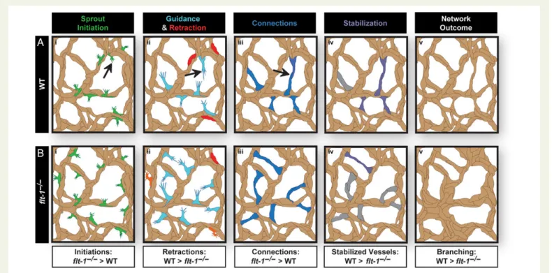
in vivo.29Endothelial cell behaviours were visualized using time-lapse confocal microscopy and ES cell lines that expressed enhanced green fluorescent protein (eGFP) driven by the vascular-specific platelet-endothelial cell adhesion molecule-1 (PECAM-1) promoter.28 Developing vessel networks derived from WT andflt-12/2ES cells were analysed during periods of robust angiogenesis. To facilitate this analysis, we defined discrete stages of branching morphogenesis that correlated with distinct endothelial cell behaviours—sprout initi-ation, sprout extension, sprout connection, and sprout stabilization (Figure1A, see Supplementary material online,Figure S1and Movie
S1). These stages were rigorously defined in this study and differ from a previously defined sprout index in several ways, including min-imum length, relation to parent vessel, and persistence.28We also followed individual sprout initiations and quantified subsequent move-ment through the stages to outcomes, which was not previously done. Some distinct behaviours, such as lumen formation, were not scored because our labelling strategy did not allow for unambiguous assess-ment of this parameter.
The rate of sprout initiations was significantly higher inflt-12/2ES cell-derived vessels than WT controls (Figure1B – D, see Supplemen-tary material online, MoviesS2 andS3), which is consistent with previ-ous observations of tip cell densities in fixed images.19New sprout
initiations are spatially and temporally coordinated via integration of several signalling pathways,19,30–32so we asked whether loss offlt-1
disrupted these signalling dynamics. We determined the space-time co-ordinates (x,y,t) of sprout initiations during live imaging of WT and
flt-12/2 vessel development (Figure2AandB). These coordinates were analysed to identify sprout initiations in close spatio-temporal proximity—specifically, a cluster was defined as a group of three or more initiations, separated by no more than300mm, and by no more than 4 h. We next calculated how many clusters would occur by chance in the absence of an underlying organizing principle using a Monte Carlo simulation (Figure2C). For each movie, we created 1000 sets of randomly generated spatio-temporal coordinates restricted by vessel geometry and movie duration and determined sprout initiation clusters using the criteria for actual initiations. Significantly, more clus-ters occurred in vessels derived from WT ES cells than expected based on chance, consistent with the concept of an underlying organizing principle, perhaps signalling-related, that regulates vessel sprout initia-tions in space and time. In contrast, sprout initiation clusters in vessels derived fromflt-12/2ES cells were no more prevalent than predicted by chance, suggesting that loss offlt-1disrupts the spatio-temporal co-ordination of blood vessel sprouting.
We next assessed sprout initiations and clustering in a different model of sprouting angiogenesis that allowed for greater temporal con-trol of signalling disruption. We used time-lapse confocal imaging to visualizeflk-1-eGFP+endothelial cells within developing blood vessels of explanted mouse embryonic skin (Embryonic Day 14.5). In thisex vivomodel of sprouting angiogenesis, we modulated VEGF-A signalling activity through application of excess VEGF-A ligand. Although the ab-solute values for sprout initiation rates and clustering differed slightly from those of ES cell-derived vessels, we found the same trends. Ex-ogenous VEGF-A caused a significant increase in sprout initiations while perturbing their spatial and temporal coordination (see Supple-mentary material online,Figure S2and MoviesS4 andS5). Taken to-gether, these data indicate that elevated VEGF-A signalling via excess ligand and genetic loss offlt-1both increase the rate of blood vessel sprout initiation and undermine the proper spatio-temporal orchestra-tion of angiogenic sprout initiaorchestra-tion, events that are predicted to in-crease the overall production of productive new branches. Because
these two models displayed similar outputs with regard to sprouting dynamics, we focused our remaining analysis on ES cell-derived blood vessels.
3.2 Blood vessel sprout retraction and
connection dynamics depend on Flt-1
activity
Because loss offlt-1undermines the guidance of blood vessel sprout,4 we reasoned that misguidedflt-12/2sprouts would retract more often and connect less often due to lack of appropriate guidance cues. To in-vestigate these hypotheses, we assessed sprout retractions and con-nections via live imaging from WT and flt-12/2 ES cell-derived vessels (Figure3A – D, see Supplementary material online, MoviesS6 –
S9). WT vessels had 32% of sprouts retract, which is consistent with the presence of collagen IV basement membrane ‘sleeves’ without ves-sels in areas of active angiogenesis.33To our surprise, only 9% of
flt-12/2vessel sprouts retracted, which was significantly less than con-trols (Figure3E – G). This result suggests that lack of Flt-1-mediated guidance allows some sprouts to avoid a normal retraction process. Moreover,flt-12/2sprouts also connected more frequently with other sprouts and vessels, as 56% of WT sprouts connected during the ob-servation period while 82% offlt-12/2vessel sprouts connected during similar observation periods (Figure3E – G), consistent within vivo obser-vations from zebrafish Flt-1 morphants.34The time between sprout ini-tiation and connection or retraction was similar between WT and
flt-12/2vessels (see Supplementary material online,Figure S3). These findings suggest that, although vessel sprouting could be time-limited in reaching an outcome due to spatial restrictions (e.g. connecting with a nearby vessel), an optimal temporal window for achieving a sprouting endpoint may exist. While this temporal regulation does not appear to require Flt-1 activity, a sprout intrinsic mechanism may control this timing. Thus, loss offlt-1prevents sprout retractions and promotes connections, which is predicted to increase vessel branching in the absence of other events.
We extended our analysis of the spatial and temporal clustering found in WT sprout initiations (Figure2) to determine if this phenom-enon had any relation to the sprouting outcome. WT sprout initiations
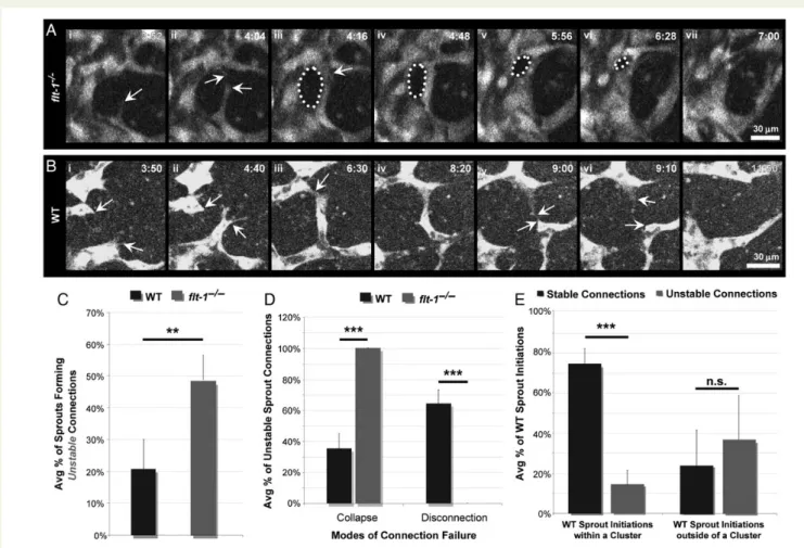
Figure 3 Flt-12/2vessel sprouts have decreased retractions and increased connections, and clustered WT sprout initiations connect more frequently. (A – D) Representative time-lapse images of developing vessel networks derived from WT andflt-12/2
ES cells expressing PECAM-eGFP in which an endothelial sprout (arrow,Ai – iiiandBi – iii) retracts into a parent vessel (AivandBiv), and two elongating sprouts (arrows,CiandDi) form a connection (arrow,Cii – iiiandDii – iii). Time (h:min), upper right. (E – G) Outcomes for each sprout initiation were assessed (WT:n¼88 from five movies,flt-12/2
:
that occurred within a cluster were approximately three times more likely to result in a connection rather than a retraction (Figure3H). However, WT sprout initiations originating outside of a cluster were just as likely to connect as they were to retract, suggesting that there are overlapping ‘hot spots’ of both sprout initiation and connection.
3.3 Flt-1 promotes the stability of new blood
vessel branches
Our system allows tracking of individual newly formed vessels over time to determine their ultimate fate.27This analysis showed that
some newly formed branches either collapsed into other vessels or broke the connection, so they ultimately did not contribute to overall branching complexity (Figure4AandB). In WT networks,20% of newly formed branches were unstable, whereas 45% of new branches formed by flt-12/2 vessels were unstable (Figure 4C).
Intriguingly, all unstableflt-12/2connections failed by vessel collapse, while unstable WT connections failed by both collapse and disconnec-tion, with disconnection being more predominant (Figure4D). These observations demonstrate that loss offlt-1destabilizes newly formed vessel connections by enhancing their probability of collapse into near-by vessels, and explains the reduction in new conduits inflt-12/2 mu-tant vessels despite increased sprout initiations and connections.
Given that analysis of WT sprout initiation clustering revealed a cor-relation between clustered initiations and sprouting outcomes (Figure3H), we asked a similar question about the relationship between sprout clustering and vessel branch stability. Clustered WT sprout in-itiations were significantly more likely to form stable connections, whereas sprout initiations outside of a cluster were just as likely to be stable as unstable (Figure4E), suggesting that constraints on sprout initiation proximity may also promote stability of nascent vessel connections.
Figure 4 Loss offlt-1destabilizes new branches via vessel collapse, and clustered WT sprout initiations have increased branch stability. (A) Represen-tative image sequences of PECAM-eGFP vessels derived fromflt-12/2ES cells. A new sprout (arrow,Ai) engages a target (top arrow,Aii) and forms a connection (arrow,Aiii). The gap area formed (dotted line,iii – vi) collapses over time (vii). (B) Representative time-lapse images of two WT vessel sprouts (top and bottom arrows,Bi – ii) that emerge and form a connection (arrow,Biii – iv) until cells within this nascent branch disconnect (Bv) and retract (Bvi – vii). Time (h:min), upper right. (C) New connections formed by WT (n¼49 from five movies) orflt-12/2(n¼107 from six movies) vessel sprouts were evaluated for stability and ultimate contribution to branching complexity, and percentages were averaged for each movie. Values are averages+SEM from five to six movies of each genotype. **P≤0.01 vs. WT usingt-test comparison. (D) The mode of failure (i.e. collapse or disconnection) for each
3.4 Flt-1 maintains vessel branches by
restricting endothelial cell movement
into avascular regions
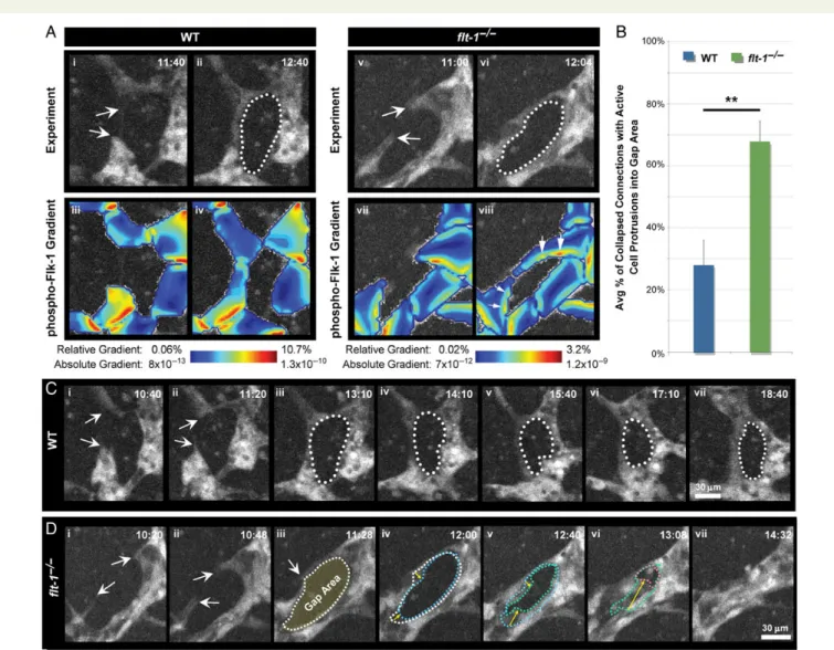
Loss offlt-1spatially alters VEGF-A signalling through the Flk-1 recep-tor.15,18Therefore, we hypothesized that newly formed branches used Flt-1 to spatially modulate VEGF-A activity and promote stabilization of new connections. We coupled live imaging observations with compu-tational modelling of VEGF-A signalling, using still-frames from movies with emerging sprouts that connected with other vessels to form new branches.18
We reconstructed the cellular architecture of each image in the simulation (Figure5Ai – iiandAv – vi), then used this architecture as the boundary condition for partial differential equations that govern diffusion of soluble molecules and the kinetics of VEGF-A ligand – receptor binding events.18The simulations showed that after sprout
fusion, overall Flk-1 signalling is higher inflt-12/2vessels than WT; in addition, the gradient in Flk-1 signalling is normally low in vessels next to avascular gaps but is elevated inflt-12/2vessels with this top-ology (Figure5Aiii – ivandAvii – viii, arrows).Immunostaining of Flk-1 in presumably recent vessel connections is consistent with these results, showing Flk-1 localization along a single side of a WT connection but encircling the entire avascular gap of aflt-12/2connection (see Supplementary material online,Figure S4). The finding that Flk-1 is mis-localized and levels increased suggests that Flt-1 influences both the levels and the spatial distribution of activated Flk-1 bound to VEGF-A. Taken together, the modelling suggests that once endothelial cells connect and form a new branch, Flt-1 dampens Flk-1 signalling at the edge of avascular zones, reducing the likelihood of collapse, and thus promoting new conduits.
Figure 5 Flt-12/2endothelial cells persistently protrude between branches. (A) Representative image sequences of vessel sprouts derived from PECAM-eGFP WT andflt-12/2ES cells as they extend (arrows,AiandAv, respectively), connect, and form gap areas (dotted lines,AiiandAvi). Time (h:min), upper right. The images provided the model geometry for partial differential equation-based simulations of pFlk-1 levels and gradients (Aiii – ivand
Avii – viii), colour-coded as indicated. Arrows, increased pFlk-1 gradients (Aviii). (B) Areas between branches scored for active endothelial cell protrusions in experimental images from WT (n¼20 from five movies) andflt-12/2(n¼50 from six movies) vessels. Percentage of collapsed connections with cell protrusions were calculated for each movie and then averaged. Values are averages+SEM from five to six movies of each genotype. **P≤0.01 vs. WT usingt-test comparison. (C) Example of stable connection and persistent gap area from vessel sprouts derived from WT ES cells (arrows,Ci – ii; dotted line,Ciii – vii). (D) Example of vessel sprouts derived fromflt-12/2ES cells (arrows,Di – ii) forming a connection and avascular gap (arrow and dotted line,
These observations led us to hypothesize that loss offlt-1increases protrusive endothelial cell activity into avascular areas, due to the ele-vated Flk-1 signalling predicted by the modelling. We examined WT andflt-12/2 ES cell-derived vessel networks where new branches had formed and asked whether subsequent collapse was correlated with persistent protrusive activity into the avascular region. In
flt-12/2vessels, 76% of regions in new branches had continuously pro-trusive endothelial cells (Figure5BandD; see Supplementary material online, MovieS10). In contrast, newly formed branches in WT vessels were often maintained (Figure5C), but when they did collapse, signifi-cantly fewer (35%) had persistently protrusive and migratory endothe-lial cells (Figure5B; see Supplementary material online, MovieS11). These data suggest thatflt-1loss promotes vessel collapse and overall reduction of new conduits by limiting persistent endothelial cell protru-sive movement into avascular regions.
4. Discussion
Most defects in blood vessel formation are defined by the final pheno-type using static images of fixed tissues. Here, we used live imaging to better understand the dynamic endothelial cell behaviours that con-tribute to vessel network formation, and we analysed the complex pro-cess of sprouting angiogenesis with respect to elemental behaviours or stages (Figure1A). These angiogenic stages are differentially affected by disruption of VEGF-A signalling through excess VEGF-A ligand or gen-etic deletion offlt-1, and an apparent paradox in increased sprout initia-tions vs. reduced numbers of new conduits inflt-12/2mutant and
VEGF-A-saturated blood vessels is explained. This dissection of stage-specific effects also suggests novel points of therapeutic intervention; for example, if the collapse of a new branch were prevented by spatial down-regulation of VEGF-A signalling in vessels surrounding avascular gaps, the overall effect of increased VEGF-A signalling in this and related vascular beds might be to increase new conduits.
Although loss offlt-1leads to an increase in tip cell formation19,35 and sprout initiations (this work), the number of new conduits formed inflt-12/2networks is reduced. Fonget al.showed that new conduit formation is significantly impaired in developing mice lackingflt-1 func-tion,17but the cellular basis for this phenotype was unclear. Tumours and wounds subjected to Notch signalling manipulations also have in-creased tip cells but fewer new conduits.22–24In the absence of live im-aging, the exact cell behaviours that are affected by these perturbations are unclear. Our live image analysis offlt-12/2mutant vessels suggests that multiple stages of angiogenesis are affected by loss offlt-1, but that reduced stabilization of new connections is dominant over effects on other stages such as sprout initiations and connections, leading to over-all reduction of new conduits (Figure6).
Our data suggest that blood vessel sprout initiations are regulated in space and time. This regulation may affect whether developing vessels contribute positively to formation of new conduits, since ectopic VEGF-A or loss offlt-1randomized the location of initiations, perhaps leading to connections in inappropriate locations that contributed to the high rate of collapse offlt-12/2mutant branches. Further, support-ing this idea is our findsupport-ing that clustered WT sprout initiations more of-ten resulted in stable connections than did sprout initiations outside of
clusters. Local cues that promote sprout clustering may also enhance sprout connection and branch stability, and perhaps sprouts connect-ing directly with one another yield more stable branches when com-pared with those fusing with an existing vessel segment. These observations are in agreement with previous studies showing that VEGF signalling is involved in spatial regulation of vascular branching morphogenesis, likely via integration with Notch signalling.19,30During
Drosophilatrachea development, branch formation is similarly spatially coordinated by fibroblast growth factor signalling that is modulated by Notch,32demonstrating that mechanisms for patterning new conduits are conserved across tissues and organisms.31Recent work suggests that, within a local area, the precise location of a vascular sprout initi-ation also depends on flow parameters,36suggesting that mechanical cues integrate with molecular signalling cues in flow-dependent mod-els. Our results highlight that tight spatio-temporal regulation of vessel sprout initiations is likely important in the formation of appropriately branched vascular networks.
Angiogenesis studies that include time-lapse analysis of blood vessel formation often focus on sprouting in stereotyped vascular beds such as zebrafish intersegmental vessels, where retraction has not been well described.37,38We show that in WT mammalian vessels, sprout initia-tions sometimes fully retract into the parent vessel. These dynamic be-haviours are consistent with reports of collagen IV ‘sleeves’ of basement membrane that remain in the absence of vessels, presumably following capillary sprout retraction or remodelling.39,40This retraction mode of network formation is also important for axonal branching and patterning, particularly during wound healing following spinal cord in-jury.41Loss offlt-1decreases the likelihood of angiogenic sprout retrac-tion and increases the connecretrac-tion rate. This is likely due to tip cells experiencing excess VEGF-A signalling, which may in turn inappropri-ately reinforce sprout extensions and connections. Thus, when VEGF-A signalling is mis-regulated, such as in solid tumours, vascular dysmorphogenesis is likely exacerbated by decreased sprout retraction and increased sprout connections.
A surprising finding was that the stability of newly formed branches is a critical stage of blood vessel development compromised by genetic loss offlt-1. Computational simulations of new branches with and with-out Flt-1 activity predicted higher levels of Flk-1 activation in areas near gaps between mutant sprouts, and this correlated with increased endo-thelial cell protrusive activity, a behaviour consistent with hyperactive Flk-1 signalling.42Previousin vivostudies in quail embryos showed that high VEGF-A reduces branching complexity by promoting vessel co-alescence,43which we also observe in the developing zebrafish liver plexus exposed to excess Vegfaa (see Supplementary material online,
Figure S5). More recently, it was shown thatin vivoblood flow through mouse yolk sac vessels also modulates branch maintenance and vessel fusion,44which represents another mechanism for further re-finement of a developing vessel plexus. In non-stereotypical vascular networks, the location of sprout connections may also be important in determining which connections are maintained and which are lost. Flt-1-mediated modulation of VEGF signalling is thus essential to regu-late sprout initiation and guidance,4and also stability of new branches, likely by regulating spatial aspects of Flk-1 signalling after a connection is formed.
Defining distinct stages of sprouting angiogenesis via live imaging shows how loss offlt-1elevates sprout initiations and connections yet ultimately leads to reduced branching complexity in our model, which mimics developmental angiogenesis in unrestrained vascular beds such as the yolk sac.19,22–24,26The finding of differential effects
offlt-1loss on distinct stages of angiogenesis also provides an explan-ation for different phenotypes associated withflt-1loss in different vas-cular beds. For example, Hoet al. recently showed that conditional loss offlt-1led to increased vascular branching in the developing mouse retina;45this observation contrasts withflt-1global knockout and VEGF-A gain-of-function phenotypes described in other tissues such as the embryonic yolk sac and mouse skeletal muscle.17,43,46We pro-pose that the collapse of new branches so prevalent in our system is prevented in the retina by the astrocyte network that stabilizes new branches independent of Flt-1, making dominant the effects offlt-1
loss on earlier angiogenic stages. In this way, a better understanding of how particular pathways affect distinct dynamic stages of blood ves-sel formation will improve predictions of therapeutic efficacy when treating a given vascular bed.
Supplementary material
Supplementary material is available atCardiovascular Researchonline.
Acknowledgements
The authors thank the Bautch Lab members for stimulating discussion during manuscript preparation.
Conflict of interest: none declared.
Funding
This work was supported by the National Institutes of Health (R01HL43174 to V.L.B.; F32HL95359 and K99HL105779 to J.C.C.; and R00HL093219 to F.M.G.), the American Heart Association (12BGIA12060154 to F.M.G.), and a Sloan Research Fellowship (to F.M.G.).
References
1. Bentley K, Franco CA, Philippides A, Blanco R, Dierkes M, Gebala V, Stanchi F, Jones M, Aspalter IM, Cagna G, Westrom S, Claesson-Welsh L, Vestweber D, Gerhardt H. The role of differential VE-cadherin dynamics in cell rearrangement during angiogenesis.Nat Cell Biol2014;16:309 – 321.
2. Siekmann AF, Affolter M, Belting HG. The tip cell concept 10 years after: new players tune in for a common theme.Exp Cell Res2013;319:1255 – 1263.
3. Logsdon EA, Finley SD, Popel AS, Mac Gabhann F. A systems biology view of blood ves-sel growth and remodelling.J Cell Mol Med2014;18:1491 – 1508.
4. Chappell JC, Taylor SM, Ferrara N, Bautch VL. Local guidance of emerging vessel sprouts requires soluble Flt-1.Dev Cell2009;17:377 – 386.
5. Lenard A, Ellertsdottir E, Herwig L, Krudewig A, Sauteur L, Belting HG, Affolter M. In vivo analysis reveals a highly stereotypic morphogenetic pathway of vascular anasto-mosis.Dev Cell2013;25:492 – 506.
6. Xu K, Cleaver O. Tubulogenesis during blood vessel formation.Semin Cell Dev Biol
2011;22:993 – 1004.
7. Dorrell MI, Friedlander M. Mechanisms of endothelial cell guidance and vascular pat-terning in the developing mouse retina.Prog Retin Eye Res2006;25:277 – 295. 8. Vieira JM, Ruhrberg C, Schwarz Q. VEGF receptor signaling in vertebrate development.
Organogenesis2010;6:97 – 106.
9. Potente M, Gerhardt H, Carmeliet P. Basic and therapeutic aspects of angiogenesis.Cell
2011;146:873 – 887.
10. Luttun A, Tjwa M, Moons L, Wu Y, Angelillo-Scherrer A, Liao F, Nagy JA, Hooper A, Priller J, De Klerck B, Compernolle V, Daci E, Bohlen P, Dewerchin M, Herbert JM, Fava R, Matthys P, Carmeliet G, Collen D, Dvorak HF, Hicklin DJ, Carmeliet P. Revas-cularization of ischemic tissues by PlGF treatment, and inhibition of tumor angiogenesis, arthritis and atherosclerosis by anti-Flt1.Nat Med2002;8:831 – 840.
11. He Y, Smith SK, Day KA, Clark DE, Licence DR, Charnock-Jones DS. Alternative spli-cing of vascular endothelial growth factor (VEGF)-R1 (FLT-1) pre-mRNA is important for the regulation of VEGF activity.Mol Endocrinol1999;13:537 – 545.
12. Waltenberger J, Claesson-Welsh L, Siegbahn A, Shibuya M, Heldin CH. Different signal transduction properties of KDR and Flt1, two receptors for vascular endothelial growth factor.J Biol Chem1994;269:26988 – 26995.
14. Hiratsuka S, Minowa O, Kuno J, Noda T, Shibuya M. Flt-1 lacking the tyrosine kinase domain is sufficient for normal development and angiogenesis in mice.Proc Natl Acad Sci USA1998;95:9349 – 9354.
15. Kappas NC, Zeng G, Chappell JC, Kearney JB, Hazarika S, Kallianos KG, Patterson C, Annex BH, Bautch VL. The VEGF receptor Flt-1 spatially modulates Flk-1 signaling and blood vessel branching.J Cell Biol2008;181:847 – 858.
16. Roberts DM, Kearney JB, Johnson JH, Rosenberg MP, Kumar R, Bautch VL. The vascular endothelial growth factor (VEGF) receptor Flt-1 (VEGFR-1) modulates Flk-1 (VEGFR-2) signaling during blood vessel formation.Am J Pathol2004;164:1531 – 1535. 17. Fong GH, Rossant J, Gertsenstein M, Breitman ML. Role of the Flt-1 receptor tyrosine kinase in regulating the assembly of vascular endothelium.Nature1995;376:66 – 70. 18. Hashambhoy YL, Chappell JC, Peirce SM, Bautch VL, Mac Gabhann F. Computational
modeling of interacting VEGF and soluble VEGF receptor concentration gradients.
Front Physiol2011;2:62.
19. Chappell JC, Mouillesseaux KP, Bautch VL. Flt-1 (vascular endothelial growth factor receptor-1) is essential for the vascular endothelial growth factor-Notch feedback loop during angiogenesis.Arterioscler Thromb Vasc Biol2013;33:1952 – 1959. 20. Wang Y, Nakayama M, Pitulescu ME, Schmidt TS, Bochenek ML, Sakakibara A, Adams S,
Davy A, Deutsch U, Luthi U, Barberis A, Benjamin LE, Makinen T, Nobes CD, Adams RH. Ephrin-B2 controls VEGF-induced angiogenesis and lymphangiogenesis.
Nature2010;465:483 – 486.
21. Jung B, Obinata H, Galvani S, Mendelson K, Ding BS, Skoura A, Kinzel B, Brinkmann V, Rafii S, Evans T, Hla T. Flow-regulated endothelial S1P receptor-1 signaling sustains vas-cular development.Dev Cell2012;23:600 – 610.
22. Ridgway J, Zhang G, Wu Y, Stawicki S, Liang WC, Chanthery Y, Kowalski J, Watts RJ, Callahan C, Kasman I, Singh M, Chien M, Tan C, Hongo JA, de Sauvage F, Plowman G, Yan M. Inhibition of Dll4 signalling inhibits tumour growth by deregulating angiogenesis.
Nature2006;444:1083 – 1087.
23. Noguera-Troise I, Daly C, Papadopoulos NJ, Coetzee S, Boland P, Gale NW, Lin HC, Yancopoulos GD, Thurston G. Blockade of Dll4 inhibits tumour growth by promoting non-productive angiogenesis.Nature2006;444:1032 – 1037.
24. Al Haj Zen A, Oikawa A, Bazan-Peregrino M, Meloni M, Emanueli C, Madeddu P. Inhib-ition of delta-like-4-mediated signaling impairs reparative angiogenesis after ischemia.
Circ Res2010;107:283 – 293.
25. Li JL, Sainson RC, Shi W, Leek R, Harrington LS, Preusser M, Biswas S, Turley H, Heikamp E, Hainfellner JA, Harris AL. Delta-like 4 Notch ligand regulates tumor angio-genesis, improves tumor vascular function, and promotes tumor growth in vivo.Cancer Res2007;67:11244 – 11253.
26. Pasula S, Cai X, Dong Y, Messa M, McManus J, Chang B, Liu X, Zhu H, Mansat RS, Yoon SJ, Hahn S, Keeling J, Saunders D, Ko G, Knight J, Newton G, Luscinskas F, Sun X, Towner R, Lupu F, Xia L, Cremona O, De Camilli P, Min W, Chen H. Endothelial epsin deficiency decreases tumor growth by enhancing VEGF signaling.J Clin Invest
2012;122:4424 – 4438.
27. Kearney JB, Bautch VL. In vitro differentiation of mouse ES cells: hematopoietic and vas-cular development.Methods Enzymol2003;365:83 – 98.
28. Kearney JB, Kappas NC, Ellerstrom C, DiPaola FW, Bautch VL. The VEGF receptor flt-1 (VEGFR-1) is a positive modulator of vascular sprout formation and branching mor-phogenesis.Blood2004;103:4527 – 4535.
29. Larina IV, Shen W, Kelly OG, Hadjantonakis AK, Baron MH, Dickinson ME. A mem-brane associated mCherry fluorescent reporter line for studying vascular remodeling
and cardiac function during murine embryonic development.Anat Rec (Hoboken)2009; 292:333 – 341.
30. Jakobsson L, Bentley K, Gerhardt H. VEGFRs and Notch: a dynamic collaboration in vascular patterning.Biochem Soc Trans2009;37:1233 – 1236.
31. Affolter M, Zeller R, Caussinus E. Tissue remodelling through branching morphogen-esis.Nat Rev Mol Cell Biol2009;10:831 – 842.
32. Ghabrial AS, Krasnow MA. Social interactions among epithelial cells during tracheal branching morphogenesis.Nature2006;441:746 – 749.
33. Mancuso MR, Davis R, Norberg SM, O’Brien S, Sennino B, Nakahara T, Yao VJ, Inai T, Brooks P, Freimark B, Shalinsky DR, Hu-Lowe DD, McDonald DM. Rapid vascular re-growth in tumors after reversal of VEGF inhibition.J Clin Invest2006;116:2610 – 2621. 34. Krueger J, Liu D, Scholz K, Zimmer A, Shi Y, Klein C, Siekmann A, Schulte-Merker S, Cudmore M, Ahmed A, le Noble F. Flt1 acts as a negative regulator of tip cell formation and branching morphogenesis in the zebrafish embryo.Development2011;138: 2111 – 2120.
35. Jakobsson L, Franco CA, Bentley K, Collins RT, Ponsioen B, Aspalter IM, Rosewell I, Busse M, Thurston G, Medvinsky A, Schulte-Merker S, Gerhardt H. Endothelial cells dynamically compete for the tip cell position during angiogenic sprouting.Nat Cell Biol2010;12:943 – 953.
36. Ghaffari S, Leask RL, Jones EA. Flow dynamics control the location of sprouting and dir-ect elongation during developmental angiogenesis.Development2015;142:4151 – 4157. 37. Blum Y, Belting HG, Ellertsdottir E, Herwig L, Luders F, Affolter M. Complex cell rear-rangements during intersegmental vessel sprouting and vessel fusion in the zebrafish embryo.Dev Biol2008;316:312 – 322.
38. Kamei M, Isogai S, Pan W, Weinstein BM. Imaging blood vessels in the zebrafish. Meth-ods Cell Biol2010;100:27 – 54.
39. Phng LK, Potente M, Leslie JD, Babbage J, Nyqvist D, Lobov I, Ondr JK, Rao S, Lang RA, Thurston G, Gerhardt H. Nrarp coordinates endothelial Notch and Wnt signaling to control vessel density in angiogenesis.Dev Cell2009;16:70 – 82.
40. Ehling M, Adams S, Benedito R, Adams RH. Notch controls retinal blood vessel mat-uration and quiescence.Development2013;140:3051 – 3061.
41. Dray C, Rougon G, Debarbieux F. Quantitative analysis by in vivo imaging of the dynam-ics of vascular and axonal networks in injured mouse spinal cord.Proc Natl Acad Sci USA
2009;106:9459 – 9464.
42. Gerhardt H, Golding M, Fruttiger M, Ruhrberg C, Lundkvist A, Abramsson A, Jeltsch M, Mitchell C, Alitalo K, Shima D, Betsholtz C. VEGF guides angiogenic sprouting utilizing endothelial tip cell filopodia.J Cell Biol2003;161:1163 – 1177.
43. Drake CJ, Little CD. Exogenous vascular endothelial growth factor induces malformed and hyperfused vessels during embryonic neovascularization.Proc Natl Acad Sci USA
1995;92:7657 – 7661.
44. Udan RS, Vadakkan TJ, Dickinson ME. Dynamic responses of endothelial cells to changes in blood flow during vascular remodeling of the mouse yolk sac.Development
2013;140:4041 – 4050.
45. Ho VC, Duan LJ, Cronin C, Liang BT, Fong GH. Elevated vascular endothelial growth factor receptor-2 abundance contributes to increased angiogenesis in vascular endo-thelial growth factor receptor-1-deficient mice.Circulation2012;126:741 – 752. 46. Ozawa CR, Banfi A, Glazer NL, Thurston G, Springer ML, Kraft PE, McDonald DM,