MBoC
|
ARTICLE
A theoretical model of cytokinesis implicates
feedback between membrane curvature and
cytoskeletal organization in asymmetric
cytokinetic furrowing
ABSTRACT During cytokinesis, the cell undergoes a dramatic shape change as it divides into two daughter cells. Cell shape changes in cytokinesis are driven by a cortical ring rich in actin filaments and nonmuscle myosin II. The ring closes via actomyosin contraction coupled with actin depolymerization. Of interest, ring closure and hence the furrow ingression are noncon-centric (asymmetric) within the division plane across Metazoa. This nonconnoncon-centricity can occur and persist even without preexisting asymmetric cues, such as spindle placement or cellular adhesions. Cell-autonomous asymmetry is not explained by current models. We combined quantitative high-resolution live-cell microscopy with theoretical modeling to explore the mechanistic basis for asymmetric cytokinesis in the Caenorhabditis elegans zygote, with the goal of uncovering basic principles of ring closure. Our theoretical model suggests that feed-back among membrane curvature, cytoskeletal alignment, and contractility is responsible for asymmetric cytokinetic furrowing. It also accurately predicts experimental perturbations of conserved ring proteins. The model further suggests that curvature-mediated filament align-ment speeds up furrow closure while promoting energy efficiency. Collectively our work un-derscores the importance of membrane–cytoskeletal anchoring and suggests conserved molecular mechanisms for this activity.
INTRODUCTION
Cytokinesis is the physical division of one cell into two. Cell shape changes during this process are accomplished by a temporary cy-toskeletal structure known as the contractile ring, which assembles
at the cell equator in anaphase (Schroeder, 1972, 1973; Mabuchi and Okuno, 1977; Rappaport, 1996; reviewed by Green et al., 2012). The contractile ring is a specialization of the cortical acto-myosin cytoskeleton, which occupies a thin volume beneath the plasma membrane (Schroeder, 1972; Clark et al., 2013). Key com-ponents of this ring include the multidomain scaffold protein anil-lin, the filament-forming septins, minifilaments of nonmuscle myo-sin II, and formin-dependent, long, unbranched actin filaments (F-actin). Actin filaments are randomly oriented at the equator at the onset of cytokinesis but become circumferentially aligned (Fish-kind and Wang, 1993; Noguchi and Mabuchi, 2001; Vavylonis et al., 2008). Actomyosin contraction coupled to actin depolymer-ization in the plane of the membrane produces contractile force tangential to the membrane, comprising small radial forces that deform the cell cortex into a furrow that ingresses to divide the cell (Schroeder, 1972).
Monitoring Editor William Bement University of Wisconsin
Received: Jun 15, 2015 Revised: Feb 11, 2016 Accepted: Feb 16, 2016
This article was published online ahead of print in MBoC in Press (http://www .molbiolcell.org/cgi/doi/10.1091/mbc.E15-06-0374) on February 24, 2016.
†Present address: Novartis Pharma AG, 4056 Basel, Switzerland.
The authors claim no conflicts of interest.
*Address correspondence to: Jian Liu ([email protected]), Amy Shaub Maddox ([email protected]).
© 2016 Dorn et al. This article is distributed by The American Society for Cell Biol-ogy under license from the author(s). Two months after publication it is available to the public under an Attribution–Noncommercial–Share Alike 3.0 Unported Creative Commons License (http://creativecommons.org/licenses/by-nc-sa/3.0). “ASCB®,” “The American Society for Cell Biology®,” and “Molecular Biology of
the Cell®” are registered trademarks of The American Society for Cell Biology.
Abbreviations used: F-actin, filamentous actin; myosin II, nonmuscle myosin II.
Jonas F. Dorna,†, Li Zhanga, Tan-Trao Phia, Benjamin Lacroixb, Paul S. Maddoxc, Jian Liud,*,
and Amy Shaub Maddoxc,*
Here we combine theoretical modeling and experiments to test this idea. Our model predicts that curvature-mediated cytoskeletal or-ganization is essential to recapitulate the observed asymmetric furrow ingression; this prediction is corroborated by experimental perturba-tions of conserved contractile ring components. The model further suggests functional roles of these proteins and indicates that the asym-metry of furrowing trades off with speed to achieve a maximum energy efficiency of cytokinesis in different cell types. Thus our model encom-passes a concept that can explain asymmetric furrowing, describes the mechanics of the contractile ring, and begins to assign molecular iden-tity to these activities. Of importance, we propose a novel contributor to contractile ring mechanics: the influence of local membrane curva-ture on cytoskeletal organization via filament–membrane attachment.
RESULTS
Model development
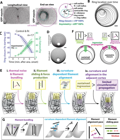
Here we present a theoretical model that aims to explain the sus-tained asymmetry of contractile ring closure in the C. elegans zygote. The model describes the mechanical contraction of the contractile ring and its coupling with the furrowing plasma membrane. The cen-tral notion of the model is that membrane curvature in the furrow af-fects the alignment of the cytoskeletal filaments in the contractile ring, which in turn governs the contractility that further drives furrow-ing (Figure 1A). We propose that this curvature-mediated feedback loop perpetuates the symmetry breaking arising from random fluc-tuations and underlies asymmetric furrow ingression (Figure 1B).
The model describes the cytokinetic furrow ingression without explicit representation of individual molecules. To discern the mech-anism underlying asymmetric furrowing, the model comprehensively integrates key components, including the cortical cytoskeleton, the membrane, and the spindle midzone. It builds upon the energy aris-ing from interactions among these key players. We first depict the model qualitatively and then formulate the quantitative model.
Although strict sarcomeric organization has not been observed in cytokinetic contractile rings, several lines of evidence support the idea that the contractile ring comprises a collection of “contractile units” (Bement and Capco, 1991; Carvalho et al., 2009; Pollard, 2010). Motivated by this evidence, we modeled the contractile ring as a circle of connected contractile segments bound to the interior of a membrane tubule, which is 50 μm long with a diameter of 30 μm (Figure 1C), similar to the dimensions of the C. elegans zy-gote. Within each segment, an ensemble of myosin II bipolar mini-filaments spans two groups of actin mini-filaments (Burns et al., 1995a,b; Yumura et al., 2008; Zhou and Wang, 2008; Vale et al., 2009), which occupy the two circumferential ends of each segment (adjacent to adjoining segments). The external, barbed ends of the actin fila-ments can be thought of as connected to the neighboring segment by actin cross-linkers, which are represented by the spring-like con-nections between the segments. Thus the ring segments resemble the sarcomeres of muscle or stress fibers. However, unlike in sarco-meres, the actin filaments within each ring segment begin with ran-dom orientation (not aligned) and align progressively with time and as the cytokinetic furrow ingresses (Figure 1C).
The model posits that actin filament alignment within each ring segment arises from two sources. First, we propose that the local membrane curvature favors the circumferential orientation of actin filaments (Figure 1A). This is because when the membrane is flat, filaments can fully attach to the membrane in any orientation (Mukhina et al., 2007; Reichl et al., 2008; Skau et al., 2011), whereas the maximization of filament-membrane binding favors circumferential filament alignment with the furrow when the cortex is curved. We termed this phenomenon curvature-dependent Furrowing is nonconcentric (asymmetric) in cells throughout
Metazoa (Reinsch and Karsenti, 1994; Rappaport, 1996; Das et al., 2003; Alsop and Zhang, 2004; Fleming et al., 2007; Maddox et al., 2007; Kosodo et al., 2008; Carvalho et al., 2009; Bourdages et al., 2014). In most cases, there exists a mechanical asymmetry within the division plane, including intercellular junctions and sub-strate adhesions, which precedes anaphase. Such features resist fur-rowing forces and are thus sufficient for asymmetric furfur-rowing (Founounou et al., 2013; Guillot and Lecuit, 2013; Herszterg et al., 2013; Morais-de-Sa and Sunkel, 2013). However, the asymmetry of ring closure can be cell autonomous, as in the Caenorhabditis ele-gans zygote (Audhya et al., 2005; Maddox et al., 2007). How does this asymmetry arise? Initially, a shallow furrow appears relatively synchronously around the division plane (Maddox et al., 2007). As cytokinesis proceeds, the furrow adopts a sharp leading edge at a singular site (Maddox et al., 2007). This feature is perpetuated such that furrowing occurs unilaterally for some time, and the center of the contractile ring is increasingly offset from the center of the divi-sion plane (Maddox et al., 2007). The cortex on the opposite side of the division plane (180° from the initial doubled-membrane feature) adopts this sharp curvature only later.
What is the physical mechanism that leads to asymmetric furrow-ing? Several theoretical models have been used to study the me-chanics of cytokinetic ring contraction (Biron et al., 2005; Wang, 2005; Zumdieck et al., 2007; Koyama et al., 2012; Mendes Pinto et al., 2012; Stachowiak et al., 2014; Turlier et al., 2014), and one describes the mechanical differences around the division plane cir-cumference when asymmetry is imposed (Sain et al., 2015). Still, none explains how cell-autonomous asymmetric closure, such as that in the C. elegans zygote, arises and persists. The prominent accumulation of ring components on the faster-ingressing side of the furrow was suggested to contribute to asymmetry (Maddox et al., 2007). However, diffusion is predicted to smooth out spatial gradients of protein distribution over the several-minute time course of furrow ingression, and thus this factor is likely insufficient to per-petuate asymmetric furrowing. It therefore remains an open ques-tion whether asymmetric furrowing causes or results from localized protein accumulation. Another potential player is the anaphase spindle midzone, which comprises bundled antiparallel microtu-bules between the segregated chromosomes, contributes to divi-sion plane specification, and concentrates many positive regulators of cytokinesis (Douglas and Mishima, 2010). Asymmetric placement of the midzone within the division plane could lead to asymmetric contact with the contractile ring and thus stabilize or amplify asym-metry. However, instead of promoting asymmetry, the midzone was implicated in attenuating asymmetry, since asymmetry decreases when the furrow contacts it (Audhya et al., 2005).
filament alignment. In addition, F-actin cross-linkers bundle filaments and promote their alignment regardless of the local membrane shape (Mukhina et al., 2007; Reichl et al., 2008; Skau et al., 2011). We further assume that filament alignment fa-cilitates filament sliding and, hence, the contractility of each ring segment. This hy-pothesis is in part supported by the obser-vation that antiparallel filament orientation potentiates contraction (Reymann et al., 2010). Consequently, contractility coupled with actin depolymerization drives seg-ment shortening and ring closure (Schro-eder, 1972; Medeiros et al., 2006; Haviv et al., 2008; Carvalho et al., 2009; Wilson et al., 2010; Mendes Pinto et al., 2012). This shortening and the associated inward movement of ring segments further drive the local membrane shape change, thus closing the curvature-mediated positive feedback loop (Figure 1B). On the other hand, aligned, cross-linked F-actin (i.e., an F-actin bundle) is much stiffer than an iso-tropic meshwork (Gardel et al., 2004; Shin et al., 2004; Claessens et al., 2006). The progressive filament alignment thus also increases the bending resistance of the contractile ring segment, thus opposing the local contractility. Although contractile ring ingression is also opposed by mem-brane resistance and a resistive force repre-senting the spindle midzone, both of these factors are of relatively low magnitude compared with that of ring rigidity (see model parameter consideration in the Sup-plemental Material). Thus furrow ingression consists of five tightly coupled subpro-cesses: filament alignment, sliding, and de-polymerization, the displacement of the spindle midzone upon contact with the ring, and membrane shape changes.
Formulation of the quantitative model
We first construct the total energy function of the contractile apparatus F, which sums the mechanical energy of the system and free energy changes arising from the order-ing of the F-actin within each rorder-ing segment:
F F F
F
Membrane mechanics Ring mechanics Mechanical energy
Phase ordering Free energy from filament alignment
= +
+ (1)
The mechanical energy includes mem-brane mechanics, the mechanics of the con-tractile ring, and the interaction between the contractile ring and the midzone spindle. We consider that filament alignment within the contractile ring is a phase transition pro-cess. The associated free energy change is FIGURE 1: Schematics of a theoretical model featuring membrane curvature–mediated
alignment order parameter ψ. First, the term a0 represents the
energetically favorable filament bundling driven by F-actin cross-linkers; a0 is a negative constant, driving filament alignment
re-gardless of local membrane shape. Second, the term ACG specifi-cally drives filament alignment via local Gaussian curvature CG
(Figure 1A), where
CG=g3−2
(
x yss s−x ys ss)(
x yzz s−x ys zz) (
− x ysz s−x ys sz)
2A represents the effective filament–membrane binding energy, which is positive (see Sections I and III in the Supplemental Material for detailed derivation). At the local furrow (negative local Gaussian curvature), the term ACG turns negative, and thus curvature-depen-dent filament alignment is favorable (Figure 1A). In addition, the term D in the phase ordering free energy represents the energy pen-alty for spatial variation in ψ, thus promoting the uniform ordering in filament alignment along the ring perimeter. The fourth-order term is positive and an entropy term that favors disorder in filament alignment.
modeled as a continuous phase transition and described by the “φ–4 theory,” which is used to characterize phase transitions in soft condensed matter such as liquid crystals (Landau and Lifshitz, 1980). We construct our model in a discrete form in order to faithfully rep-resent actomyosin contractility (see Section I in the Supplemental Material for detailed reasons and Table 1 for term definitions). Later we will elaborate on each energy contribution.
Equation 2 describes the membrane mechanical energy, which is Helfrich-like membrane energy in its continuum limit, and includes contributions of bending energy, surface tension, and osmotic pres-sure. Throughout the model, a cylindrical coordinate system is used. The membrane tubule is divided into Nz + 1 parts along the z-axis (the pole-to-pole axis) and Ns segments in the radial direc-tion, where s stands for the sth membrane patch in the radial direc-tion (Figure 1C). Under this coordinate system, the posidirec-tion of the membrane patch r s z t
(
, ,)
=(
x s z t y s z t(
, , ,) (
, ,)
)
is represented as a function of s, z, and time t. From the differential geometry, themean curvature of the local membrane is C
g1 x y x y
2
M ss s s ss
3
1.5
(
)
= −
x y x y x y x y x x y y x y x y
1 z2 z2 s2 s2 zz s s zz 2 s z s z sz s s sz
(
+ +) (
+ +)
(
−) (
− +)(
−)
Here g s z t3
(
, ,)
=x2s+ys2+(
x ys z−x yz s)
2. Finally, all of the expressions of derivatives are in discrete form; for example, the partial derivative of x with regard to s is xs=12(
x s(
+ −1) (
x s−1)
)
.We have
F g s z t, , C 12 x y xy P
s Ns
z Nz Nz
M s s
Membranemechanics 3 1 2 2
1 2
Bendingenergy from meancurvature Surface tension
0 Osmoticpressure
∑
∑
(
)
(
)
= κ + σ + −
= =−
(2)
F
f s t l s t r s t r s t r s t f s t l s t f s t l s t
K g s t l s t g s t C K r s t r t
1
2 , , 1, 1, , 1, 1, 1, 1,
1
2 , , 12 , 12 ,
s Ns
s Ns
R m
Ring mechanics Contraction
1
1 2
2
Ring segment connectivity 1
3 2 2
Ring bending energy
2 2
Interaction between ring and midzone spindle
∑
∑
{
(
)
}
(
)
(
)
(
)
( ) ( ) (
) (
)
( )
(
) (
)
(
) (
)
( ) ( )
( )
( )
( )
= ⋅ + − − − ⋅ + + + − − + − + κ + −
+ + + + − − = =
(3)
Equation 3 characterizes the contractile ring mechanics, includ-ing the contraction, connectivity, bendinclud-ing, and rinclud-ing–midzone spin-dle interaction. Here f s t l s t+( , ) ( , )+ and f s t l s t−( , ) ( , )− are the internal
contractile forces on the left and right ends for the sth segment, respectively (Figure 1D). In addition, f s t+( , )+f s t–( , ) 0= and
f s+( )=f s−( )=f s( ), where f(s) has the unit of force per length and
l s t+( , )=l s t–( , )=12l s t( , ), where l(s, t) is the length of the actin
fila-ments overlapping with the myosin II heads within the ring seg-ment. Because the ring is constitutively attached to the membrane at the equator (z= 0), the model dictates that the position of the sth ring segment is the same as that of the corresponding mem-brane patch. The position of the ring segment is similarly represented as
r s z t
(
, ,)
=(
x s z(
, , 0, ,= t y s z) (
, =0,t)
)
which is reduced to r s t
( )
, =(
x s t y s t( ) ( )
, , ,)
for simplicity. Here g s z2(
, =0,t)
=xs2+y2s, and g s t2( )
, is the end-to-end length of thesth ring segment (Figure 1D). In addition, because actin cross-linkers connect the ring segments, force balance dictates that this mismatch within the ring segment could stretch or compress these cross-linkers between the neighboring ring segments (see Sections II and III in the Supplemental Material for detailed parameter derivation together with references). A mismatch between the myosin II minifilament
length g s t2
( )
, and actin filament length l(s, t) (Figure 1D) could thusincur an energy penalty, approximated by 1 K g s t( , ) l s t,
2 1
(
2 −( )
)
2 in the model. The local curvature of the ring is CR=(
x yss s−x ys ss)
/g21.5, and the position of the spindle midzone is denoted as rm.F
D s s t, a AC s t, B s t,
s Ns
Free energy from filament alignment
2
0 G 2 4
1
∑
{
( ) (
) ( )
( )
}
= ψ + + ψ + ψ
=
(4)
and κ = κ3 3(0)ψ . Here f(s) is assumed to be proportional to (ψ + |ψ|): only the organization in which filaments are antiparallel with pointed ends facing toward each other (ψ> 0) can confer productive myosin II contraction; f0 is the maximum contractile force per
unit length; and R0 is the original radius of the ring. The α term Critically, the local filament ordering affects the contractility f(s)
and the stiffness of the ring segment κ3. Specifically,
f s f0 s s 1 R r s t, rm 0
(
)
(
)
( )
= ⋅ ψ( )
+ ψ( )
⋅ − α( )
−Term Definition
A Coefficient of curvature-dependent free energy change for filament alignment a0 Curvature-independent free energy change for filament alignment
α Inhibitory effect on contractility by the spindle midzone
B Coefficient of the fourth-order term in the filament alignment free energy change associated with entropy
CG Gaussian curvature of the local membrane
CM Mean curvature of the local membrane
CR Local curvature of the contractile ring
D Energy penalty for spatial variation in the order parameter ψ
Eattachment Attachment energy (free energy) when a filament is fully attached to the membrane
F Total energy of the furrowing system
FMembrane mechanics Energy contribution of membrane mechanics
FPhase ordering Free energy change from filament alignment
FRing mechanics Energy contribution of ring mechanics
f(s) Internal contractile forces for the sth ring segment (sum of forces on both sides of the segment, f+ and f-)
f0 Maximal contractile force per unit length
g2(s) End-to-end length of the sth ring segment g3(s, z) Surface area of the local membrane patch at (s, z)
K1 Spring constant of the linkage between adjacent ring segments
K2 Spring constant of midzone-ring repulsion potential
κ1 Membrane bending modulus
κ3 Bending modulus of contractile ring
l(s) Length of actin filaments overlapping with myosin II heads in the sth ring segment (sum of overlap on both sides of the segment, l+ and l-)
λ1 Viscous drag coefficient for ring contraction
λ2 Viscous drag coefficient for filament shrinkage
λ3 Viscous drag coefficient for filament alignment
λ4 Viscous drag coefficient for spindle midzone displacement
NS Number of segments in the radial direction
NZ Number of membrane tubule sections along the z-axis
P0 Osmotic pressure
ψ(s) Order parameter for filament alignment within the sth ring segment r s( ) Position of the midzone spindle in (x, y, z) coordinates
rm Position of the sth ring segment in (x, y, z) coordinates
s Spatial coordinate along the radial direction of the ring or the membrane tubule
σ Membrane surface tension
x Cartesian coordinate along x-axis
y Cartesian coordinate along y-axis
z Cartesian coordinate along z-axis
ξ White Gaussian noise in filament alignment level
Because the shrinkage of actin filament length l(s) is an active process, it is assumed to be directly driven by the local contraction (see Section I in the Supplemental Material for detailed theoretical considerations). The λi (i= 1–4) in the following equations are the effective viscous drag coefficients for ring contraction and membrane shape change (λ1), filament shrinkage (λ2), filament
align-ment (λ3), and midzone spindle
displace-ment (λ4), respectively:
r s z t
t r s z tF
, ,
, ,
1
(
)
(
)
λ ∂ ∂ = −δ δ (5)
l s t
t f s t g s t
, , ,
2
( )
(
( )
)
2( )
λ ∂ ∂ = − ψ (6)
s t
t Fs t s t
,
, ,
3
( )
( ) ( )
λ ∂ψ∂ = − δδψ + ζ (7)
r s z t
t r s z tF
, ,
, ,
m
m
4
(
)
(
)
λ ∂ ∂ = −δ δ (8)
To calculate furrowing dynamics, we nu-merically integrate Eqs. 5–8 over time from the initial state in which filament orientation is random and contraction force is zero, the midzone is centered on the origin, and the membrane tubule is at mechanical equilib-rium. We consolidated all of the random-ness of the system into the phase ordering process in Eq. 7 by a white Gaussian noise
(
ζ( )
s t, =0 and ζ(
s t1 1,) (
ζ s t2, 2)
s s t t
02
(
1 2) (
1 2)
)
= ζ δ − δ − . At each time
step, the model reports the state of the sys-tem by the position of each membrane patch and midzone spindle and the posi-tion, the length, and the filament ordering level of each ring segment.
The key model result of asymmetric fur-rowing is robust against variations of the model parameters (Supplemental Figure S1). It emerges as a natural consequence of the curvature-dependent filament align-ment, largely independent of specific model parameter values.
The theoretical model recapitulates asymmetric furrowing of the
C. elegans zygote when contraction is faster than filament alignment
We first tested whether our model based on a simple mechanical feedback loop can re-capitulate experimentally observed kinetics and geometry of cytokinesis. We performed high-resolution three-dimensional time-lapse imaging of C. elegans zygotes and mea-sured contractile ring size and position over time (Figure 2, A–C, and Supplemental Movie S1; Dorn et al., 2010; Bourdages et al., 2014). Our model was able to recapitulate three aspects of the kinetics of cytokinesis in vivo (Figure 2, C and D): initiation timing, closure represents the chemical inhibitory effect on contraction from the
spindle midzone (Miller and Bement, 2009).
The dynamics of the system is governed by the functional deriva-tive of the total energy by variations of the position (r) and the fila-ment ordering (ψ) of the ring segment/corresponding membrane patch, and the position of the spindle midzone (rm; see Eqs. 5–8).
longer filaments are aligned, they share more bundling cross-links, which inhibit sliding.
This agreement between theory and experiments suggests that sustained asymmetry requires furrow ingression (which is mediated by contraction) to be dominant over circumferential furrow propaga-tion (driven by filament alignment).
Conserved contractile ring proteins govern the kinetics and asymmetry of furrow ingression
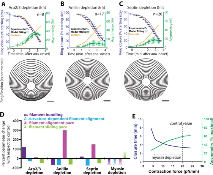
The conserved structural contractile ring components myosin II, anil-lin, and the septins are proposed to contribute to contraction, cyto-skeletal cross-linking, and anchoring of the cytoskeleton to the membrane. These activities could influence both the energy and the dynamics of the contractile apparatus (Figure 3A) and hence the feedback among filament alignment, contractile force, and mem-brane curvature. To further test our model predictions and function-ally annotate contractile ring proteins, we next analyzed the changes in the four critical parameters required to fit the phenotypes result-ing from the depletion of these conserved proteins. The four critical parameters are 1) the free energy gain from curvature-independent filament alignment, a0 in Eq. 1; 2) the free energy gain from
curva-ture-dependent filament alignment, A in Eq. 1; 3) the viscous drag coefficient for ring contraction, λ1 in Eq. 2; and 4) the viscous drag
coefficient for filament alignment, λ3 in Eq. 4 (Figure 2G).
Anillin is a multidomain protein implicated in coupling the acto-myosin cytoskeleton to the plasma membrane directly (Liu et al., 2012) or via the septins (Piekny and Maddox, 2010). The septins form hetero-oligomeric complexes, filaments, and sheets in associa-tion with the plasma membrane (Kinoshita et al., 2002; Mostowy and Cossart, 2012). Septins directly bind anillin and myosin II and create curved F-actin bundles (Kinoshita et al., 2002; Joo et al., 2007; Mavrakis et al., 2014). Our model predicts that reducing membrane–cytoskeleton attachment decreases the free energy gain from curvature-dependent filament alignment (a smaller A in Eq. 1), increases the rate of filament alignment due to reduced membrane friction (a smaller λ3), and decreases contraction pace (a
smaller λ1), since anchoring is needed to translate contractility into
furrow ingression (Figure 3, B and D). By decreasing the energy in-centive for curvature-mediated filament alignment, the reduction in membrane–cytoskeleton attachment compromises curvatumedi-ated positive feedback. Consequently the membrane curvature re-sulting from furrowing does not influence filament alignment locally. As a result, curvature-independent filament alignment dominates, circumferential filament alignment is more uniform, and furrowing is more symmetric (Figure 3, B and C, solid lines).
Indeed, depletion of anillin or the septins results in concentric ring closure (Figure 3, B and C; Maddox et al., 2007). We fitted the model to three aspects of experimental ring closure dynamics and geometry: initiation timing, duration of closure, and asymmetry. Fitting the model to measurements from cells depleted of anillin or of the septins required varying the four critical model parameters (Figure 3D; see later discussion). As predicted for decreased cyto-skeleton–membrane coupling, curvature-dependent filament alignment was made less energetically favorable (smaller A) and the filament alignment pace was increased (smaller λ3). The
neces-sary decrease in the filament sliding rate (larger λ1) could be
con-ferred by a reduction in cytoskeleton–membrane attachment. This model result demonstrates the necessity of curvature-mediated positive feedback for asymmetric furrow ingression. In addition, these results support the idea that anillin and the septins contribute to cytokinesis by coupling actomyosin to the membrane (Piekny and Maddox, 2010; Saarikangas and Barral, 2011; Liu et al., 2012). duration, and nonconcentricity. According to our model, actin
fila-ments are initially randomly oriented (ψ= 0 everywhere). The model assumes that a small and constant driving force (a0 < 0 in Eq. 4)
promotes circumferential filament alignment. Thermal fluctuations, however, seed the initial heterogeneity in ψ along the perimeter (Figure 2Ei). As more filaments become locally aligned, actomyosin contraction becomes more efficient (Figure 2Eii). The resulting stron-ger contraction drives a deeper furrow ingression, the larstron-ger Gauss-ian curvature of which locally promotes a greater filament alignment that in turn facilitates even more local contractions (Figure 2Eiii). Thus the local curvature mediates positive feedback between the cytoskeletal organization that facilitates force generation and the re-sulting furrow ingression, amplifying asymmetry.
For the initial asymmetric furrow to be perpetuated, our model predicts that the pace of filament sliding (and thus ring contraction) is faster than the filament alignment pace. This is because when the fur-row starts to ingress locally, the membrane curvature of the adjacent region will gradually increase, in turn locally promoting filament align-ment. Thus filament alignment, contraction, and furrowing will spread circumferentially from the initial furrowing site to the entire ring (Figure 2Eiv). Only when the rate of filament alignment is much slower than that of ring contraction can the initial asymmetry be amplified and sustained far into furrow ingression (Figure 2F). We thus found that four of the model parameters were critically important in dictating the dynamics of ring contraction: the free energy gains from filament bun-dling and for curvature-dependent filament alignment and the rates of filament alignment and sliding (Figure 2G). More specifically, whereas the free energy gains for filament bundling and curvature-dependent filament alignment (a0 and A in Eq. 4) promote ring
clo-sure, closure dynamics is slowed by the associated viscous drag coef-ficients of the dynamic processes (λ1 in Eq. 5 and λ3 in Eq. 7).
The nature of contractile ring actin influences furrow asymmetry
The model predicts that when contraction is slow relative to filament alignment, the initial ingression propagates more quickly around the cell circumference and eliminates the initial asymmetry (Figure 2F). To test this model prediction in vivo, we depleted the protein ARX-2, a member of the well-characterized, conserved Arp2/3 complex, which regulates the actin cytoskeleton in many contexts (Firat-Kara-lar and Welch, 2011) and was implicated in negative regulation of contractility during cytokinesis (Moulding et al., 2007; Canman et al., 2008) but whose depletion or mutation does not grossly alter contractile events in the zygote (Severson et al., 2002). We reasoned that perturbing this complex would alter the equilibrium between dendritic and unbranched F-actin; the latter is more amenable to bundling and sliding (Yang et al., 2012). In the model, this perturba-tion corresponds to a comparatively faster rate of filament align-ment (a smaller viscous drag coefficient λ3 in Eq. 7), which results in
on anillin recruitment (Supplemental Figure S2C) likely facilitate bundling when septins are depleted.
Myosin II is believed to drive contractile ring closure by actomyo-sin filament sliding or processively cross-linking depolymerizing ac-tin filaments (Green et al., 2012; Ma et al., 2012; Mendes Pinto et al., 2012). Consequently myosin II inhibition could be expected to reduce the rate of contraction (i.e., larger viscous drag coefficient λ1
or, equivalently, smaller contraction force f0) and thus cause slower,
more-symmetric furrowing (Figure 3E). Instead, although partial myosin II inhibition by depletion of the myosin light chain activator Rho kinase predictably delays furrow initiation and slows ingression, it in fact increases furrow asymmetry (Piekny and Mains, 2002; Mad-dox et al., 2007). In addition, depletion of NOP-1, a condition also believed to decrease activity of Rho effectors, results in markedly asymmetric furrowing (Tse et al., 2012). Our model suggests that increased asymmetry can result from an exaggerated increase in the rate of contraction relative to that of filament alignment (smaller Of interest, anillin and septin depletions have opposite effects
on furrow initiation timing: septin depletion allows earlier initiation, whereas anillin depletion causes a delay (Maddox et al., 2007; Figure 3, B and C). To recapitulate the kinetics as well as the asym-metry results of the two perturbations, we made opposite altera-tions of one model parameter. To fit anillin depletion ring closure kinetics, we made filament bundling less energetically favorable (a0 in Eq. 4 is less negative; Figure 3D), indicating that anillin’s F-actin– bundling activity (Field and Alberts, 1995; Maddox et al., 2005; Dorn et al., 2010) is important for its contributions to ring closure dynamics. Indeed, anillins bundle F-actin in vitro (Supplemental Figure S2A; Field and Alberts, 1995; Kinoshita et al., 2002). In con-trast, an increase in free energy gain from filament bundling (a0 in Eq. 4 is more negative) is necessary to recapitulate the earlier initia-tion resulting from septin depleinitia-tion (Figure 3D). The reducinitia-tion of membrane binding–based friction, overabundance of myosin II (Supplemental Figure S2B; Maddox et al., 2007), and lack of effect
relative to that of filament alignment. In sum, due to the increased asymmetry seen after myosin II inhibition, our model sug-gests dual roles for myosin II in cytokinesis: driving contractility and bundling actin.
The geometry of furrowing is largely intrinsic to the cortex
The spindle midzone comprises antiparallel bundled microtubules and lies in the center of the division plane. A nonconcentric furrow encounters the midzone when it is approxi-mately half closed (Figure 4A). On impact, the midzone may attenuate the asymmetry of the furrow by physically resisting its in-gression or it may boost asymmetry because it harbors activators of contractility. Our model predicts that the asymmetry of furrow ingression is largely intrinsic to the cell cortex since the forces exerted by the contractile ring are much larger than those holding the midzone in place (Supplemental Material). In silico, the ring closes asymmetrically even in the absence of resistive forces representing the spindle midzone (Figure 4, B and C). In-deed, the midzone is readily displaced upon impact with the furrow in control cells (Figure 4A). To test whether the midzone attenuates asymmetry, we depleted the microtubule bundler SPD-1, eliminating midzone micro-tubule bundles but not blocking cytokinesis completion (Verbrugghe and White, 2004; Figure 4B and Supplemental Figure S3, A and B). Furrow asymmetry is slightly higher in SPD-1–depleted cells than in controls (Figure 4, B and C, and Supplemental Figure S3A), suggesting that the midzone is a weak barrier to furrow ingression. Therefore the midzone is not necessary for asymmetry.
The furrow can initiate asymmetrically but terminate close to the center of the divi-sion plane (it can recenter) after perturba-tion of cortical proteins (Figure 3). Although our model suggests that asymmetry is intrin-sic to the cortex, recentering could result from the balance of forces between the cor-tex and midzone. We directly tested whether recentering requires resistance by the zone or can occur in the absence of mid-zone bundles. We combined thorough de-pletion of SPD-1 with a cortical perturbation that elicits recentering (partial depletion of anillin; Supplemental Figure S3, C and D). Alteration of the cortical cytoskeleton was thus sufficient to dictate the geometry of ring closure (recentering), even in the absence of a midzone barrier (Figure 4D and Supplemental Figure S3D). Therefore we conclude that ring geometry is primarily dictated by cortical players.
Asymmetric furrowing increases energy efficiency
We previously showed that reduction of active myosin II causes failure of symmetric cytokinesis, even though asymmetric furrowing can succeed in this condition (Maddox et al., 2007). We thus λ1/λ3; Figure 3, D and E). One way in which myosin II inhibition
could cause this imbalance is if myosin II contributes not only to contraction, but also to filament alignment. In support of this idea, myosin II exists in the contractile ring as bipolar filaments (Yumura et al., 2008; Zhou and Wang, 2008; Vale et al., 2009; Beach et al., 2014) that robustly bundle actin in vitro (Reymann et al., 2010; Tho-resen et al., 2011). In addition, myosin II, like other actin bundlers, could slow filament sliding and hence contraction by generating friction (Janson et al., 1992; Mukhina et al., 2007). Partial inhibition of myosin II would thus be predicted to reduce friction and impede filament alignment and therefore increase the contraction speed
FIGURE 4: The spindle midzone is a weak barrier to contractile ring ingression. (A) The ingressing furrow and spindle midzone are visible in a C. elegans embryo expressing a GFP-tagged plasma membrane probe (Audhya et al., 2005) and GFP-tagged tubulin. The furrow displaces the midzone microtubules from the center of the division plane. (B–D) Left, schematics illustrating perturbations of the midzone (pink), contractile ring (yellow), and chromosome (blue) segregation. Vertical dashed lines denote normal centroids of control daughter nuclei. Center, averaged, aligned positions of the contractile ring at 5% closure intervals for control C. elegans
cytokinetic furrow ingression in different cell types, where speed trades off with en-ergy efficiency.
DISCUSSION
A novel model for cytokinesis implementing membrane curvature– dependent cytoskeletal organization explains asymmetric furrowing
Here we investigate the physical mechanism of cytokinetic ring closure. Our model based on mechanical feedback among filament alignment, contraction, and membrane cur-vature recapitulates not only the kinetics of cytokinesis but also its asymmetry, a phe-nomenon that is ubiquitous but unexplained in its cell-autonomous instances. Central to the feedback is curvature-dependent fila-ment alignfila-ment (Figure 1A). Furthermore, protein-based mem-brane–cytoskeleton linkers (e.g., anillin and the septins) are essential for this feedback, as they govern the relationship between the paces of contraction and filament alignment and consequently the ance between furrow ingression and furrow propagation. This bal-ance, rather than differences in overall mechanism, likely accounts for the observed differences in the geometry of ring closure among cell types.
Of interest, while not consistently observed, a subset of controls contains cells that display spiraling movement of the cytokinetic ring center in addition to asymmetric furrowing (Supplemental Figure S4). To date, our theoretical model has not been able to recapitulate this large-scale spiral movement of the ring center. This is because the model describes membrane as an elastic sheet and imposes that the ring segment and the membrane patch to which it attaches share the same spatial position. The observed spiral movement re-flects the ring rotation, which in reality likely entails dissociation be-tween the ring segment and the local membrane and/or rearrange-ments of the associated membrane patches in the plane of the membrane. The present model cannot describe either of these two events sufficient for ring rotation. In particular, the in-plane rear-rangement of membrane patches requires the lateral movement of lipids in the membrane plane. It is hydrodynamic in nature, which cannot be faithfully described by the elastic illustration in the model, which assumes that membrane patches do not rearrange their rela-tive position. This intriguing spiraling phenomenon thus warrants future investigation.
The model allows functional annotation of conserved contractile ring proteins
The simplicity of our model and absence of protein-based agents allow an unbiased investigation of protein function. Because we found that the mechanics of cytokinesis is largely intrinsic to the cortical cytoskeleton, we focused on annotating several conserved ring components—anillin, septins, and myosin II—which have been proposed to have multiple, overlapping functions. Fitting the model to the kinetics of three independent experimental readouts (initia-tion timing, closure dura(initia-tion, and asymmetry) upon protein pertur-bations allows us to make specific mechanistic hypotheses about the roles of these ring components in membrane–cytoskeleton cou-pling, filament bundling, and contractility.
Anillin, a large multidomain protein that binds many cytokinetic ring components, has engendered myriad hypotheses for its mode of action (Piekny and Maddox, 2010). Our model suggests that hypothesized that symmetric furrowing requires more energy than
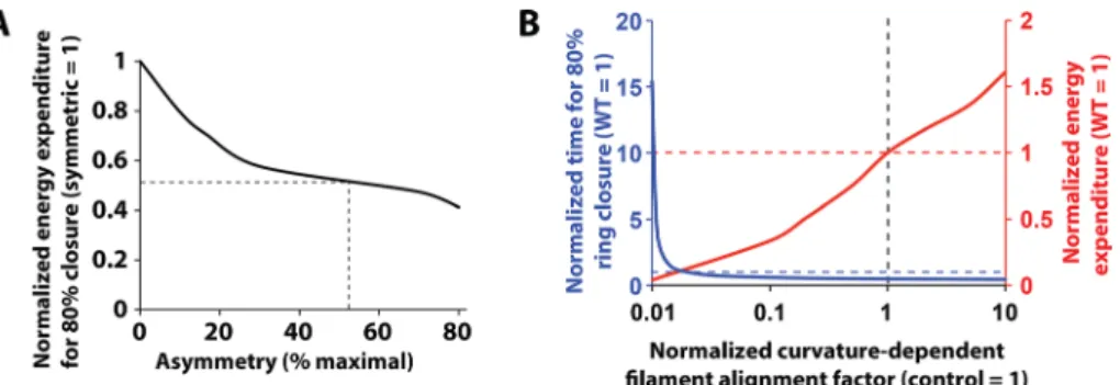
asymmetric closure. We tested this hypothesis computationally by integrating the total energy expenditure for contractile ring closure in scenarios of different relative rates of filament sliding and align-ment while keeping other model factors unchanged. That is, we numerically calculated the work done during furrowing by integrat-ing the drivintegrat-ing force of filament contraction (the right-hand side of Eq. 1) times the displacements of the ring over time. We found that asymmetric furrow closure is ∼50% more energy efficient than sym-metric furrow closure (Figure 5A). Although filament alignment facili-tates contraction, it also increases the rigidity of the cortical cyto-skeleton and consequently the bending resistance within the contractile ring. In a symmetric contractile ring, where alignment is uniformly high, ingression thus costs more energy. This suggests that the relationship between the paces of filament alignment and of sliding, which dictates the asymmetry of furrow ingression, deter-mines the energy efficiency of cytokinesis.
Energy efficiency of furrow ingression trades off with speed
Energy efficiency is unlikely to be the only factor that influences cell behavior. In embryos, speed is of the essence, cell cycles are short, and cell division events are fast, as compared with their counterparts in somatic cells. We leveraged our model to test whether removing the constraint of closure speed could reveal a more energy-efficient mode. We reduced curvature-dependent filament alignment (i.e., decreasing the value of A in Eq. 4 while keeping all of the other parameters the same) such that curva-ture-mediated positive feedback is eliminated (while keeping all other factors unchanged). With decreased A, the energy effi-ciency of ingression increases (Figure 5B, red trace). Decreasing the curvature-dependent filament alignment factor A by 100 times from that for the control causes ring closure to take more than six times longer than in the C. elegans zygote (Figure 5B, blue trace), longer than the entire C. elegans embryonic cell cycle (Deppe et al., 1978) but fully compatible with the kinetics of cyto-kinesis in somatic cells. In this mode, ring closure is relatively sym-metric (Figure 5A). In cells, this likely corresponds to a broad con-tractile apparatus such as that of mammalian and Drosophila cultured cells, which yields a very small Gaussian curvature at the furrowing site. The model further reveals that, although the en-ergy gain from curvature-dependent filament alignment greatly increases furrowing speed, it also increases energy cost (Figure 5B, red trace). The model thus suggests that the strength of cur-vature-dependent filament alignment dictates different modes of
(Shlomovitz and Gov, 2008) or amphipathic helices or that form curved polymers, such as septins (Kinoshita et al., 2002; Tanaka-Takiguchi et al., 2009). These factors could thus take part in driving cytokinesis by promoting the positive feedback loop we propose here.
Myosin II has been shown to contract more powerfully in re-sponse to mechanical load (Cremo and Geeves, 1998; Veigel et al., 2003; Kovas et al., 2007). Increased filament alignment due to bun-dling or curvature-dependent filament alignment would allow en-gagement of more myosin II heads on actin and thus more load. This could in turn lead to more powerful myosin II contraction, po-tentially myosin II filament growth (Ren et al., 2009), and deeper in-gression. Therefore load-sensitive contractility would participate in the same feedback loop we propose.
One model to explain different modes of cytokinesis
Asymmetric furrowing occurs in most, but not all, popular model species. Most prominently, budding and fission yeast exhibit sym-metric ring closure (Bi et al., 1998; Wu et al., 2003). This behavior is predicted by our model: within the parameter ranges tested, when the diameter of the cell is <5 μm, circumferential propagation of the initial asymmetric furrow completes so quickly that furrowing be-comes symmetric (Supplemental Figure S1C). In addition, circumfer-ential furrow propagation may be favored by cytoskeletal arrange-ment; in budding and fission yeasts, the septin and actomyosin cytoskeletons, respectively, become circumferentially aligned be-fore the onset of ring closure (Vrabioiu and Mitchison, 2006; Vavylo-nis et al., 2008). According to our model, this phenomenon corre-sponds to filament bundling outpacing filament sliding, which results in symmetric furrowing.
Asymmetric furrowing is an energy-efficient way to perform fast cytokinesis, according to our model. Speed is very important in em-bryonic systems. Indeed, many species’ embryos exhibit asymmet-ric furrowing. Whereas in the C. elegans zygote the mechanical feedback loop may be sufficient to initiate asymmetric furrowing, in other systems, such as the ctenophore zygote, supplementary mechanisms including eccentric spindle placement account for asymmetry. For epithelial cells in a monolayer, mechanical resistance by apical junctional complexes biases the effectiveness of equatorial contractility to the basolateral cell surface (reviewed by Bourdages and Maddox, 2013). Similarly, remnant substrate adhesions likely locally attenuate furrowing upward from the coverslip during asym-metric furrowing in cultured human and Drosophila cells (Bourdages et al., 2014). In all of these cases, the feedback loop we propose likely contributes to the maintenance of asymmetry. In future work, it will be interesting to examine furrowing kinetics and geometry in somatic cultured cells in mechanical isolation. Given the conserva-tion of the structural proteins implicated in our positive feedback loop, we expect that in such cells, furrowing will be measurably asymmetric even in the absence of extrinsic cues.
MATERIALS AND METHODS
See the Supplemental Experimental Procedures for further consid-erations of the theoretical model.
C. elegans maintenance and sample preparation
C. elegans were maintained according to standard procedures at 20°C. Strains used were JJ1473 (Munro et al., 2004; Carvalho et al., 2009) and OD122 (Dorn et al., 2010). See Supplemental Table S1 for full genotypes. Embryos were dissected from gravid hermaphro-dites and mounted under coverslips on agarose cushions as in Gonczy et al. (1999).
anillin contributes to actomyosin filament alignment, likely via its F-actin–bundling domain but also possibly by linking F-actin with ac-tive myosin II (Field and Alberts, 1995; Straight et al., 2005). Model fitting also suggests that anillin contributes to cytoskeleton–mem-brane linkage. Indeed, anillin bears a lipid-binding PH domain (Liu et al., 2012) and can bind the septins (Kinoshita et al., 2002). An important role in cytoskeleton–membrane linkage may explain the division plane instability in anillin-depleted cultured mammalian and Drosophila cells (Piekny and Maddox, 2010). Although these roles are not unexpected, our work suggests that this particular behavior is a major factor in how anillin contributes to contractile ring function.
Septins’ roles in metazoan cytokinesis are poorly defined and hypothesized based on their in vitro activities and roles elsewhere. As linear and grid-like polymers, they could aid cytoskeleton align-ment. As membrane associated proteins, they could link the cyto-skeleton to the membrane. In most organisms, many septin proteins and isoforms are expressed and likely form multiple combinatorial complexes (Sellin et al., 2011). In C. elegans, a single heterotetra-meric species is believed to be formed from the only two septin proteins (John et al., 2007), and septin function can be completely removed with a single molecular perturbation (Nguyen et al., 2000). Our model suggests that septins are important for membrane–cyto-skeletal linkage. This proposition is consistent with the role of septins in bleb retraction (Gilden et al., 2012).
Our model suggests that myosin II not only provides contractility, but it also contributes significantly to actin cross-linking and bun-dling. This result likely relates to the fact that the myosin II in con-tractile ring is in the form of bipolar minifilaments (Yumura et al., 2008; Beach et al., 2014). Myosin II’s cross-linking activity has been implicated in providing the driving force for cytokinetic furrowing (Yumura et al., 2008; Mendes Pinto et al., 2012; Beach et al., 2014). Going forward, it will be important to uncover the relative contribu-tions of myosin II’s multiple activities.
Together these results demonstrate the power of our model in annotating the functions of conserved contractile components.
Further considerations of curvature-mediated positive feedback
The key factor of our model is that the curvature of the furrow is both the cause and the result of contractile ring constriction and furrow ingression, that is, curvature-mediated positive feedback. With a minimal model describing the essential elements of the system, we recapitulated furrow asymmetry with a positive feedback loop medi-ated by curvature-dependent filament alignment. This feedback concept is based on the energy consideration that actin filaments maximize their attachment to the membrane and free energy is re-duced when filaments orient circumferentially along the furrow (Figure 1A). The model assumes that the actin filaments are straight throughout their length but could buckle with a 300-nm radius of curvature (Murrell and Gardel, 2012). Consequently they could par-tially conform to the high curvature at the furrowing site (likely <150-nm radius of curvature) and align transversely across the furrow. Nevertheless, filament buckling is energetically unfavorable. Thus our proposed configuration of straight actin filaments with circum-ferential alignment has a relatively lower free energy and is more favorable. Future work will be aimed at precisely measuring furrow curvature and its relationship to local cytoskeletal orientation.
Live-cell imaging
A Nikon TE-2000 inverted microscope equipped with a swept field real-time confocal module (Prairie Technologies [Bruker], Middle-ton, WI) and CoolSNAP HQ2 charge-coupled device camera (Pho-tometrics, Tucson, AZ) was used. The use of the 70-μm slit, a 60× oil-immersion objective (with 2 × 2 binning), and a 200-ms exposure time was kept constant for all embryos of a given strain.
RNA interference
Protein depletion by RNA-mediated interference (RNAi) was per-formed as done previously (Maddox et al., 2005, for injection; Kamath et al., 2001, for feeding). Bacterial strains for RNAi feeding were obtained from a whole-genome RNAi library (Kamath et al., 2003). See Supplemental Table S2 for primer sequences.
Image and data analysis
Ring geometry was determined using cyanRing (for cytokinesis anal-ysis of the contractile ring; Dorn et al., 2010; Bourdages et al., 2014), a semiautomated software tool written in Matlab (MathWorks, Natick, MA). cyanRing allows rectangular cropping with arbitrary ori-entation of the equatorial region of the embryo. This region is ro-tated and maximum projected to produce a view of the contractile ring along the spindle axis in the posterior direction. The user then defines the cell outline, as well as the outline of the contractile ring, by selecting at least three points, through which best-fit circles are drawn. Ring geometry is then calculated as shown in Supplemental Figure 2A.
Not all depletions exhibited 100% penetrance. To characterize the depletion phenotype rather than the degree of penetrance, we did not include data for cells that were indistinguishable from con-trols. Including these data with those of affected cells would have yielded a population average that does not reflect either the pertur-bation or the control. In addition, outlier data points were dis-carded, such as an apparent increase in the size of the contractile ring to more than the cell size, that are nonphysiological and most likely due to human error in annotating images. To avoid stylizing the data, the analysis pipeline does not allow reannotating the images.
To average data, the time of anaphase onset was determined by visual inspection of the data and set to zero. This assignment has an error of up to 10 s (our sampling frequency). Owing to this error, averaging per time point would result in a population average for closure with an artifactually low slope. By contrast, ring size mea-surements are comparatively more reliable: the maximum speed of ring closure is very similar among cells within a condition. To avoid introducing error by averaging per time point, we instead averaged at specific intervals of relative ring closure. Because ring closure oc-curs smoothly, we used linear interpolation to find the time at which specific ring closure percentages were reached and calculated aver-age and SD for 5% ring-closure intervals (5, 10,..., 90, 95%) using custom, publically available code (ch.mathworks.com/matlabcen-tral/fileexchange/27134-plot-average-line). Because the values for ring closure percentage have been imposed, no uncertainties are reported for them. Data for asymmetry and time were averaged across curves, and their experimental variability was calculated as the SD.
The 5% closure intervals of cell-biological and theoretical data averages were plotted as series of circles with the measured or cal-culated radii, respectively. The displacement between the center of the cell or a cytokinetic ring time point and the previous time point (the nonconcentricity) was given an arbitrary, consistent direction (downward from the center of the cell).
ACKNOWLEDGMENTS
We thank Kerry Bloom, Julie Canman, and Ewa Paluch for critical reading of the manuscript. Carlos Patino Descovich and all members of the Maddox labs provided discussion and support. Jean-Claude Labbé provided feeding RNAs. James Fethiere (Institute for Research in Immunology and Cancer, Montreal, Canada) provided purified protein fragments. We acknowledge the thoughtful and constructive input from the anonymous referees. J.F.D. was supported by a post-doctoral fellowship from the Swiss National Science Foundation. This work was supported by National Institutes of Health Grant GM102390 to A.S.M. J.L. is supported by the intramural research program of the National Heart, Lung, and Blood Institute at the National Institutes of Health.
REFERENCES
Alsop GB, Zhang D (2004). Microtubules continuously dictate distribution of actin filaments and positioning of cell cleavage in grasshopper sper-matocytes. J Cell Sci 117, 1591–1602.
Audhya A, Hyndman F, McLeod IX, Maddox AS, Yates JR 3rd, Desai A, Oegema K (2005). A complex containing the Sm protein CAR-1 and the RNA helicase CGH-1 is required for embryonic cytokinesis in Cae-norhabditis elegans. J Cell Biol 171, 267–279.
Beach JR, Shao L, Remmert K, Li D, Betzig E, Hammer JA 3rd (2014). Nonmuscle myosin II isoforms coassemble in living cells. Curr Biol 24, 1160–1166.
Bement WM, Capco DG (1991). Analysis of inducible contractile rings sug-gests a role for protein kinase C in embryonic cytokinesis and wound healing. Cell Motil Cytoskeleton 20, 145–157.
Bi E, Maddox P, Lew DJ, Salmon ED, McMillan JN, Yeh E, Pringle JR (1998). Involvement of an actomyosin contractile ring in Saccharomyces cerevi-siae cytokinesis. J Cell Biol 142, 1301–1312.
Biron D, Alvarez-Lacalle E, Tlusty T, Moses E (2005). Molecular model of the contractile ring. Phys Rev Lett 95, 098102–098104.
Bourdages KG, Lacroix B, Dorn JF, Descovich CP, Maddox AS (2014). Quan-titative analysis of cytokinesis in situ during C. elegans postembryonic development. PLoS One 9, e110689.
Bourdages KG, Maddox AS (2013). Dividing in epithelia: cells let loose dur-ing cytokinesis. Dev Cell 24, 336–338.
Burns CG, Larochelle DA, Erickson H, Reedy M, De Lozanne A (1995a). Single-headed myosin II acts as a dominant negative mutation in Dictyo-stelium. Proc Natl Acad Sci USA 92, 8244–8248.
Burns CG, Reedy M, Heuser J, De Lozanne A (1995b). Expression of light meromyosin in Dictyostelium blocks normal myosin II function. J Cell Biol 130, 605–612.
Canman JC, Lewellyn L, Laband K, Smerdon SJ, Desai A, Bowerman B, Oegema K (2008). Inhibition of Rac by the GAP activity of centralspin-dlin is essential for cytokinesis. Science 322, 1543–1546.
Carvalho A, Desai A, Oegema K (2009). Structural memory in the contractile ring makes the duration of cytokinesis independent of cell size. Cell 137, 926–937.
Claessens MMAE, Bathe M, Frey E, Bausch AR (2006). Actin-binding pro-teins sensitively mediate F-actin bundle stiffness. Nat Mater 5, 748–753. Clark AG, Dierkes K, Paluch EK (2013). Monitoring actin cortex thickness in
live cells. Biophys J 105, 570–580.
Cremo CR, Geeves MA (1998). Interaction of actin and ADP with the head domain of smooth muscle myosin: implications for strain-dependent ADP Release in smooth muscle. Biochemistry 37, 1969–1978. Das T, Payer B, Cayouette M, Harris WA (2003). In vivo time-lapse imaging
of cell divisions during neurogenesis in the developing zebrafish retina. Neuron 37, 597–609.
Deppe U, Schierenberg E, Cole T, Krieg C, Schmitt D, Yoder B, von Ehren-stein G (1978). Cell lineages of the embryo of the nematode Cae-norhabditis elegans. Proc Natl Acad Sci USA 75, 376–380.
Dorn JF, Zhang L, Paradis V, Edoh-Bedi D, Jusu S, Maddox PS, Maddox AS (2010). Actomyosin tube formation in polar body cytokinesis requires Anillin in C. elegans. Curr Biol 20, 2046–2051.
Douglas ME, Mishima M (2010). Still entangled: assembly of the central spin-dle by multiple microtubule modulators. Semin Cell Dev Biol 21, 899–908. Field CM, Alberts BM (1995). Anillin, a contractile ring protein that cycles
from the nucleus to the cell cortex. J Cell Biol 131, 165–178.
Maddox AS, Lewellyn L, Desai A, Oegema K (2007). Anillin and the septins promote asymmetric ingression of the cytokinetic furrow. Dev Cell 12, 827–835.
Mavrakis M, Azou-Gros Y, Tsai FC, Alvarado J, Bertin A, Iv F, Kress A, Brasselet S, Koenderink GH, Lecuit T (2014). Septins promote F-actin ring formation by crosslinking actin filaments into curved bundles. Nat Cell Biol 16, 322–334.
Medeiros NA, Burnette DT, Forscher P (2006). Myosin II functions in actin-bundle turnover in neuronal growth cones. Nat Cell Biol 8, 216–226.
Mendes Pinto I, Rubinstein B, Kucharavy A, Unruh JR, Li R (2012). Actin de-polymerization drives actomyosin ring contraction during budding yeast cytokinesis. Dev Cell 22, 1247–1260.
Miller AL, Bement WM (2009). Regulation of cytokinesis by Rho GTPase flux. Nat Cell Biol 11, 71–77.
Morais-de-Sa E, Sunkel C (2013). Adherens junctions determine the apical position of the midbody during follicular epithelial cell division. EMBO Rep 14, 696–703.
Mostowy S, Cossart P (2012). Septins: the fourth component of the cytoskel-eton. Nat Rev Mol Cell Biol 13, 183–194.
Moulding DA, Blundell MP, Spiller DG, White MR, Cory GO, Calle Y, Kempski H, Sinclair J, Ancliff PJ, Kinnon C, et al. (2007). Unregulated actin polymerization by WASp causes defects of mitosis and cytokinesis in X-linked neutropenia. J Exp Med 204, 2213–2224.
Mukhina S, Wang YL, Murata-Hori M (2007). Alpha-actinin is required for tightly regulated remodeling of the actin cortical network during cytoki-nesis. Dev Cell 13, 554–565.
Munro E, Nance J, Priess JR (2004). Cortical flows powered by asymmetrical contraction transport PAR proteins to establish and maintain anterior-posterior polarity in the early C. elegans embryo. Dev Cell 7, 413–424. Murrell MP, Gardel ML (2012). F-actin buckling coordinates contractility and
severing in a biomimetic actomyosin cortex. Proc Natl Acad Sci USA 109, 20820–20825.
Nguyen TQ, Sawa H, Okano H, White JG (2000). The C. elegans septin genes, unc-59 and unc-61, are required for normal postembryonic cyto-kineses and morphogenesis but have no essential function in embryo-genesis. J Cell Sci 113, 3825–3837.
Noguchi T, Mabuchi I (2001). Reorganization of actin cytoskeleton at the growing end of the cleavage furrow of Xenopus egg during cytokinesis. J Cell Sci 114, 401–412.
Piekny AJ, Maddox AS (2010). The myriad roles of anillin during cytokinesis. Semin Cell Dev Biol 21, 881–891.
Piekny AJ, Mains PE (2002). Rho-binding kinase (LET-502) and myosin phosphatase (MEL-11) regulate cytokinesis in the early Caenorhabditis elegans embryo. J Cell Sci 115, 2271–2282.
Pollard TD (2010). Mechanics of cytokinesis in eukaryotes. Curr Opin Cell Biol 22, 50–56.
Rappaport R (1996). Cytokinesis in Animal Cells, Cambridge, UK: Cam-bridge University Press.
Reichl EM, Ren Y, Morphew MK, Delannoy M, Effler JC, Girard KD, Divi S, Iglesias PA, Kuo SC, Robinson DN (2008). Interactions between myosin and actin crosslinkers control cytokinesis contractility dynamics and mechanics. Curr Biol 18, 471–480.
Reinsch S, Karsenti E (1994). Orientation of spindle axis and distribution of plasma membrane proteins during cell division in polarized MDCKII cells. J Cell Biol 126, 1509–1526.
Ren Y, Effler JC, Norstrom M, Luo T, Firtel RA, Iglesias PA, Rock RS, Robinson DN (2009). Mechanosensing through cooperative interac-tions between myosin II and the actin crosslinker cortexillin I. Curr Biol 19, 1421–1428.
Reymann AC, Martiel JL, Cambier T, Blanchoin L, Boujemaa-Paterski R, Théry M (2010). Nucleation geometry governs ordered actin networks structures. Nat Mater 9, 827–832.
Saarikangas J, Barral Y (2011). The emerging functions of septins in metazo-ans. EMBO Rep 12, 1118–1126.
Sain A, Inamdar MM, Julicher F (2015). Dynamic force balances and cell shape changes during cytokinesis. Phys Rev Lett 114, 048102.
Schroeder TE (1972). The contractile ring. II. Determining its brief existence, volumetric changes, and vital role in cleaving Arbacia eggs. J Cell Biol 53, 419–434.
Schroeder TE (1973). Actin in dividing cells: contractile ring filaments bind heavy meromyosin. Proc Natl Acad Sci USA 70, 1688–1692.
Sellin ME, Sandblad L, Stenmark S, Gullberg M (2011). Deciphering the rules governing assembly order of mammalian septin complexes. Mol Biol Cell 22, 3152–3164.
Fishkind DJ, Wang YL (1993). Orientation and three-dimensional organiza-tion of actin filaments in dividing cultured cells. J Cell Biol 123, 837–848. Fleming ES, Zajac M, Moschenross DM, Montrose DC, Rosenberg DW,
Cowan AE, Tirnauer JS (2007). Planar spindle orientation and asymmet-ric cytokinesis in the mouse small intestine. J Histochem Cytochem 55, 1173–1180.
Founounou N, Loyer N, Le Borgne R (2013). Septins regulate the contrac-tility of the actomyosin ring to enable adherens junction remodeling during cytokinesis of epithelial cells. Dev Cell 24, 242–255. Gardel ML, Shin JH, MacKintosh FC, Mahadevan L, Matsudaira P, Weitz
DA (2004). Elastic behavior of cross-linked and bundled actin networks. Science 304, 1301–1305.
Gilden JK, Peck S, Chen YC, Krummel MF (2012). The septin cytoskeleton facilitates membrane retraction during motility and blebbing. J Cell Biol 196, 103–114.
Gonczy P, Schnabel H, Kaletta T, Amores AD, Hyman T, Schnabel R (1999). Dissection of cell division processes in the one cell stage Caenorhabditis elegans embryo by mutational analysis. J Cell Biol 144, 927–946. Green RA, Paluch E, Oegema K (2012). Cytokinesis in animal cells. Annu
Rev Cell Dev Biol 28, 29–58.
Guillot C, Lecuit T (2013). Adhesion disengagement uncouples intrinsic and extrinsic forces to drive cytokinesis in epithelial tissues. Dev Cell 24, 227–241.
Haviv L, Gillo D, Backouche F, Bernheim-Groswasser A (2008). A cytoskele-tal demolition worker: myosin II acts as an actin depolymerization agent. J Mol Biol 375, 325–330.
Herszterg S, Leibfried A, Bosveld F, Martin C, Bellaiche Y (2013). Inter-play between the dividing cell and its neighbors regulates adherens junction formation during cytokinesis in epithelial tissue. Dev Cell 24, 256–270.
Janson LW, Sellers JR, Taylor DL (1992). Actin-binding proteins regulate the work performed by myosin II motors on single actin filaments. Cell Motil Cytoskeleton 22, 274–280.
John CM, Hite RK, Weirich CS, Fitzgerald DJ, Jawhari H, Faty M, Schlapfer D, Kroschewski R, Winkler FK, Walz T, et al. (2007). The Caenorhabditis elegans septin complex is nonpolar. EMBO J 26, 3296–3307.
Joo E, Surka MC, Trimble WS (2007). Mammalian SEPT2 is required for scaffolding nonmuscle myosin II and its kinases. Dev Cell 13, 677–690.
Kamath RS, Fraser AG, Dong Y, Poulin G, Durbin R, Gotta M, Kanapin A, Le Bot N, Moreno S, Sohrmann M, et al. (2003). Systematic functional analysis of the Caenorhabditis elegans genome using RNAi. Nature 421, 231–237.
Kamath RS, Martinez-Campos M, Zipperlen P, Fraser AG, Ahringer J (2001). Effectiveness of specific RNA-mediated interference through ingested double-stranded RNA in Caenorhabditis elegans. Genome Biol 2, 1–10.
Kinoshita M, Field CM, Coughlin ML, Straight AF, Mitchison TJ (2002). Self- and actin-templated assembly of mammalian septins. Dev Cell 3, 791–802.
Kosodo Y, Toida K, Dubreuil V, Alexandre P, Schenk J, Kiyokage E, Attardo A, Mora-Bermudez F, Arii T, Clarke JD, et al. (2008). Cytokinesis of neu-roepithelial cells can divide their basal process before anaphase. EMBO J 27, 3151–3163.
Kovas M, Thirumurugan K, Knight PJ, Sellers JR (2007). Load-dependent mechanism of nonmuscle myosin 2. Proc Natl Acad Sci USA 104, 9994–9999.
Koyama H, Umeda T, Nakamura K, Higuchi T, Kimura A (2012). A high-reso-lution shape fitting and simulation demonstrated equatorial cell surface softening during cytokinesis and its promotive role in cytokinesis. PLoS One 7, e31607.
Landau LD, Lifshitz EM (1980). Statistical Physics, Oxford, UK: Butterworth Heinemann.
Liu J, Fairn GD, Ceccarelli DF, Sicheri F, Wilde A (2012). Cleavage furrow organization requires PIP(2)-mediated recruitment of anillin. Curr Biol 22, 64–69.
Ma X, Kovacs M, Conti MA, Wang A, Zhang Y, Sellers JR, Adelstein RS (2012). Nonmuscle myosin II exerts tension but does not translocate actin in vertebrate cytokinesis. Proc Natl Acad Sci USA 109, 4509– 4514.
Mabuchi I, Okuno M (1977). The effect of myosin antibody on the division of starfish blastomeres. J Cell Biol 74, 251–263.