Black Carbon Loading in Sputum and Bronchoalveolar Lavage Macrophages as a Biomarker of Air Pollution Exposure
Anna Tillery
Honors Thesis ENHS
Department of Environmental Sciences and Engineering Gillings School of Global Public Health
ACKNOWLEDGEMENTS
This project would not be possible without the Center for Environmental Medicine,
Asthma, and Lung Biology. I would like to thank Dr. Neil Alexis for the support and guidance
offered throughout the project. In addition, thank you to all of whom work in Dr. Alexis’s lab,
specifically Heather Wells and Ceolamar Ways for providing assistance and advice where
necessary within and outside of this project. The support and encouragement of everyone
ABSTRACT
Previous literature has proven that black carbon (BC) can be used as a biomarker for
long-term personal exposures to particulate matter air pollution from measurements of
particulates within airway macrophages (Macs). Macs can be collected by, Bronchoalveolar
Lavage (BAL) and Sputum. Sputum samples are preferred within research because they are
inexpensive and less-invasive compared to BAL. Due to the differences in location of Macs
collected in the lungs from the two collection types, there is the potential for differential BC
deposition and uptake by Macs. To fill this gap in research, we are interested in observing the
differences in BC content between Macs derived from sputum samples versus BAL through
image analysis to determine if both are equally viable forms of collection for use as a biomarker
for air pollution exposure through BC assessment. ANOVA testing showed that there were little
significant differences between BC content in Macs collected from Sputum and BAL. Because
results have shown no significant differences in BC uptake between Sputum and BAL Macs,
Table of Contents
INTRODUCTION...5
METHODOLOGY ...7
SUBJECTS AND SAMPLE COLLECTION ... 7
BLACK CARBON ... 9
DATA ANALYSIS ... 10
RESULTS ... 11
DISCUSSION ... 12
CONCLUSIONS ... 17
FUTURE DIRECTIONS ... 17
APPENDIX ... 19
TABLES ... 19
FIGURES ... 20
INTRODUCTION
Black carbon (BC) is a byproduct of fossil fuel combustion and a component of particulate
matter (PM) air pollution, usually comprising PM of 2.5 µm and below.1,2 Exposure to BC in air
pollution is associated with an amalgam of adverse health outcomes such as chronic respiratory
diseases and lung cancers. Additionally, BC is thought to be a sufficient biomarker of exposure to
fossil fuel (such as diesel and gasoline fumes) and other PM air pollution.3,4 It has been previously
determined that BC content in airway macrophages is effective for assessing personal exposure to
PM air pollution in cases of long-term exposures (either months or years).5 Simple air-quality
monitoring at fixed locations only conveys external exposures to air pollution within a certain mile
radius, while biomonitoring and assessment of effective biomarkers indicates internal and personal
exposures. Because of this, biomonitoring provides a deeper understanding of a subject’s
individual exposure to air pollution and its associated risks.
Macrophages (Macs) either reside on the airway lumen or below the epithelial cell barrier
of the lung and are the first response of immune defense.6 Phagocytosis of PM by Macs is rapid
and occurs to completion within 24 hours of exposure.6 Particulates in air pollution generally range
from 2-10 µm in diameter and have shown to deposit preferentially in the bronchial airways over
alveolar airways.7 It has been previously determined that phagocytes (macrophages, neutrophils,
monocytes) residing in the bronchial airways exhibit increased inflammatory cell function,
specifically increased levels of surface-receptors mediating phagocytosis, compared to those in the
alveolar airways, but it has not been examined in relation to BC uptake whether these cells function
differently.7
samples have difficulties and benefits for collection of samples and ease of measurement of BC.
PM deposits differently within the lung depending on several factors including its size. Ultrafine
PM (<2.5 µm) deposits further within the lungs which has led to BAL macrophages exhibiting
smaller size distributions of phagocytized particles than sputum macrophages.5 BAL samples have
been shown to contain more homogenous samples in terms of macrophage phenotype and yield
greater numbers of macrophages than sputum samples.7 Sputum induction samples the surfaces of
the bronchial airways whereas BAL samples derive from the peripheral, alveolar airways; because
of this, BAL and Sputum samples acquire different kinds of Macs, either from the large, central
airways for Sputum or from the distal airways for BAL.8 BAL is an invasive procedure that
requires the use of anesthesia within a hospital suite compared to the less-invasive sputum
induction procedure which uses hypertonic saline solution and can be performed safely by a trained
professional outside of hospitals. Historically, BAL collection has provided more reliable
measurements and a greater number of Macs while Sputum induction is easier and more
cost-efficient, although the methods have not yet been compared for their use in BC measurements.5
The purpose of this study is to compare BC in Sputum and BAL derived Macs in order to
assess the efficacy of each method for biomarker application to PM exposure. This assessment will
give insight into whether BAL and sputum are equally effective techniques for examining BC
content of macrophages. Additionally, we will be able to determine if bronchial macrophages
exhibit different BC uptake than alveolar macrophages. In order to accomplish this, stained BAL
and sputum cytospin slides from the same subject were assessed for macrophage BC content using
light microscopy and Image J software. We examined the difference in BC content and deposition
METHODOLOGY
SUBJECTS AND SAMPLE COLLECTION
Subjects were selected from the SubPopulations and Intermediate Outcome Measures in
COPD Study (SPIROMICS). SPIROMICS collected samples, including sputum and
bronchoalveolar lavage, from COPD patients at several institutions in the United States. UNC
Chapel Hill Center for Environmental Medicine, Asthma, and Lung Biology (CEMALB)
participated in SPIROMICS as a central sputum slide reading core. Sputum slides and BAL
cytospin slides from the same subject were acquired from various institutions participating in the
SPIROMICS bronchoscopy sub-study and were examined for BC. Twenty subjects’ matched
sputum and BAL slides were assessed for macrophage BC. Subjects were excluded if they did not
participate in both Sputum and BAL collection.
Sputum induction and processing was performed according to the SPIROMICS manual of
operation (MOP V5). Sputum induction was performed using a Devilbiss Ultra-Neb 99 Ultrasonic
Nebulizer, and a spirometer was used to assess forced expiratory volume (FEV1). Two induction
protocols were employed depending on the post bronchodilator baseline FEV1. If the post
bronchodilator FEV1 of the subject was greater than 1 liter, the induction procedure was
performed. The induction procedure followed this protocol: 15 mL of 3% saline solution in the
nebulizer for 2 minutes, then spirometry was run to determine if the subject was able to continue,
if so, the nebulizer was continued for 5 minutes. After induction, the subject attempted to cough
up sputum until they could no longer do so. Spirometry was repeated, and if the subject remained
stable, the process was repeated with 15 mL of 4% saline solution. The subject attempted to cough
attempted to cough up the resulting sputum without scraping their throat, and the sample was sent
for processing.
If the post bronchodilator FEV1 was less than 1 liter, the nebulizer was filled with 15 mL
of 0.9% saline solution. Spirometry was performed at 1, 2, 5, and 7 minutes, gathering the induced
sputum at the end of the 7 minutes. The subject always attempted to cough up the induced sputum
without scraping their throat. At the spirometry checks, if the FEV1 fall from baseline was between
10-19%, the procedure could continue with the same saline concentration. If the FEV1 fall from
baseline was < 10%, the saline concentration could be increased to the next highest concentration.
Sputum processing occurred using the whole sample. The sample was weighed and 0.1%
dithiothreitol (DTT) was added equaling 4 times the sputum weight. Sample was vortexed and
tumbled for 15 minutes, then ethylenediaminetetraacetic acid (EDTA) was added in the same
volume of DTT previously added. Sample was tumbled for 5 minutes and filtered through a 53µm
nylon mesh filter. Cells were spun down at 500xg for 10 minutes and 1-2 mL of Hank’s Balanced
Salt Solution (HBSS) then counted. Sample was adjusted to a concentration of 1.0*10^6 cell per
mL of HBSS media and slides were prepared with 60 µL of sample and spun at 450 rpm for 6
minutes. Slides were stained with Hema 3 stain set, mounted with Cytoseal and coverslips and
stored at room temperature.
Bronchoscopy sample collection occurred after the subject was treated with anesthesia.
The bronchoscope was safely inserted, and the airways of the subject were deemed normal enough
to continue. The BAL was performed in the lower lobe bronchus. In the first segment, sterile saline
was injected in 2x40cc and then 1x50cc. If less than 15cc of fluid was returned, the 1x50cc wash
was not performed. Then, the bronchoscope was moved to another segment and the washes were
processing. The BAL fluid was centrifuged at 500xg for 10 minutes and the supernatant was stored.
The cell pellet was resuspended in 10 mL of Roswell Park Memorial Institute (RPMI) media.
Hemacytometry was performed to determine cell count and viability; 4 cytospin slides were
prepared after the sample was adjusted to a concentration of 1.0*10^6 cell per mL of media. The
slides were prepared with 60 µL of sample and spun at 450 rpm for 6 minutes. They were then
fixed with 95% ethanol and stored at room temperature.
BLACK CARBON
BC was measured using NIS Elements Imaging Software Basic Research 5.00.00. Pictures
of macrophages were taken on a Life Technologies evos XL core microscope. For each subject, a
minimum of 50 macrophages were photographed and read on each of their sputum and BAL slides.
If 50 macrophages were not found on the slide, the subject slide was included if total macrophages
counted and photographed exceeded 30. Macrophages were included for measurement if they had
an intact cell wall and features easily discernable on a microscope. BC was measured manually,
measurements included carbon deposits in the nucleus and the cytoplasm following the protocol
previously described in Jary et.al.9 NIS Elements recorded the total macrophages counted, the BC
area, in µm2, per macrophage [Objects Area (OA)], the number of BC deposits found in each
macrophage [Number Objects (NO)], the % of BC occupying the interior of the macrophage
[Objects Area Fraction (OAF)], and the average internal area, in µm2, of each macrophage
measured [ROI Area]. Objects area fraction was calculated as follows: OAF = OA/ROI Area x
100%. Black carbon deposits were included if they were completely or partially inside the Mac
DATA ANALYSIS
Mean, standard deviation, minimum, and maximum values were measured for each
parameter. Statistical analysis was performed using GraphPad Prism 8 for Windows 64-bit. A
one-way ANOVA was run with an assumption of a Gaussian distribution of residuals with an alpha
level 0.05 and a Tukey post-Hoc analysis. The data were log transformed and an ANOVA test
with the same assumptions and post-Hoc analysis was run. Data presented in graphs and figures is
the non-transformed data. Pearson and Spearman correlation tests were also run on the total
RESULTS
Black Carbon (BC) was found in airway macrophages in all 20 subjects assessed, in both
sputum and BAL slides. Figure 1 shows examples of phagocytized BC in Macs from a Sputum
and BAL sample from one subject. Mean, standard deviation, minimum value and maximum value
measured for total macrophages, object area fraction, object area, number of objects, and
macrophage area for Sputum measurements are found in Table 1: Summary Characteristics of
Sputum Measurements and BAL measurements are found in Table 2:Summary Characteristics of
BAL Measurements from NIS Elements BC Measurements. This data is presented in graph form
in Figure 2.
The ANOVA test found significant differences at alpha level 0.05 with a p value less than
0.001. In the post-Hoc analysis, Sputum ROI Area was significantly different than BAL ROI Area.
The remaining significant differences were between parameters of the same source, Sputum or
BAL correlating with another parameter from Sputum or BAL; these significant differences were
not of interest because we were not interested in looking to how the parameters compared within
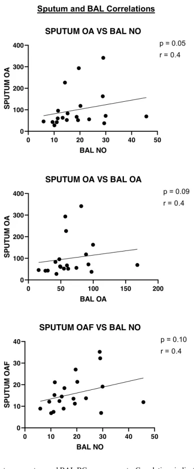
the same type of collection (i.e. Sputum OA to Sputum OAF). In terms of correlations, we found
that Sputum OA and BAL NO were significantly correlated (R = 0.4, p = 0.05) and significant
trends were observed between Sputum OA and BAL OA (R = 0.4, p = 0.08) and Sputum OAF and
DISCUSSION
The goal of this experiment was to ascertain whether Sputum and BAL are equally
effective methods of data collection for use of BC as a biomarker for air pollution exposure. In
previous literature, Sputum and BAL have been used independently for air pollution exposure
assessment by BC but it has never been determined if they are both viable. This assessment needed
to be done to better understand how BC deposits within the lungs and whether Macs derived from
Sputum induction or BAL can both be used. The data gathered suggest that both forms of collection
are equally viable for BC assessment and can be used in place of one another. This is advantageous
for future research as Sputum induction is quicker, cheaper, and less invasive than BAL so it is a
preferred method for human studies.
The data demonstrate that on average Sputum macrophages are larger than BAL
macrophages, and BC occupies a larger absolute internal area (µm2) of sputum macrophages versus
BAL macrophages. However, when macrophage size was normalized by calculating the percent
BC occupancy, we found that Sputum and BAL macrophages take up relatively similar amounts
of BC. This was further shown when the number of BC deposits did not significantly differ
between sputum and BAL macrophages. From this information, we know that BC can penetrate
deep into the alveolar airways and can be phagocytosed by alveolar macrophages residing there.
Another implication is that functionally, sputum macrophages are not inferior to BAL
macrophages in terms of BC uptake, supporting earlier work by Alexis et al., that showed sputum
macrophages had greater phagocytic capability than autologous alveolar macrophages.7
Additionally, there were no other significant differences found between any parameters measured
viable when considering BC uptake and measurement meaning that either could be used for
assessment of BC as a biomarker for future studies.
Sputum OA and BAL NO were significant at an alpha of 0.05 indicating that as sputum
OA increases, BAL NO also increases. Sputum OAF and BAL NO were trending towards
significance, and there could be a positive correlation between the amount of BC in Sputum Macs
and the NO found in BAL AM. All of these correlations show moderate correlations (Figure 3),
although there are apparent outliers in the data, these correlations indicate that BC in Sputum and
BAL are related to each other and increase at the same rates. Correlation tests also revealed that
Sputum and BAL object areas trended towards significance. This correlation is logical because of
the object areas similarity in absolute values, it would indicate that as Sputum BC area increased,
BAL BC area also increased. This could show that BC uptake scales similarly in upper airway
macrophages as well as alveolar macrophages and reveal how Macs take in carbon. This also
indicates that Sputum Macs do not prevent BC from penetrating deep into the alveolar region of
the lung, also supporting previous studies that have shown differential BC deposition within the
lungs.10 The correlation data simply shows that Sputum and BAL BC content are related to each
other in the sense that within subjects, higher content in Sputum Macs is associated with higher
content in BAL Macs as well. This provides further evidence supporting Sputum and BAL as
equally viable methods. As more subjects are included in the study, it should become apparent
whether these correlations get stronger or grow further apart to understand if these correlations say
something about BC in the lungs. It is also important to remember that these samples were not
collected on the same day, and without ambient air pollution data these comparisons are not ideal.
BC uptake. While it has already been determined that sputum macrophages function comparably
to BAL macrophages and Sputum can be used to collect biomarkers for disease, it has not been
previously assessed whether this holds true for BC uptake as well.3,8 The data collected in this
study have shown that BC uptake between Sputum and BAL macrophages shows no significant
differences within the same subject, indicating that Sputum could be used in place of BAL.
Because BAL is a much more expensive and invasive method to use for data collection, it is
preferred to use Sputum as a method of data collection, especially for large sample sizes. Our data
suggest that Macs in the bronchial and alveolar regions may function with the same capacity with
regards to BC uptake, so data collected from Sputum and BAL methods should give the same
information. Although, it is important to remember that samples were not taken on the same day
so there is the potential for differing amounts of BC within the lungs. Samples are independent of
each other so order of collection would not matter, solely the exposure to air pollution. This is
important because it has been found that Macs produce reactive oxygen species in a
dose-dependent manner to their BC loading.11 Understanding how BC is taken up in Macs in relation to
air pollution exposure is the first step to learning more about the health effects of BC on the body
and how this relates to a person’s overall health.
Varying sizes of PM deposit differently throughout the lungs. It has been found previously
that PM with diameter of 3 µm preferentially deposits in the larger, proximal regions of the lungs
while ultrafine PM (~1 µm) deposits throughout the lungs.10,12 It has also been shown that about
80% of BC in the air is considered ultrafine PM while the remaining 20% is around 2-3 µm.13 It
was thought originally that the differential deposition patterns would affect the rates of BC uptake
within the lungs due to varying amounts present in the proximal and alveolar regions of the lungs,
the large airways do not prevent BC from penetrating in the deep, alveolar region of the lung. This
supports previous research stating that BC PM in ambient air pollution is comprised of different
sizes. While BC may deposit differentially throughout the lungs, amount of BC does not seem to
affect Macs uptake regardless of whether they were BAL or Sputum derived.
Although these results are promising for use of Sputum BC as a biomarker for air pollution
exposure, it is important to remember the subjects used in this study were COPD patients and not
healthy. It has been previously determined that BC is taken up in healthy subjects and can be used
as a biomarker for air pollution; it has also been found that Sputum macrophages can be hyper
functional compared to BAL macrophages.3,7 If COPD patient’s macrophages are hyper functional
in relation to BC uptake, this could affect their total lung function and potentially make the patient
sicker. If unhealthy COPD subjects Macs focus too heavily on BC uptake rather than the full range
of pathogens invading their lungs, there is a potential for them to get infections easier. We need to
be able to understand how BC is affecting healthy and sick subjects and if BC uptake is differential.
Sputum and BAL samples taken from subjects were not collected on the same day.
Therefore, this limitation hinders our ability to accurately assess whether BC concentrations within
the subject’s lungs were the same. Because of this limitation, it is important to move on with the
project and gather ambient air pollution data for each subject during the time of collection for each
sample. An additional limitation to this study is the small sample size. Only 20 subjects were
included in this study, while data collection continues for the project as a whole and more subjects
are included, we will get a clearer understanding of the true population distribution of BC content.
Additionally, BC was measured manually and the process was not computerized. This adds
Although this approach may have certain advantages over previous literature using computerized
CONCLUSIONS
In summary, there have been no significant differences found between Sputum derived
Macs and BAL derived Macs. Summary characteristics and analyses have shown that Sputum and
BAL only significantly differ in size of macrophages found in the areas collected but that has not
seemed to have an effect on capacity of Macs to load BC. This indicates that Sputum collection is
equally as effective for deriving BC biomarker information as BAL collection for COPD patients
although this should be investigated further to understand how this relates to PM air pollution
exposure and whether this relationship differs with healthy subjects.
FUTURE DIRECTIONS
In the future, we will look to assess how this information relates to the air pollution
exposure measurements for each subject on the days of their bronchoscopy/ sputum induction. It
has previously been found that BC content in Macs is a better indicator of adverse health effects
to the subject rather than PM mass in the air but the relationship has not been further investigated.14
This information could tell us more about carbon uptake rates and correlations between the level
of exposure and BC in sputum/BAL measurements, and provide further proof that BC in
macrophages can be used as a biomarker for air pollution exposure. We would want to investigate
whether BC uptake differs with air pollution exposure and if uptake rates differ with amount of
BC in the body. This relationship has been investigated in regards to proximity to traffic patterns,
and it has been found that increased proximity is associated with higher amounts of BC content in
Macs, although this has not been investigated for simple ambient air pollution levels.15 This
In addition, it would be interesting to see if this data is different for healthy subjects versus
COPD subjects. We could look to see if there is a difference between carbon uptake in
macrophages between the two types of subjects to determine if COPD patients are actually more
prone to infections due to air pollution exposure. It has previously been shown that exposure to
diesel exhaust particles inhibits immune functions due to the increased demand on macrophages
phagocytosis to remove the particles which could support the hypothesis for BC affecting immune
function in healthy and COPD subjects.16 If the BC uptake is differential for COPD and healthy
patients further investigation would need to be done to determine if air pollution exposure plays a
role in the overall health of COPD patients more so than healthy patients. Due to COPD patient’s
already reduced lung function we would be interested in knowing whether BC has a greater health
effects to them over healthy subjects.
Additionally, it has been shown that PM deposition within the lungs is increased in the
lungs of COPD patients over healthy, non-smoking patients.17 A previous study investigated the
effects of BC loading in children with asthma compared to healthy children and found that with
more severe cases of asthma, BC loading by Macs decreased.18 This could indicate a difference
between COPD and healthy patients in terms of their Macs ability to load BC. It should be
investigated to see whether this increased deposition of BC affects Macs phagocytosis and/or plays
a role in the increased likelihood of infection of COPD patients. Previously, it has been found that
BC/PM in COPD patients has greater retention within the lungs compared to that of healthy
patients.19 Retention of BC also increases with disease severity of COPD subjects indicating that
more health effects associated with PM in ambient air pollution would be seen in more severely
affected patients.19 The differences in lung/ immune function and BC deposition between healthy
APPENDIX
TABLES
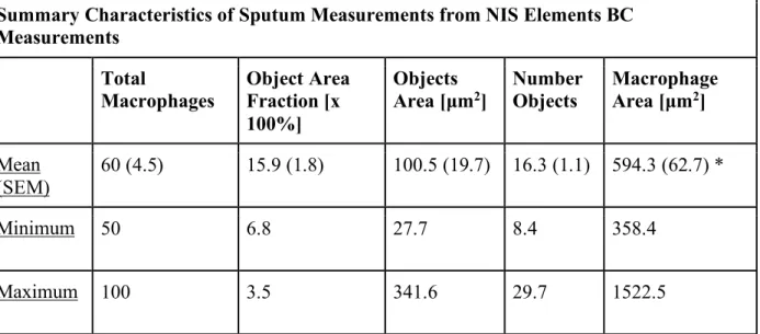
Summary Characteristics of Sputum Measurements from NIS Elements BC Measurements Total Macrophages Object Area Fraction [x 100%] Objects
Area [µm2] Number Objects Macrophage Area [µm2]
Mean
(SEM) 60 (4.5) 15.9 (1.8) 100.5 (19.7) 16.3 (1.1) 594.3 (62.7) *
Minimum 50 6.8 27.7 8.4 358.4
Maximum 100 3.5 341.6 29.7 1522.5
Table 1. Summary characteristics of sputum black carbon measurements recorded from NIS Elements software.
Measurements include the mean measurement for the 20 subjects along with the standard error of the mean, minimum, and maximum values. Total Macrophages indicates the average total number of Macs measured on the slide, Object Area Fraction indicates the average percent area cover of BC in the Macs, Objects Area indicates the average area of BC per Mac, Number Objects indicates the average number of BC deposits per Mac, and Macrophage Area indicates the average area of each Mac.
*Indicates a statistically significant difference in compared measurements from Sputum to BAL
Summary Characteristics of BAL Measurements from NIS Elements BC Measurements
Total
Macrophages Object Area Fraction [x
100%]
Objects
Area [µm2] Number Objects Macrophage Area [µm2]
Mean (SEM)
49.4 (0.6) 15.8 (1.5) 64.8 (7.6) 19.1 (2.2) 417.3 (22.3) *
Minimum 39 5.3 15.9 5.9 246.9
Maximum 51 2.9 168 45.7 641.3
Table 2. Summary characteristics of BAL black carbon measurements recorded from NIS Elements software.
FIGURES
Figure 1. Sputum (A and B) and BAL (C) macrophages from one COPD patient enrolled in SPIROMICS
showing phagocytized BC. Arrows point to BC deposits.
A. B.
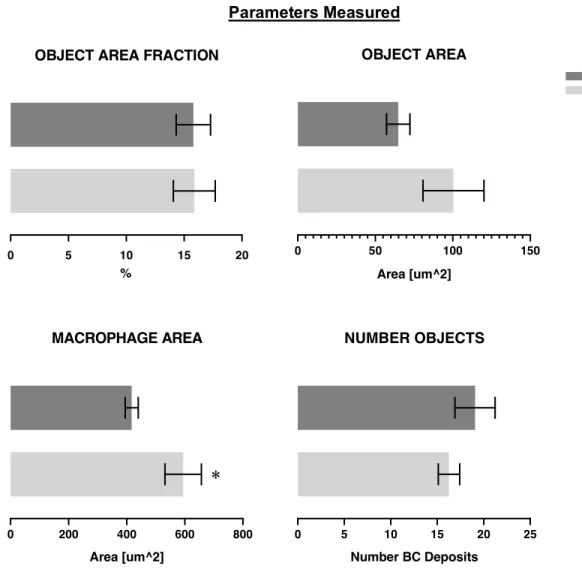
Parameters Measured
Figure 2. Mean of parameters measured with standard error of the mean. Parameters shown are the Object Area
Fraction, Object Area, Macrophage Area, and Number of Objects.
*Indicates a statistically significant difference between parameters
0 5 10 15 20
OBJECT AREA FRACTION
%
0 50 100 150
OBJECT AREA
Area [um^2]
BAL SPUTUM
0 200 400 600 800
MACROPHAGE AREA
Area [um^2]
0 5 10 15 20 25
NUMBER OBJECTS
Number BC Deposits
Sputum and BAL Correlations
Figure 3. Correlations between sputum and BAL BC measurements. Correlations indicate how BC uptake in
0 10 20 30 40 50
0 100 200 300 400
SPUTUM OA VS BAL NO
BAL NO SP U T U M O A
r = 0.4 p = 0.05
0 50 100 150 200
0 100 200 300 400
SPUTUM OA VS BAL OA
BAL OA SP U T U M O A
p = 0.09 r = 0.4
0 10 20 30 40 50
0 10 20 30 40
SPUTUM OAF VS BAL NO
BAL NO SPU T U M O A F
REFERENCES
1. Kulkarni N, Pierse N, Rushton L, Grigg J. Carbon in Airway Macrophages and Lung
Function in Children. N Engl J Med. 2006;355(1):21-30. doi:10.1056/NEJMoa052972.
2. Grigg J. The health effects of fossil fuel derived particles. Arch Dis Child.
2002;86(2):79-83. doi:10.1136/ADC.86.2.79.
3. Bai Y, Bové H, Nawrot TS, Nemery B. Carbon load in airway macrophages as a
biomarker of exposure to particulate air pollution; a longitudinal study of an international
Panel. Part Fibre Toxicol. 2018;15(1):14. doi:10.1186/s12989-018-0250-8.
4. Fireman EM, Lerman Y, Ganor E, et al. Induced Sputum Assessment in New York City
Firefighters Exposed to World Trade Center Dust. Environ Health Perspect.
2004;112(15):1564-1569. doi:10.1289/ehp.7233.
5. Bai Y, Brugha RE, Jacobs L, Grigg J, Nawrot TS, Nemery B. Carbon loading in airway
macrophages as a biomarker for individual exposure to particulate matter air pollution —
A critical review. Environ Int. 2015;74:32-41. doi:10.1016/J.ENVINT.2014.09.010.
6. Geiser M. Morphological aspects of particle uptake by lung phagocytes. Microsc Res
Tech. 2002;57(6):512-522. doi:10.1002/jemt.10105.
7. Alexis N, Soukup J, Ghio A, Becker S. Sputum Phagocytes from Healthy Individuals Are
Functional and Activated: A Flow Cytometric Comparison with Cells in Bronchoalveolar
Lavage and Peripheral Blood. Clin Immunol. 2000;97(1):21-32.
doi:10.1006/CLIM.2000.4911.
8. Alexis NE, Lay JC, Zeman KL, Geiser M, Kapp N, Bennett WD. In vivo particle uptake
2006;34(3):305-9. Jary H, Rylance J, Patel L, Gordon SB, Mortimer K. Comparison of methods for the
analysis of airway macrophage particulate load from induced sputum, a potential
biomarker of air pollution exposure. BMC Pulm Med. 2015;15(1):137.
doi:10.1186/s12890-015-0135-7.
10. Brand P, HÄUßINGER K, Meyer T, et al. Intrapulmonary Distribution of Deposited
Particles. Vol 12. Mary Ann Liebert, Inc. Pp; 1999. www.liebertpub.com. Accessed
February 25, 2019.
11. Aam BB, Fonnum F. Carbon black particles increase reactive oxygen species formation in
rat alveolar macrophages in vitro. Arch Toxicol. 2007;81(6):441-446.
doi:10.1007/s00204-006-0164-3.
12. Kim CS, Hu SC, Dewitt P, Gerrity TR. Assessment of regional deposition of inhaled
particles in human lungs by serial bolus delivery method. J Appl Physiol.
1996;81(5):2203-2213. doi:10.1152/jappl.1996.81.5.2203.
13. Krecl P, Johansson C, Targino AC, Ström J, Burman L. Trends in black carbon and
size-resolved particle number concentrations and vehicle emission factors under real-world
conditions. Atmos Environ. 2017;165:155-168. doi:10.1016/J.ATMOSENV.2017.06.036.
14. Janssen NAH, Hoek G, Simic-Lawson M, et al. Black Carbon as an Additional Indicator
of the Adverse Health Effects of Airborne Particles Compared with PM 10 and PM 2.5.
Environ Health Perspect. 2011;119(12):1691-1699. doi:10.1289/ehp.1003369.
15. Momtaz SM, Mehdipour P, Dadvand P, et al. Environmental and behavioral determinants
affecting the association of airway macrophages carbon load with distance to major roads
and traffic density. Chemosphere. 2018;217:680-685.
16. Yang HM, Antonini JM, Barger MW, et al. Diesel exhaust particles suppress macrophage
function and slow the pulmonary clearance of Listeria monocytogenes in rats. Environ
Health Perspect. 2001;109(5):515-521. doi:10.1289/ehp.01109515.
17. Möller W, Felten K, Sommerer K, et al. Deposition, Retention, and Translocation of
Ultrafine Particles from the Central Airways and Lung Periphery. Am J Respir Crit Care
Med. 2008;177(4):426-432. doi:10.1164/rccm.200602-301OC.
18. Brugha RE, Mushtaq N, Round T, et al. Carbon in airway macrophages from children
with asthma. Thorax. 2014. doi:10.1136/thoraxjnl-2013-204734.
19. Ling SH, McDonough JE, Gosselink J V., et al. Patterns of Retention of Particulate Matter
in Lung Tissues of Patients With COPD. Chest. 2011;140(6):1540-1549.