THE ASSOCIATIONS BETWEEN FOOTBALL EXPOSURE, CONCUSSION HISTORY
AND PLAYING POSITION ON CEREBRAL WHITE MATTER INTEGRITY AND
NEUROCOGNITIVE PERFORMANCE
By
Allen Anthony Champagne
Senior Honors Thesis
Department of Exercise and Sport Science University of North Carolina at Chapel Hill
2015
Approved by:
Kevin M. Guskiewicz (Supervisor)
Jason P. Mihalik
Michael Clark
ABSTRACT
ALLEN CHAMPAGNE: The associations between football exposure, concussion history and playing position on cerebral white matter integrity and neurocognitive performance
(Under the direction of Kevin M. Guskiewicz)
Diffusion tensor imaging (DTI) has emerged as an important tool for quantitative analysis of white matter (WM) integrity following sport-related concussions. The purpose of this research was to investigate the variances in WM integrity (defined by fractional anisotropy (FA) and
medial diffusivity (MD)) and neurocognitive performances in retired college and professional
football athletes based on concussion history, duration of playing career, and playing position.
MRI scans and neurocognitive test scores from 32 former college and 31 former age-matched
professional players (avg age=58.5 SD=3.7) were compared. A permuted, voxel-wise 3x2
ANOVA was performed on the WM skeleton to investigate the main and interaction effects on
WM integrity. Threshold-free cluster enhancement (TFCE) was used to identify clusters of
significantly different FA or MD and post-hoc univariate analyses were used to determine the
direction of interaction effects. A priori alpha level was set at 0.05 after correction for multiple
comparisons. Differences in FA were observed in 3 clusters in the forceps minor and genu of the
corpus callosum for the concussion by position interaction. Post-hoc analysis of the peak voxels
within each cluster revealed consistently lower FA in non-speed athletes with 3+ concussions as compared to those with 01 concussions (Cohen’s d: 0.89, 0.95, and 1.29; P<0.05). No clear differences in neurocognitive abilities were identified. Our results suggest a history of multiple concussions is associated with lower FA in former non-speed position players compared to speed players, particularly in frontal white matter tracts. We did not observe main effects of football exposure, suggesting that without concussive injuries, added football exposure does not account
for variances in white matter integrity and neurocognitive abilities. A limitation of these results
TABLE OF CONTENTS
LIST OF TABLES ... V LIST OF FIGURES ... VI
CHAPTER I ... 1
1.1 GOAL OF THE WORK ... 1
1.2 BACKGROUND AND SIGNIFICANCE ... 1
1.3 SPECIFIC AIMS/RESEARCH QUESTIONS ... 3
1.4 OPERATIONAL DEFINITIONS ... 5
1.5 DELIMITATIONS ... 6
1.6 ASSUMPTIONS AND LIMITATIONS ... 7
1.7 DIRECTION OF THE THESIS ... 7
CHAPTER II ... 8
2.1 INTRODUCTION ... 8
2.2 NEUROANATOMY ... 10
2.2A GENERAL OVERVIEW OF THE CEREBRAL CORTEX ... 10
2.2B THE REGIONAL FUNCTIONALITY OF THE CEREBRAL CORTEX ... 11
2.2C THE LIMBIC SYSTEM ... 12
2.3 CONCUSSIONS ... 13
2.3A NEUROPHYSIOLOGY OF CONCUSSIONS ... 13
2.3B BIOMECHANICS OF CONCUSSIONS ... 14
2.3C ANATOMICAL VULNERABILITY OF CEREBRAL TISSUES ... 16
2.4 SYMPTOMATOLOGY OF CONCUSSIONS ... 17
2.4A SHORT-TERM NEUROLOGICAL SYMPTOMS ... 17
2.4B LONG-TERM NEUROLOGICAL SYMPTOMS ... 18
2.4C PREVALENCE TO NEUROLOGICAL AND NEUROPSYCHIATRIC CONDITIONS ... 19
2.5 NEUROCOGNITIVE TESTING AND CONCUSSIONS ... 20
2.6 NEUROIMAGING AND TRAUMATIC BRAIN INJURIES ... 20
2.6A FUNCTIONAL MAGNETIC RESONANCE IMAGING (FMRI) ... 20
2.6a(1) Description and overview ... 20
2.6a(2) Abnormal activation patterns related to concussions ... 21
2.6B DIFFUSION TENSOR IMAGING (DTI) ... 22
2.6b(1) Description and overview ... 22
2.6b(2) Tractography and specific networks of interest ... 25
2.6b(3) Concordance with fMRI readings ... 26
2.7 CONCLUSION AND DIRECTION FOR FUTURE RESEARCH ... 28
CHAPTER III ... 29
3.1 SUBJECTS ... 29
3.1A PARTICIPANTS ... 29
3.1B RECRUITMENT ... 29
3.1C INCLUSION CRITERIA ... 30
3.1D EXCLUSION CRITERIA ... 30
3.1E STRATIFICATION ... 31
3.1e(1) Concussion history and football exposure ... 31
3.2 MEASUREMENTS AND INSTRUMENTATION ... 33
3.2A CONCUSSION HISTORY ... 33
3.2B CONTACT EXPOSURE INDEX ... 33
3.2C MAGNETIC RESONANCE IMAGING ... 34
3.2c(1) Structural Images ... 34
3.2c(2) Diffusion Tensor Imaging (DTI) ... 34
3.2D NEUROCOGNITIVE SELECTED TEST BATTERY ... 35
3.3 TESTING PROCEDURES ... 39
3.3A SETTING ... 39
3.4 DATA ANALYSIS ... 39
3.4A MAGNETIC RESONANCE IMAGING ... 39
3.4a(1) Preprocessing steps ... 39
3.4a(2) Tract-Based Spatial Statistics (TBSS) ... 40
3.4a(3) Voxelwise Statistical Analysis ... 41
3.4B NEUROCOGNITIVE TESTS ... 42
3.4b(1) Statistical Analysis ... 42
CHAPTER IV ... 45
4.1 MAGNETIC RESONANCE IMAGING: DIFFUSE TENSOR IMAGING (AIM 1) ... 45
4.1A STRUCTURAL DIFFERENCES IN FRACTIONAL ANISOTROPY (FA) ... 45
4.1B STRUCTURAL DIFFERENCES IN MEDIAL DIFFUSIVITY (MD) ... 47
4.2 NEUROCOGNITIVE TEST PERFORMANCES (AIM 2) ... 47
4.3 THE RELATIONSHIP BETWEEN STRUCTURAL WHITE MATTER DIFFERENCES AND NEUROCOGNITIVE PERFORMANCES (EXPLORATORY AIM) ... 48
CHAPTER V ... 49
5.1 DISCUSSION ... 49
5.2 LIMITATIONS ... 53
5.3 CONCLUSION ... 54
LIST OF TABLES
TABLE 3.1: FOOTBALL EXPOSURE AND CONCUSSION HISTORY ... 31
TABLE 3.2: PARTICIPANTS' FOOTBALL PLAYING POSITION ... 32
TABLE 3.3: STRATIFICATION BY FOOTBALL EXPOSURE, CONCUSSION HISTORY AND PLAYING POSITION ... 32
TABLE 3.4: INSTRUMENTATION, MEASUREMENTS AND THEIR ROLES. ... 38
TABLE 3.5:ADJUSTED SAMPLE SIZES USED FOR 3-WAY ANOVA BY TASKS ... 43
TABLE 3.6: DATA ANALYSIS PLAN ... 44
TABLE 4.1: DEMOGRAPHICS (MEAN AND STANDARD DEVIATION) ... 56
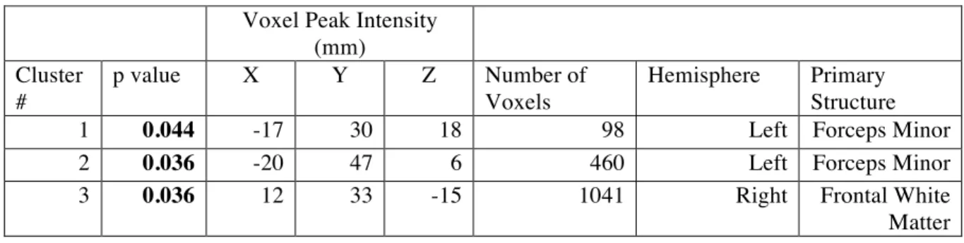
TABLE 4.2: 2X2X2 ANOVA SIGNIFICANT CLUSTER LOCATIONS FROM RANDOMISE TFCE ANALYSIS (CONCUSSION HISTORY-POSITION). SIGNIFICANCE P VALUE SET AT P<0.05. ... 57
TABLE 4.3: MEAN WEIGHTED CONTACT EXPOSURE HOURS (STANDARD DEVIATION) FOR POST-HOC IMAGING ANALYSES ... 58
TABLE 4.4: MEAN FA VOXEL (STANDARD DEVIATION) FOR POSITION-CONCUSSION HISTORY SIGNIFICANT CLUSTERS FROM TFCE ANALYSIS ... 59
TABLE 4.5: POST-HOC 2X2 ANOVA F-VALUES (P-VALUES) FOR MAIN EFFECTS AND INTERACTIONS OF CONCUSSION HISTORY-POSITION. SIGNIFICANCE P VALUE SET AT P<0.05. ... 60
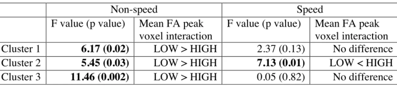
TABLE 4.6: POST-HOC UNIVARIATE ANALYSIS F-VALUES (P-VALUES) FOR CONCUSSION HISTORY-POSITION INTERACTION AND DIRECTION OF SIGNIFICANT FA DIFFERENCES. SIGNIFICANCE P VALUE SET AT P<0.05. ... 61
TABLE 4.7: POST-HOC UNIVARIATE ANALYSIS F-VALUES (P-VALUES) FOR POSITION PLAYED-CONCUSSION HISTORY INTERACTION AND DIRECTION OF SIGNIFICANT FA DIFFERENCES. SIGNIFICANCE P VALUE SET AT P<0.05. ... 62
TABLE 4.8: 2X2X2 ANOVA F-VALUES (P-VALUES) FOR THE MAIN EFFECTS AND INTERACTION OF EXPOSURE, CONCUSSION HISTORY AND POSITION VARIABLES. SIGNIFICANCE P VALUE SET AT P<0.05. ... 63
TABLE 4.9: SAT MEAN SCORES (STANDARD DEVIATIONS) FROM 3-WAY ANOVA ANALYSIS. ... 64
TABLE 4.10: POST-HOC 2X2 ANOVA F-VALUES (P-VALUES) OF SAT SCORES 3-WAY INTERACTION BETWEEN EXPOSURE, POSITION AND CONCUSSION HISTORY (EXPXC). SIGNIFICANCE P VALUE SET AT P<0.05. ... 65
LIST OF FIGURES
FIGURE 2.1: THE THREE DIMENSIONAL ELLIPSOID MODEL USED FOR DIFFUSION TENSOR ... 23 FIGURE 2.2: DIFFUSION TENSOR ELLIPSOID SHAPES WITH VARIOUS MAGNITUDES OF EIGENVALUES
... 24
FIGURE 4.1: DIFFUSE DIFFERENCES IN FRACTIONAL ANISOTROPY (FA) INTERACTION BETWEEN CONCUSSION HISTORY AND PLAYING POSITION VARIABLES ... 66 FIGURE 4.2: DIFFUSE DIFFERENCES IN FRACTIONAL ANISOTROPY (FA) INTERACTION BETWEEN
CONCUSSION HISTORY AND PLAYING POSITION VARIABLES OVERLAID ON FORCEPS MINOR (LIGHT BLUE) ... 67 FIGURE 4.3: POST-HOC INTERACTIONS FROM ANOVA ANALYSIS BETWEEN CONCUSSION HISTORY
AND PLAYING POSITION VARIABLES IN CLUSTERS (1-3) DEFINED BY TFCE ANALYSIS ... 68 FIGURE 4.4: POST-HOC 2-WAY (PLAYING POSITION AND CONCUSSION HISTORY) ANOVA FOR MEAN SAT SCORES IN COL ONLY EXPOSURE GROUP ... 69 FIGURE 4.5: POST-HOC 2-WAY (PLAYING POSITION AND CONCUSSION HISTORY) ANOVA FOR MEAN
SAT SCORES IN COL+NFL ONLY EXPOSURE GROUP ... 70
CHAPTER I: INTRODUCTION
1.1 Goal of the work
The purpose of this research is to investigate the long-term effects of concussive traumatic brain injuries, football exposure and playing position on white matter integrity and neurocognitive performances in former professional and college football athletes. It is intended, via the use of diffusion tensor imaging and neurocognitive testing, to determine the degree of neuroanatomical and neurocognitive variance between those two populations based on concussion history, duration of playing career, and playing position. Conclusions about the clinical use of brain-imaging techniques to assess concussive injuries and the predictability of the structural integrity changes are expected from this project.
1.2 Background and Significance
history of concussions, different levels of football exposure and positions with the aim of investigating the contribution of these potential predispositions to differences in white matter integrity and cognitive aptitudes over time. As such, the proposed study represents the first of its kind.
1.3 Specific Aims/Research Questions
1.3a Primary aim - Investigate the integrity of the white matter tracts in National Football
League (COL+NFL) and college (COL) football retired players with respect to concussion
history, football exposure, and playing position.
1. Are there significant differences between the integrity of the white matter tracts, defined
by fractional anisotropy (FA) and medial diffusivity (MD), observed in diffusion tensor images of National Football League and college football retired players based on years of football exposure, position played and concussion history?
(1) Variable analysis:
I. Independent: Years of football exposure, playing position and
concussion history.
II. Dependent: The integrity of the white matter tracts (defined by FA and
MD). (2) Hypotheses:
I. Research hypothesis: We hypothesize that significant differences in
white matter integrity, more specifically, decreased FA and increased MD, will be observed in the high concussion history group and the speed position group.
II. Statistical hypothesis:
1. Null (Ho): There will be no significant differences between white
matter integrity across the groups.
2. Alternate (HA): There will be significant differences in white
1.3b Secondary aim: Investigate the effects of concussion history, football exposure and
playing position on neurocognitive performances of National Football League and college
football retired players.
1. Are there significant differences between the performances of National Football League
and college retired football players on the selected neurocognitive battery tests based on football exposure, playing position and concussion history?
(1) Variable analysis:
I. Independent: Years of football exposure, playing position and
concussion history.
II. Dependent: Performance on selected neurocognitive battery tests. (2) Hypotheses:
I. Research hypothesis: We hypothesize that the high concussion history
group will demonstrate greater deficits in neurocognitive tests performance compared to the other groups.
II. Statistical hypothesis:
1. Null (Ho): There will be no significant differences in performance
on the neurocognitive testing across the groups.
2. Alternate (HA): There will be significant differences in
1.3c Exploratory aim: Investigate the relationship between neurocognitive performances of
National Football League and college football retired players and observed structural
differences in white matter integrity.
1. If there are significant differences between the performances on the neurocognitive tests,
do the observed differences in cerebral white matter integrity (from DTI) correlate with cognitive domains where athletes have shown performance deficits?
(1) Variable analysis:
I. Independent: The structural differences in white matter integrity
(expected decrease in FA and increase in MD) observed from years of football exposure, position played and concussion history.
II. Dependent: The performance on neurocognitive tests. (2) Hypotheses:
I. Research hypothesis: We hypothesize that there will be a correlation
between regions of significant differences in white matter integrity (decreased FA and increased MD) and deficits in neurocognitive test performances involving such damaged regions.
II. Statistical hypothesis:
1. Null (Ho): There will be no significant correlation between the
performances on neurocognitive tests and region-specific structural differences in white matter integrity (decreased FA and increased MD).
2. Alternate (HA): There will be significant correlations between the
performances on neurocognitive tests and region-specific structural differences in white matter integrity (decreased FA and increased MD).
1.4 Operational Definitions
Concussion History: Subjects will be split into two groups: A ‘LOW’ concussion history group,
with players who have reported none or a single concussion injury (0-1), and a ‘HIGH’ concussion history group, with players who have reported three or more concussive injuries (≥3). Football Exposure: Determined by the subjects’ number of years of football played and the weighted number of hours from both practices and games. Subjects will be divided into two groups: Players who have played college football only (‘COL’) and players who have played both college and professional (NFL) football (‘COL+NFL’).
Sub-concussive head impact: Repeated head trauma, none-concussive (that does not result in a
concussion), that may also contribute to the development of neurodegenerative diseases such as CTE [6].
White matter integrity differences: Linear rearrangement of the axonal cytoskeleton caused from stretch-induced axonal damage (swelling, disconnection, retraction) within the cerebral white matter detected and quantified by diffusion tensor imaging [7].
1.5 Delimitations
1. This study recruitment was delimited by the fact that data was already collected, which
restricted what could be done and future analyses. The previous recruiting team selected all selecting variables.
2. Participants are aged between 50-65 years (N=63) and consist of 32 former college
players and 31 National Football League retired players.
3. The previously recruited population of college and professional retired players limits our
future positional analysis.
4. Subjects were randomly assigned an order of testing conditions in order to control for
procedural bias within the order of test taking.
5. No cognitively impaired subjects were included in the study in order to provide insightful
results form the neurocognitive performance tests.
6. LOW concussion group was defined with individual who have suffered none or a single
concussive injury.
7. HIGH concussion group was defined with individual who have suffered of three or more
1.6 Assumptions and Limitations
1. Assumption: The data collected by the DTI and fMRI technicians is valid and reliable. 2. Assumption: The scanner used to collect the DTI and fMRI images were properly
adjusted and functional.
3. Assumption: Subjects were honest in answering all questions and in adhering to the
inclusive and exclusive criteria required for this study.
4. Assumption: DTI analyzing tools used in this study, such as TBSS, are reliable and
accurate.
5. Assumption: Subjects gave maximal effort when preforming their neurocognitive tests. 6. Limitation: No baselines were used to compare the potential changes in white matter
integrity and neurocognitive performances.
7. Limitation: Subjects may have experienced learning curve practice effects as a result of
repetitive testing and/or from previous exposure to concussion assessment tools.
1.7 Direction of the thesis
CHAPTER II: LITERATURE REVIEW
2.1 Introduction
Defined as a complex pathophysiological process induced by rapid biomechanical forces applied directly or indirectly to the head, concussions are functional brain injuries that result in tissue alterations, which lead to physiological and neurological complications [5]. In 2011, the Centers for Disease Control estimated that 1.6 to 3.8 million concussions occurred in sports and recreational activities [8]. Such is still an under-estimate, although it includes an expected number of individuals who suffered of concussive injuries and did not seek medical assistance. American football, among all other sports played in the US, has the greatest number of traumatic brain injuries, but also the largest participation rates [9]. Because of its popularity and such high rates, traumatic brain injuries sustained in football players deserve deeper examination in order to allow coaches, athletic trainers and physicians to better assess and manage head injuries.
The recent boom in research on sport-related concussive injuries has allowed the field of sports medicine to improve its knowledge of the psychological and neurocognitive effects of those injuries, which has led to many positive changes both on and off the football fields. The improvements in the ways physicians and athletic trainers manage concussed athletes is an indication that the findings provided by research are being put into good use. Consequences of this increase in public knowledge has also led public and private organizations as well as state legislatures to implement management protocols of sport related concussions [10]. Along with this, raised awareness on concussions has affected other areas of sports, such as equipment design and rule revisions. Such have shown to be effective when for example looking at the recent decline in the number of concussions in the National Football League (NFL) following the changes in kickoff rules [11].
impacts to the head or actual concussions sustained over a football career weighs more into exposing players to long-term health issues like working memory problems, neuropsychiatric conditions and diffuse axonal injuries in the white matter (WM), which has recently been found to be a common pathological feature in the retired population of former football players who have suffered of traumatic brain injuries [12]. It is therefore anticipated that through the use of techniques like functional magnetic resonance imaging (fMRI) and diffusion tensor imaging (DTI), researchers will be able to deepen their understanding of the acute and chronic effects of concussions at a neuroanatomical and structural level. Such improvements would be clinically significant by allowing physicians and other healthcare professionals to better educate their players on the risks that they face in the long term after sustaining concussive injuries. Additionally, such findings may lead to supplementary changes in rules by the governing bodies in order to further protect the players’ health.
2.2 Neuroanatomy
2.2a General Overview of the Cerebral Cortex
The brain is the main structure affected by concussive injuries [13]. Fueled by the cerebrovascular blood supply, originating from internal carotid and vertebral arteries, the main regions of the brain are the medulla, pons, cerebellum, midbrain, thalamus, basal ganglia and cerebral cortex. Separated by sulci, the cerebral cortex is divided into five main lobes: the Frontal, the Parietal, the Temporal, the Occipital and the insular lobes. Previous work on head injuries has found significant changes in regions of white and gray matter (GM) volumes, where patients with history of concussions showed cerebral tissue atrophy [14]. This global atrophy was observed with overall changes in the brains’ volume along with more specific affected areas such as the anterior cingulate and the left cingulate gyrus isthmus. Such observations may suggest vulnerability of the frontal regions of the brain to long-term structural changes, which functionally, is mainly responsible for cognitive and motor functions. Regional changes are thought to affect athletes’ abilities to accomplish certain cortical tasks such as motor planning and cognitive processing [15, 16]. Further changes in neurocognitive functions such as speed of information processing, attention, memory, and reaction time have also been related to traumatic brain injuries [17]. Additionally, evidences have linked concussive injuries and neurocognitive functions deficits to neural substrates such as alterations in distributed network connections within the cortex [18].
2.2b The regional functionality of the Cerebral Cortex
To understand the symptomatology of sport related concussions and their neurological effects on the cognitive abilities of athletes, it is important to take a more detailed look at the way our brain is topographically divided, and at how cortical functions are related to the cerebral anatomy. At the microarchitectural level, cerebral tissues consist of gray and the white matter; gray matter is primarily comprised of neuronal cell bodies, whereas white matter consists largely of neuron extensions referred to as axons [20]. Major pathways of the white matter are called tracts (e.g. the corpus callosum is the white matter tract connecting the two cerebral hemispheres). At a macroarchitectural level, the division of the brain into lobes is subdivided into main regions (i.e. the Brodman’s areas), which are known to be involved with a number of functions.
associated with attention, behavior regulation and motivation, and social cognition. Damaged in this area are usually accompanied with impaired explicit memory and lack of behavioral self-initiation [21].
The complex neural interconnections of those areas within the brain are key to proper functionality of the nervous system. These interactions can be structurally and functionally delineated based on white matter tracts projections, and concurrent regional activation, respectively. As shown by previous studies in this field, the neural recruitment process is not localized. For instance, neural mechanisms responsible for working memory functions involve complicated networks between regions like the prefrontal cortex and the rest of the brain, which are necessary for the conservation of normal behaviors [22]. Furthermore, concussions have recently been associated with functional inefficiencies in episodic memory network and different neural recruitment patterns. These findings are thought to be caused by changes in white matter integrity as a result of multiple sport-related concussions [23]. Such outcomes are the foundations that drive the continuation of research about the neuronal changes following concussions.
2.2c The limbic system
moods, reflected pathophysiology that was consistent with limbic-frontal neural activation changes [27]. Furthermore, those symptoms of depression experienced by patients following head trauma seem to closely share neural mechanisms found in diagnosed major depressive disorder (MDD) [27].
In vivo exploration of the brain using diffusion tensor imaging (DTI), which allows for identification of changes in white matter integrity, has revealed that symptomatic patients suffering of mTBI show declines in FA of limbic regions such the bilateral sub-genual and perigenual anterior cingulated cortex, the bilateral posterior cingulate cortex, the bilateral amygdala and the parahippocampal gyrus [26]. Other findings, comparing a control and a mild depression symptom group, have also targeted differences of gray matter concentration in the medial frontal and temporal regions along with the left parahippocampal gyrus [27]. These post-concussive regional changes in the limbic system may provide partial evidence to help explain changes in emotional behaviors experienced by patients who suffer and have suffered of traumatic brain injuries [27].
2.3 Concussions
2.3a Neurophysiology of Concussions
Concussions have been established as one of the most common form of traumatic brain injuries (TBI) across the world [28]. It is important, however, to note that not all cases of mild TBI (mTBI) are concussive, meaning that some cases of mTBI do not result in concussions. One understands that the complex interrelated chemistry between the cellular and vascular changes following concussive impacts, caused by biomechanical sheering forces within the cranial vault, is responsible for the triggering of a multilayered chain of neuro-pathophysiological reactions in the brain tissues [28, 29]. Ionic shifts, abnormal energy metabolism, diminished cerebral flow, and abnormal neurotransmission have all been identified acutely following concussion. This sequence of events is known as the “neurometabolic cascade” of concussion and is initiated by the mechanical stretching and shearing of neural axons leading to deregulated influx of Ca2+
and efflux of K+
N-methyl-D-aspartate (NMDA) receptors, the uncontrolled flux of ions induces further depolarization (with more influx of Ca2+
ions) and the suppression of neurons with glucose hypometabolism [29-31]. The increase activity of the active pumps working to restore the membrane ionic balance is energetically demanding. High glucose consumption, through increased glycolysis, causes more Ca2+
ions influx into the mitochondria of the neural cells, which eventually disrupts the oxidative metabolic and anaerobic glycolytic reactions of the cells leading to acidosis (decrease in pH) and edema (swelling) [29-31]. For several days following a concussive injury, levels of intracellular magnesium appear to decline substantially. Magnesium is used for production of adenosine-triphosphate (ATP), along with initiation of protein synthesis and balance of the cellular membrane potential [30].
As it will be seen later in this chapter, diffusion tensor imaging has been identified as a new tool to asses and identify changes in axonal integrity involved sports-related concussive and sub-concussive brain injuries [32]. Along with the triggering of neurophysiological reactions like the one described above, DAI have also shown to cause neurofilament compaction and microtubule disassembly. This disorganization, resulting from axons stretching, seems to trigger even more progressive disassembly of microtubules leading to axons breakage and axonal swelling [29]. Such micro-changes in the structural connectivity of the axons in the white matter can be quantified using DTI techniques. DTI tracts the water diffusion along the axonal parallel tract arrangement and provides a quantified analysis of the directionality along with changes in integrity of the white matter as a function of spatial locations [33-35]. Therefore, DTI is expected to become a significant structural analysis tool in the field of concussion research.
2.3b Biomechanics of Concussions
sides were most common in leading to concussive injuries [36]. As discussed above, such impacts transmit strain and shear forces through the white matter tracts causing alterations of the axonal integrity [29]. Symptoms most often reported by players following a concussion are headache, balance/dizziness, slowing down of mental reasoning, concentration, sensitivity to light and memory loss, which anatomically match the association areas of the brain related to the frontal lobe [3, 38].
More findings in this field have failed to identify a specific threshold force at which a certain impacts would guarantee a concussive injury. Although one would expect that a greater impact would increase the likelihood of concussions, work by Mihalik and al. [39] reports that it is not actually the case. The collected data showed that only 0.35% of studied impacts with forces greater than 80 g resulted in concussions.
examine differences in white matter structural differences and neurocognitive performances between retired speed and non-speed players (both college and professional).
2.3c Anatomical vulnerability of cerebral tissues
Because of the nature of head injuries, some regions of the brain tend to be more commonly affected than others. While only reported as preliminary evidence, findings from a small group of concussed athletes have shown increased neuropsychological deficits in athletes who had sustained head injuries following impacts to the crown of the head [39]. This provides an interesting bridge between the biomechanical factors of concussive impacts and the vulnerability of cerebral brain tissues. Further work in this field has also confirmed that post-concussive symptoms are associated with regional changes in brain activations [43].
Recent development of brain imaging techniques such as DTI have shown that contact sport athletes are exposed to changes in fractional anisotropy (FA) and medial diffusivity (MD) in the cerebral white matter [33]. Although not limited to the following, certain cerebral regions such as the corpus callosum, the external capsule, the inferior fronto-occipital and corticospinal tracts, the inferior fronto-occipital fasciculus, as well as smaller regions of the superior/posterior corona radiata have shown consistent structural changes from TBI under DTI analysis [33]. Furthermore, changes in axonal structures and white matter connectivity not only resulted from concussive impacts but also from repeated hits to the head sustained by contact athletes, which is now known to contribute to the cumulative long term changes in the integrity of the white matter observed in football players [54]. Although this topic will be revisited later in this chapter, further understanding of such microstructural changes in the integrity of the white matter is expected to provide more information about the vulnerability and changes in functionality of cerebral tissues following concussions.
2.4 Symptomatology of Concussions
2.4a Short-term neurological symptoms
Acute symptoms of concussive injuries such as headache, dizziness, concentration problems, fatigue and memory loss have already been mentioned in this chapter. An important distinction that needs to be made with regards to sport-related concussions is that acute loss of consciousness is not usually seen in athletes, and that post-traumatic amnesia is most often very brief [30]. Furthermore, it is also known that overall symptom duration is typically 3.5 days and that most athletes reach a full asymptomatic point within a week following their head injury. This is known as the recovery period [3]. Even if an athlete is asymptomatic, it does not imply full recovery. There may be persistent microstructural changes in cerebral tissues, which are important when discussing the long-term effects of concussions.
concussive impact within that time frame, which would normally be considered non-lethal for the athlete, could in fact cause irreversible cell damages leading to drastic unpredicted increase in intracranial hypertension and death. The phenomenon is known as the “second impact syndrome” and has involved mainly sport-related activities [28].
Acute molecular changes within the brain have also been identified with concussive injuries. Following the acceleration-deceleration forces applied to the brain, beta amyloid and tau proteins tend to accumulate on neuronal bodies within hours of the injury [55]. Tau is a normal axonal protein that promotes microtubule assembly and stability, and exists in six different isoforms each containing potential levels of phosphorylation. This is clinically significant since hyper-phosphorylated tau proteins are known to impair axonal transport disrupting neuronal and synaptic functions. Hyper-phosphorylated tau is a hallmark feature of Alzheimer’s disease (AD) [29]. Additional findings have shown that the APOE gene plays a role in the pathophysiological response to head injuries [56]. Following head injuries in animal studies, the APOE-mediated lipid transport system is stimulated [56]. Greater deposition of beta-amyloid proteins were observed with possession of the APOE e4 following traumatic brain injuries [57] and in AD brains [58]. As it will be discussed later, such information has led researchers to look into former athlete’s prevalence to neuropsychiatric disease and the contribution of previous head injury exposure.
2.4b Long-term neurological symptoms
The long-term repercussions of concussive injuries on retired football athletes and the correlations between neurocognitive dysfunctions and the integrity of white matter in the brain have garnered considerable attention recently. Findings have revealed potential late-life memory problems and risk of mild cognitive impairment along with potential early onsets of AD [1]. Retired professional players with a history of concussion showed declined mental health and cognitive functioning along with higher rates of memory problems. Additional dementia-related symptoms were also hypothesized to be linked with concussion history and repetitive impacts to the head sustained by football players [1].
functions [59]. Research on aging former NFL players compared to healthy controls found that additional cognitive deficits such as difficulties in naming and word finding were more common in the retired athletes [60]. Such deficits are thought to be associated with abnormal changes in white matter integrity, presence of deep white matter lesions and alterations of regional cerebral blood flow [60]. This leaves questions on whether or not football exposure and/or concussion history plays a major role in causing retired NFL players to experience greater cognitive impairments or depression as they age when compared to the general population.
2.4c Prevalence to neurological and neuropsychiatric conditions
The increased risks of neurological and neuropsychiatric conditions as a result of head injuries is a controversial topic of research as scientists debate on whether or not CTE is a distinct pathology [2]. First witnessed in a 51-year-old boxer suffering from delayed posttraumatic dementia with AD pathological changes [61], the search for the long-term neurodegenerative effects of concussive injuries is still incomplete. Recent imaging research has shown that athletes suffering from depression symptoms as a result of concussive injuries display reduced activation of the DLPFC and striatum, and “attenuated deactivation” of the MTL and frontal regions [27]. These findings indicated losses of gray matter in these cerebral regions. Additionally, neurodegenerative mortality in retired NFL players is three times higher than the general population in the United States, and that it is even greater (four times) more specifically for two major neurodegenerative conditions: Alzheimer disease (AD) and amyotrophic lateral sclerosis (ALS) [2]. Although unable to determine the cause of those correlations, the research seems to suggest that having a history of concussions is associated with an increased risk of neurologic disorders.
vulnerability of cerebral tissues discussed in section 2.3c. AD, ALS and CTE patients often share similar symptoms, which has led researchers in this field to hypothesized that some causes of death from AD and ALS, as reported on the death certificates, may have actually been caused by CTE, which is still not recognized as a distinct cause of death in the current ICD (International Classification of Disease) revisions [2]. Hopes are that advances in DTI techniques will deepen our understanding of the interrelations between the degradation of white matter and cognitive dysfunctions along with the prevalence to neurodegenerative conditions observed in retired football players.
2.5 Neurocognitive Testing and Concussions
Research on the relationship between concussion and neurocognitive performance in college football players have found significant associations between history of concussions and long-term deficits of certain cerebral domains [62]. More specifically, domains of executive functioning, speed of information processing and areas associated with self reported symptoms were identified in individuals who have sustained two or more concussions. This is significant for this research since concussion history is a main variable in looking at white matter integrity of former football players. This work will look for deficits in neurocognitive outcomes in retired players and a potential coincidence with alterations in white matter integrity from the DTI analyzes. Future findings may allow for new combinations of tests, such as specific neurocognitive and neuroimaging examinations, in order to better predict regional anatomical structural changes and network connectivity alterations as a result of concussive injuries along with better understanding their effects on the cognitive state of injured/recovered athletes [17].
2.6 Neuroimaging and traumatic brain injuries
2.6a Functional magnetic resonance imaging (fMRI)
2.6a(1) Description and overview
which is an indirect measure of neuronal activity [64]. Cognitive tasks and associated stimuli can be presented during a scanning session to determine neuronal activity in response to the task. A typical experimental paradigm used for fMRI analysis of working memory is the N-back task, which involves identification and location of verbal and nonverbal stimuli. Consistent activation of the frontal and parietal cortical regions have been shown using this test [65]. Advantages of fMRI in clinical neuroscience are that it is noninvasive, relatively widely available in both medical and university settings, and has high spatial resolution [66].
The use of neuroimaging techniques has been explored clinically to assess cognitive alterations. Such deficits in cognitive performances were observed on tasks associated with memory, naming and word finding in aging former NFL players when compared to non-depressed and healthy subject groups [60]. The use of fMRI has also recently been used to map human functional networks, which has confirmed hypotheses about the changing functionality of the brain as adults age. Aging is associated with changes in properties of the brain functional networks, which confirmed previous findings on the differences of “default-mode networks” between children and adults [67]. Such findings suggest that the natural process of aging along with the maturation of the brain may account for quantifiable changes in neurological functions and network topology. These findings should therefore be considered in this current study while carrying DTI analyses, as they could account for some of the predicted changes in white matter integrity and structural connectivity of former college and professional players.
2.6a(2) Abnormal activation patterns related to concussions
significant differences in their performance on computerized tests of executive functions, when compared to a control group, their fMRI analysis revealed “pronounced abnormalities in functional activation within the dorsal frontoparietal network” [68]. There appears to be a trend where the underlying functional and structural neurological changes are common long-term consequences of repetitive traumatic brain injuries. More importantly, those changes do not seem to be reflected in neurocognitive performances, which indicates the need for imaging analyses when managing concussed athletes. It was also suggested that this “compensatory mechanism” and over-activation of the brain was in place to counter the structural changes associated with repetitive concussive impacts [68].
The number of previous concussions was identified as a potential factor for differences in neuro-functional activity with regards to episodic memory. Although performing similarly to an age-matched adult group behaviorally, fMRI data analysis of former NFL players showed that high concussion group (three or more) exhibited different patterns of activation with relational memory processing (memory for the relationships between elements) [23]. As proposed by the researchers, these findings suggests that repeated number of concussive injuries and neurological trauma may be a factor in causing inefficient changes in neural recruitment, which seems to have an effect on memory tasks in the long term. Further findings on this topic are consistent with these conclusions showing that concussion history may contribute to greater differences in neural recruitment during working memory task performance when compared to weighted football exposure in both college and NFL retired players [4].
2.6b Diffusion Tensor imaging (DTI)
2.6b(1) Description and overview
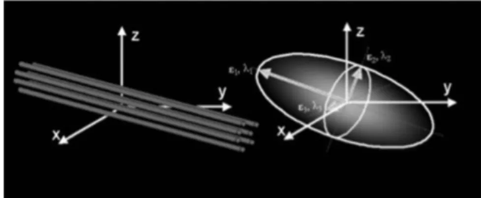
directions. This unique property of white matter is exploited by DTI analysis to tract and quantify three-dimensional directionality and integrity as a function of spatial location [33-35]. Structural connectivity patterns can be derived from whiter matter tractography where trajectories of fibers of interest can be three-dimensionally reconstructed and segmented using continuous tracking algorithm [35, 73]. The two most frequently used DTI markers used by this field of research are trace and anisotropy of the diffusion tensor [35]. The diffusion tensor uses a symmetric 3x3 matrix, which represent the 3D properties of the diffusion of water molecules based on a Gaussian model [74]. Three pairs of orthogonal eigenvectors (ε1, ε2, ε3) can be calculated from each diffusion tensor of imaging voxels using matrix diagonalization [34]. Such vectors are ordered by the magnitude of their eigenvalues (λ1>λ2>λ3) and represent the direction and magnitude of diffusivity within biological tissues; ε1, the largest diffusion magnitude in each voxel, is the dominant fiber direction [34]. The spatial arrangement of the diffusion tensor is visualized using the ellipsoid model, which is defined by the three eigenvectors as seen in Figure 1 [35].
Figure 2.1: The three dimensional ellipsoid model used for diffusion tensor
Various magnitudes of eigenvalues change the shape of the ellipsoid. The trace of the diffusion tensor (Tr) is the sum of the diagonal elements (D) of the diffusion tensor. It is a measure of the magnitude of diffusion and is rotationally invariant [35]. The apparent diffusion coefficient (also called mean diffusivity or MD) acts as an averaged index of water molecules mobility and diffusivity. It is obtained by dividing the trace by three (MD=Tr/3), which is equivalent to the three-eigenvector values of the ellipsoid model:
Changes in MD are suggested to correlate pathologically with myelin loss and/or axonal injuries [35]. Fractional anisotropy (FA) on the other hand is more commonly reported as an indicator for white mater integrity. Diffusion anisotropy measures the degree to which the diffusivities are function of the diffusion-weighted encoding directions [35]. FA is the most widely used measure of anisotropy and is described by the formula below [35]:
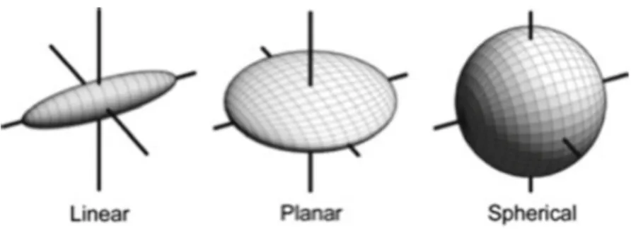
When diffusion is isotropic, with diffusivity equal in all directions (λ1=λ2=λ3), the ellipsoid is reduced to a sphere. Abnormalities and deficits in the white matter such as loss and destructive lesions of axons have values approaching 0. Contrarily, if diffusion is anisotropic, the highly directional white matter has a FA value approaching 1 (1 being completely anisotropic diffusion), which defines the theoretical FA range between 0 and 1 based on the three eigenvalues of the ellipsoid [35]. Figure 2 provides a visual for the various shapes of the ellipsoid based on relative magnitudes of eigenvalues [34]:
Figure 2.2: Diffusion tensor ellipsoid shapes with various magnitudes of eigenvalues
It is important to note that studies have disagreed concerning the direction of changes in FA and MD in early phase of recovery following concussive injuries [69]. While some reported increases in FA and decreases in MD [75, 76], other works reported opposite trends with decreased FA and increased MD [77, 78]. Acknowledging the fact that this current work will be focusing on long-term effects of concussive impacts and repetitive head injuries, those conflicting results are less of interest. What is more important, and also consistent with research, is that individuals with persistent cognitive impairment in later phases of recovery showed decreased FA [79], and increased MD [80], which are the methodological guidelines that will be used to conduct our DTI analyses. An additional note about previous DTI findings in neurology is that region-specific analysis of DTI differences have been proposed on verbal and memory tasks performed by populations suffering from neuropsychiatric diseases such as schizophrenia or parkinsonism, among others [16]. Cerebral areas such as the uncinate fasciculus, anterior corona radiata, forceps minor, and superior longitudinal fasciculus seem to show meaningful differences on DTI analyzes.
Final comments about the general overview of DTI regard the tract-based spatial statistics (TBSS) that will be used to conduct voxel-wise analysis on the cerebral white matter tracts [81]. Under the assumption that maximum FA is found at the center of the white matter tracts, TBSS generates a three-dimensional skeleton of the white matter using a non-linear FA volumes registration tool (FNIRT) which transforms all subjects’ FA into a common space (MNI) [82]. These averaged spatial arrangement allow for statistical voxel-wise analysis of the differences in white matter integrity [34, 71] between the subdivided groups.
2.6b(2) Tractography and specific networks of interest
network analysis of individuals with sport-related concussions reported lack of structural integrity of the left temporal lobe, which typically host association and projection fiber tracts running anterior-posterior and superior-inferior within the white matter [71]. Affected regions included the anterior-posterior oriented long associations fibers from the inferior fronto-occipital fasciculus, which connects the frontal and occipital lobes, as well as from the inferior longitudinal fasciculus, connecting the temporal and occipital lobes. Such findings, and others with similar conclusions [32], correlate with neurobehavioral impairments experienced by individuals post-injury. Further work on white matter anatomy determined that only association and commissural fibers were involved in cortico-cortical networks, whereas projection fibers were typically involved in connecting the cortex to non-cortical structures such as the brainstem and the thalamus [83].
2.6b(3) Concordance with fMRI readings
The brain’s structural and functional connectivity seem to be intimately related, sharing different correlations with neurocognitive deficits observed in post-traumatically brain-injured patients. Structural disconnections measured using DTI were found to correlate with traumatic brain injury severity, which provided evidences for an underlying relationship between the gravity of the initial injury and long-term white matter alterations [84].
Considering that both fMRI and DTI contribute to our understanding of diffused axonal injuries resulting from traumatic brain trauma, it will be important to continue to look for such relationships linking the two methods in order to advance our knowledge in this field.
2.6c Limitations and Complications
Both fMRI and DTI techniques have their own limitations based on the type of signal that is collected for analyses. For instance, although functional neuroimaging offers insights on neuroanatomical functionality, the selection of task paradigms, in addition to the alterable brain-behavior plasticity limits the breath of individual study conclusions and comparability of results in the literature [15]. More limitations on fMRI reports include its dependency on metabolic demands of neuronal activity, which itself depends on the density of capillary networks and their locations. Such interdependence is suggested to limit the spatial resolution and quantitative comparisons of different areas of the cortex, which differ in cerebrovascular supplies and therefore their ability to be detected by hemodynamic-based signal [85]. Highly vascularized area such as the frontal cortex, which will be the focus of this work, should reveal relatively high spatial resolution reducing this limitation. DTI is limited by factors such as thermal and physiological image noises along with misregistration errors in images resulting from eddy currents and head motions at the time of the capture. Further limitations in DTI, although inevitable considering the type of analysis conducted, are the averaging of tissues such as gray matter, white matter and cerebrovascular fluid into large voxels and the many cerebral areas known to contain significant amount of fiber crossing. Such considerable fiber crossing within one voxel hinders one’s ability to correctly identify white mater abnormalities since the micro-alterations are usually undetected [35].
limitations to this research are the age and target groups that have been mentioned in earlier studies, which usually vary between college and professional athletes, and others.
2.7 Conclusion and Direction for future Research
In summary, as mentioned earlier, the neurological effects of concussions have been intensely studied as well as the biomechanics of impacts leading to these injuries. The recovery periods of injured athletes at all levels, from high school to professional as well as the neurocognitive ways to test for these traumatic brain injuries have also been deeply investigated. The next step in the research is to deepen our understanding of the relationship between anatomical and neurocognitive changes occurring from these brain injuries, which will allow us to find ways to better prevent them, in addition to improve our treatments. Recently, various procedures have been put into place with specific focus on behavioral changes of playing athletes as well as rule changes, which have allowed experts to observe a significant reduction in the rate of concussive injuries. The use of brain imaging techniques, like the fMRI and DTI, will allow experts in field to further improve their understanding of concussions as well as to develop more specific and accurate methods to diagnose injured athletes.
CHAPTER III: METHODOLOGY
3.1 Subjects
3.1a Participants
Recruited participants in this study compiled a total of thirty-two former collegiate football players who played a minimum of three years of college football (“COL”; M=58.63; sd=3.663; all male) and thirty-one former National Football League (NFL) players who played a minimum of five seasons of professional football (“COL+NFL”; M age=58.12; sd=3.74; all male). An additional former professional football player from the high concussive group underwent all neurocognitive and genotyping assessments but had to be removed from the dataset because of his invalid magnetic resonance imaging results. All participants were given written informed consent prior to participating in the study, which were in accordance with the requirements of the Institutional Review Board at the University of North Carolina at Chapel Hill.
3.1b Recruitment
Former collegiate football players were recruited from the North Carolina, Virginia, and South Carolina regions surrounding Chapel Hill, NC. A sufficient pool of subjects was extracted to match with the recruited retired NFL players. The sport information directors and athletic trainers of Division I football programs within this tri-state region were contacted to identify graduates of those respective programs who lived in these areas. Identified individuals were then sent a letter from the research team inquiring if they would be willing to participate in the research study. Interested participants were then further assessed on the same screening instruments and then invited to campus for further studies. Former college and professional football players were matched on age, education level, and position played. The following inclusive and exclusive criteria were determined prior to recruitment of the subjects.
3.1c Inclusion Criteria
− Must be a retired American football player at either the college or professional level.
− Must be 50 years of age and older.
− Subject stratified in the college group were required to have played a minimum of 3 years of college football.
− Subject stratified in the professional group were required to have played a minimum of 5 years in the NFL (non-kicker/punter).
− Subjects stratified in the HIGH concussion history group were required to have a sustained and reported three or more concussions (≥3) during their football career.
− All subjects had recent self-reported memory problems.
− Report of below average response on at least two of the following four questions on the mild cognitive impairment survey instrument: Questions 1, 2, 3 and 7. In cases where both the retiree and the intimate partner or close relative returned a survey, the average of the two scores on the four previous highlighted questions was used.
3.1d Exclusion Criteria
− Former kickers or punters.
− Conditions unsafe for magnetic resonance scanning, such as cardiac pacemaker, epicardial pacemaker leads, cochlear implants or claustrophobia.
− Functional Activities Questionnaire (FAQ) score below 8 (indicative of dementia).
3.1e Stratification
3.1e(1) Concussion history and football exposure
Selected subjects were subdivided into four groups based on both concussion history and football exposure. The following groups are defined below with their respective inclusion criteria:
1) (COL+NFL)-LOW: A sample of 16 retired professional football players. - Minimum of 5 years of NFL experience;
- Mild memory impairment reported and confirmed on MCI survey;
- Reported none or single concussion (0-1) injury during professional football career.
2) (COL+NFL)-HIGH: A sample of 15 retired professional football players. - Minimum of 5 years of NFL experience;
- Mild memory impairment reported and confirmed on MCI survey; - Reported 3 or more (≥3) concussions during professional football career 3) (COL)-LOW: A sample of 16 former collegiate football players.
- Minimum of 3 years of collegiate experience;
- Mild memory impairment reported and confirmed on MCI survey;
- Reported none or single concussion (0-1) during collegiate football career. 4) (COL)-HIGH: A sample of 16 former collegiate football players.
- Minimum of 3 years of collegiate experience;
- Mild memory impairment reported and confirmed on MCI survey; - Reported 3 or more (≥3) concussions during collegiate football career.
Table 3.1: Football exposure and concussion history
Football Exposure
COL COL+NFL
Concussion History LOW (0-1) 16 16
3.1e(2) Positional
This project also plans to stratify subjects based on primary football positions played in college and in the professional football league. Such stratification is inspired from a recent study on neurodegenerative causes of death in NFL players by Lehman et al. published in 2012 [2]. The recruited players will be sub-divided into two positional groups: ‘speed’ (Quarterbacks, Running backs, Halfbacks, Fullbacks, Wide Receivers, Thigh ends, Defensive backs, Safeties, Linebackers) and ‘non-speed’ (All Offensive and Defensive linemen). Table 3.2 below summarizes the sub-division:
Table 3.2: Participants' football playing position
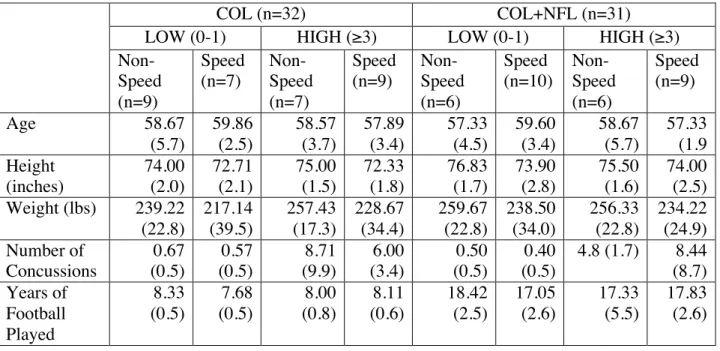
The next table (Table 3.3) summarizes all three variables of interest and the stratification used to conduct the three-way ANOVA.
Table 3.3: Stratification by football exposure, concussion history and playing position
Football Exposure Concussion
History
Position N (COL) N (COL+NFL) N (Total)
LOW (0-1) Non-speed 9 6 15
Speed 7 10 17
Total 16 16 32
HIGH (≥3) Non-speed 7 6 12
Speed 9 9 18
Total 16 15 31
Stratified Groups
College: COL (n=32) NFL: COL+NFL (n=31)
Total
Speed (QB, RB, HB, FB, WR, TE, DB, ST, LB)
16 19 35
Non-speed (All OL and DL) 16 12 28
3.2 Measurements and Instrumentation
3.2a Concussion history
Concussion history was obtained from the data collected from the retirees’ general health survey. Once selected subjects were finally invited to campus, the concussion history was further confirmed through interviewing the subjects and getting access to any available medical documentation provided by the subject. Following confirmation of the concussion history, each subjects was assigned to the HIGH and LOW concussion sub-groups. Permission to review any available pertinent clinical records was obtained from each retiree. In most cases, such records were hardcopy files stored by team physicians, neurologists, and athletic trainers. High ability to recall concussion history was observed in the subject and this because such injuries are significant to the athletes in influencing their ability to play. Data on concussive injuries other than those that occurred during the professional playing career were also collected. These include concussions from motor vehicle crashes, or from pre-professional play. We have noted that the data obtained on professional playing career concussions tend to be of high quality and this will form our main focus.
3.2b Contact Exposure Index
Research staff tracked subjects’ football career histories from high school level continuing through college and all the way to professional career by conducting structured oral interviews. For each year of their football career, participants provided information about: primary position played (i.e., quarterback, offensive line, running back, defensive line, defensive back, linebacker, wide receiver, special teams); number of games in the pre-season, regular season, and post-season; percent of time that they played in games; number and length of contact practices. The exposure history variable was then created from this information [4].
The following formula was used to calculate the number of practice contact hours for each year: (# pre-season practice sessions/week * # pre-season weeks * # hours/pre-season practice session) + (# regular season practice sessions/week * # regular season weeks * # hours/regular season practice session) + (# season practice sessions/week * # post-season weeks * # hours/post-post-season practice session)
The number of games hours was then calculated using this following formula:
(# pre-season games * % of time active in pre-season games * 1 hour) + (# regular season games * % of time active in regular season games * 1 hour) + (# post-season games * % of time active in post-season games * 1 hour)
Finally, the sum of both the number of hours for practices and games per year throughout participants’ career was used to create their total contact exposure [4].
3.2c Magnetic Resonance Imaging
3.2c(1) Structural Images
Magnetic resonance images were acquired using a Siemens Trim-Trio 3-T scanner. The subjects’ heads were held in place in the scanner using cushions and a headrest. Following an initial localizing scan, T1 weighted images were obtained. Covering the entire brain with a voxel size of 1x1x1 mm3, T1 weighted structural scans for anatomical visualization were obtained with the following scanner parameters: 160 slices at TR/TE/TH/=1750msec/4.38msec/1mm. DTI series of images were taken following the T1 scans (details below).
3.2c(2) Diffusion Tensor Imaging (DTI)
A spin echo diffusion tensor weighted sequence was used to acquire the images. A baseline image and 64 directional images were acquired at an isotropic resolution of 1x1x1 mm3
. The baseline image was taken without a diffusion gradient (b = 0) and the remaining six images were taken with b = 1000s/mm2
. As outlined below, tensor maps, fractional anisotropy and mean diffusivity will be the metrics of interest computed from the raw DTI data. All diffusion tensor images will be transferred to a workstation for data analysis.
MD is used as an averaged index of water molecules’ mobility and diffusivity within white matter. It is obtained by dividing the trace (Tr) of the diffusion tensor in three (MD=Tr/3) in order to get the three-eigenvector values of the ellipsoid model used for diffusion tensor 3x3 spatial matrix [35].
highly isotropic white matter tracts whereas FA values approaching 1 signify highly directional white matter. Recalling from chapter 2, the FA formula is outlined below [35]:
3.2d Neurocognitive Selected Test Battery
The selected neurocognitive test battery for this research project was carefully chosen in order to cover as many of the cognitive domains as possible. Find below a description of each selected test and an accompanied reasoning for their selection. Table 3.4 below summarizes the selected test battery along with more specific information about expected regions of interests affiliated with each test.
hits and correct passes, immediate and delayed. Correct responses from VBM and VIM are summed to generate a composite memory or memory domain score. The highest score one can attain is 120; the lowest is 60. Scores below 60 suggest willful exaggeration.
For this specific test battery, only the delayed memory score were used to conduct the statistical analysis since delayed forgetting seams to be more symptomatic in patient with history of traumatic brain injuries [87]. A corrected recognition score was generated for both the VERM and the VISM (VERM_cr and VISM_cr). The converted scores were obtained using the formulas below:
VERM_delayed_CR=[(verm_delayed_correct_hits)−(15−(verm_delayed_correct_passes))] /15
VISM_delayed_CR=[(virm_delayed_correct_hits)−(15−(virm_delayed_correct_passes))] /15
Symbol-Digit Coding (SDC) – Coding has been a component of the Wechsler Intelligence Scales since 1944 (Digit Symbol Substitution, DSST). The Symbol Digit Modalities Test (SDMT) is a variant of the Wechsler DSST, but the position of symbols and digits is reversed. The clinical and psychometric properties of the SDMT are similar to those of the DSST. Although the SDMT may be a “harder” test, and thus more sensitive to neurotoxicity, performance on the SDMT and the DSST are highly correlated. The SDC in CNS Vital Signs draws from a reservoir of 32 symbols. Each time the test is administered, the program randomly chooses eight new symbols to match to the eight digits. Scoring is the number of correct responses generated in 2 minutes. The total of right and left taps from the FTT and total correct responses on the SDC generates a composite score for “psychomotor speed.” Thus the SCD correct scores were selected in this battery for their ability to provide a cognitive measure of processing and fine motor speed.
change at random. For one presentation, the rule is to match the figures by shape, for another, by color. This goes on for 90 seconds. The goal is to make as many correct matches as one can in the time allowed. The scores generated by the SAT are: number correct, errors, and response time in milliseconds. There is not a precise parallel to the SAT in the compendium of conventional neuropsychological tests, although Trails B and the Wisconsin Cart Sort are considered to be tests of shifting attention. Thus, the correct SAT correct and response time scores were selected in this battery to provide a measure of complex attention, cognitive flexibility and executive functioning as well as reaction time.
Finger Tapping Test (FTT) – The FTT is one of the most commonly used tests in neuropsychology, because of its simplicity and reliability, and because it generates relevant data about fine motor control, which is based on motor speed as well as kinesthetic and visual-motor ability. The FTT is believed to be one of the most sensitive neurocognitive tests for determining brain impairment. In CNS Vital Signs, the FTT is a very simple test. Subjects are asked to press the Space Bar with their right index finger as many times as they can in 10 seconds. They do this once for practice, and then there are three test trials. The test is repeated with the left hand. In this selected test battery, the scores of the non-dominant hand were used to provide a measure of fine motor speed as more variations in those scores would be expected due less immunity to fatigue [88] and a slower tapping rate [89].
Table 3.4: Instrumentation, measurements and their roles
Measure Role
Concussion History Self-Administered Questionnaire
Stratification tool
Contact Exposure Index Exposure history including games and practices
Stratification tool
Playing Position Self-Administered Questionnaire
Stratification tool
Diffusion-Tensor Imaging (DTI)
Fractional anisotropy (FA) White matter integrity
Mean diffusion (MD) White matter integrity
CNS Vital signs tests Delayed VERM and VISM corrected recognition score
Provides measure of verbal and visual memory.
Symbol-Digit Coding (SDC)
Provides measure of psychomotor speed.
The Shifting Performance Test (SAT)
Provides measure of subject’s ability to shift from one instruction set to another quickly and accurately.
Finger Tapping Test (FTT) – Non dominant hand
Provides measure of fine motor speed (kinesthetic and visual-motor ability). Non-verbal Reasoning test Provides measure of
3.3 Testing Procedures
3.3a Setting
All subjects were assessed at UNC-Chapel Hill’s Biomedical Imaging Research Center, and The Matthew Gfeller Sport-Related TBI Research Center. Study participants were evaluated as part of a two-day visit in which they were assessed for neurocognitive status, brain imaging findings, ApoE4 genotyping, and depressive status. A pre-visit telephone interview conducted by researchers at the Center for the Study of Retired Athletes established background demographics regarding their previous football exposure (involving both sub-concussive and concussive impacts) and prior medical history, including sport-related and non-sport-related concussions sustained during their career.
3.4 Data Analysis
3.4a Magnetic Resonance Imaging
3.4a(1) Preprocessing steps
All acquired neuroimaging data were processed under supervision of our image analysis expert, Dr. Shen, who has applied his techniques to study various brain diseases [90, 91]. All pre-processing steps were performed with the FMRIB Diffusion Tool Box (FDT; FMRIB Centre, University of Oxford, UK). The pre-processing steps for the neuroimaging analyses included (1) alignment of all modality images (including T1, and DTI) of the same subject by rigid registration [92], (2) removal of extra-cranial tissues (skull-stripping) [93], and (3) fitting of the diffusion tensors at each voxel of the corrected data to check for quality. The end outputs (the FA and L2 images) were carried into the TBSS protocol prior to the voxelwise analysis.