Introduction
Phospholipase C g1 (PLCg1) is a protein found highly expressed in human T-cells that plays critical roles in cellular signal transduction, with roles in pathways ranging from DNA transcription to innate immune responses.1 PLCg1 is directly activated by tyrosine phosphorylation mediated by numerous tyrosine kinases to catalyze the
hydrolysis of phosphatidylinositol 4,5-bisphosphate (PIP2), a phospholipid found enriched in the plasma membrane. Cleavage of PIP2 by PLCg1 generates membrane-bound diacylglycerol (DAG) and cytosolic inositol
1,4,5-trisphosphate (IP3), both of which act as second messengers in many signal transduction pathways via stimulation of protein kinase C (PKC) and the mobilization of intracellular calcium, respectively. These signaling cascades downstream of PLCg1 control various processes including embryonic development and cell migration.2,3
In addition, aberrant regulation of these pathways can result in diseases like cancer. Notably, PLCg1 has been
shown to harbor mutations in patients diagnosed with adult T-cell leukemia/lymphoma (ATL).4
ATL is an aggressive T-cell cancer associated with infection by human T-cell lymphotrophic virus type 1 (HTLV-1) and further associated with a poor prognosis for patients that contract the disease. However, only a fraction of individuals infected with HTLV-1 develop ATL and the latency period between infection and disease is typically decades.4,5 These observations indicate that additional mechanisms must also contribute to the development of ATL. Recent work has demonstrated that ATL patients often harbor mutations in components of the T-cell receptor (TCR) signaling pathway. In fact, PLCg1, which plays a major role in proximal TCR signaling (Figure
1), is the most frequently mutated gene, with mutations in PLCg1 identified in 36% of patients with ATL.4 Interestingly, point mutations in PLCg1 span the entirety of the protein (Figure 2), suggesting the possibility of
multiple mechanisms for dysregulation of PLCg1 activity. Despite evidence suggesting that PLCg1 can contribute
to cancer phenotypes, for example by driving cellular transformation,6 little research into how cancer-associated mutations in PLCg1 affect its lipase activity has been conducted. To define the effects of various cancer-linked
mutations in PLCg1 on its activity, we have conducted experiments transiently over-expressing mutant forms of
PLCg1 in HEK293 cells to measure accumulation of inositol phosphates (IPs), a read-out of PLCg1 phospholipase
mutant isozymes had an increase in lipase activity compared to the wild-type enzyme under basal conditions, as well as in the context of receptor-dependent activation. These data strongly suggest that PLCg1 is an oncogene
that drives ATL pathogenesis.
Methods Cloning
Wild-type Rattus norvegicus PLCg1 was expressed in a modified pcDNA3.1 vector that incorporates an HA epitope tag at the N-terminus of the protein. To introduce point mutations into PLCg1, primer-mediated
mutagenesis was conducted using polymerase chain reaction. PLCg1 was synthesized in two fragments and
subcloned into the modified pcDNA3.1 vector backbone using Gibson Assembly cloning according to the manufacturer’s protocol (New England BioLabs). The products of the reaction were transformed into DH5a
Escherichia coli and selection was performed on agar plates containing 75 µg/mL carbenicillin. Plasmid DNA was isolated and analyzed by BamHI restriction digest to confirm correct assembly of products. Plasmids with the correct digestion pattern had their open reading frames fully sequenced by automated di-deoxy sequencing. Mutant forms of PLCg2 were cloned using the same process.
Cell Culture
HEK293 cells were maintained in Dulbecco’s Modified Eagle Medium (DMEM) supplemented with 10% (v/v) fetal bovine serum (FBS) and 1X antibiotic-antimycotic (AA). Cells were grown in a humidified incubator maintained at 37ºC and 5% CO2.
Quantification of Inositol Phosphate Species
Accumulation of IPs was determined using a four-day protocol. On day one, HEK293 cells were plated at 75,000 cells per well in supplemented DMEM in 12-well tissue culture plates and incubated at 37 ºC, 5% CO2. Each well contained a total volume of 1 mL. On day two, the cells were transfected using Continuum transfection reagent according to the manufacturer’s protocol (Gemini Biosciences) with an expression vector encoding wild-type or mutant forms of PLCg1 (100 ng) and an empty expression vector (200 ng) to keep the amount of DNA per well
DMEM containing [3H]myo-inositol such that each well received 1 µCi of [3H]myo-inositol. In addition, 10mM LiCl was included in the labeling medium to block IP degradation and IP species were allowed to accumulate for ~16 hours. On day four, media were removed and cells were lysed by the addition of 750 µL of 50 mM formic acid. After a 45 min incubation on ice, the lysates were neutralized with 250 µL of 150 mM ammonium hydroxide and applied to Dowex AGI-X 200-400 mesh anion-exchange columns. Columns were washed with 10 mL of 50 mM ammonium formate and IPs were eluted into 4 mL of scintillation fluid using 2 mL of 1.2 M ammonium formate/0.1 M formic acid. The mixture was shaken until clear and IP accumulation was quantified by liquid scintillation counting.
A similar protocol was used to quantify PLCg1 activity downstream of the epidermal growth factor receptor
(EGFR). However, for these experiments, expression vectors for PLCg1 (200 ng) and EGFR (100 ng) were
co-transfected and cells were treated with an isotonic, buffered solution containing 20 nM recombinant EGF (Invitrogen) plus 10 mM LiCl for 30 min prior to lysis with formic acid.
Basal phospholipase activity of PLCg1 mutants
All of the mutant forms of PLCg1 found in ATL had increased basal phospholipase activity, as compared to the
wild-type enzyme after transient over-expression in HEK293 cells (Figure 3). This increase in activity ranged between 8- and 2155-fold over the activity of wild-type PLCg1. Given this broad spectrum of basal activities, the
mutant forms of PLCg1 can be divided into three different groups: those mutations that result in a relatively small
(<50 fold) increase in basal PLCg1 activity (R48W, E952K, Q641K, E856A, Q606R, I854E, F1167E, D625Y,
R753H, R687W), a moderate (50<fold increase<1000) increase (M1166R, I486E, V501E, F1167I, P867R, D1169G), and a large (>1000 fold) increase (Y509E, D1165H, E1163K, S345F) in phospholipase activity relative to wild-type PLCg1. Mutants of PLCg1 with moderate to high increases in basal activity are found across multiple
domains of PLCg1, suggesting they may operate by multiple different mechanisms.
Three of the four point mutations in PLCg1 found most frequently in ATL (D1165H, 6%; E1163K, 5%; S345F,
the context of ATL, these mutant forms of PLCg1 function as oncogenes that drive disease pathogenesis.
Interestingly, the R48W mutation is also frequently found in ATL (14%),2 but shows an only 8-fold increase in activity, much less than the other common ATL point mutations. The correlation between frequency of mutations in ATL and the basal activity of PLCg1 remain unclear. It is also unknown whether a threshold level of PLCg1
activity is necessary to induce cellular transformation and experiments exploring this concept are proposed in the future directions.
EGFR-stimulated activity of PLCg1 mutants
Epidermal growth factor receptor (EGFR) is a receptor tyrosine kinase (RTK) that phosphorylates tyrosine residues on its downstream effectors, like PLCg1. In the case of PLCg1, tyrosine phosphorylation directly
stimulates phospholipase activity in response to activation of EGFR by its ligand, EGF. By quantifying phospholipase activity in the presence or absence of EGF, we determined how the mutant forms of PLCg1 respond
to receptor-dependent activation.
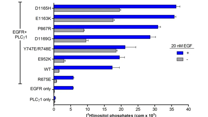
HEK293 cells were transiently co-transfected with EGFR and wild-type or mutant forms of PLCg1, and
challenged with 20 nM EGF for 30 min (Figure 4). Cells over-expressing PLCg1 alone showed no
EGF-dependent increase in IPs, indicating that these cells do not endogenously express EGFR. The growth factor dependent increase in IP production in cells over-expressing EGFR alone is attributable to the presence of endogenous PLCg1. Co-expression of EGFR and PLCg1 resulted in an ~10-fold increase in IP accumulation. As
an additional control, EGFR was co-expressed with PLCg1(R675E). Our lab has previously shown this mutant
form of PLCg1 is refractory to activation by EGFR7; as expected, IP accumulation in cells expressing
PLCg1(R675E) was essentially identical to cells expressing EGFR alone.
In the absence of EGF, the basal activity levels among the mutant forms of PLCg1 were consistent with the relative
levels of basal activity observed after the overnight incubation with LiCl. That is, the relative activity level of each mutant form of PLCg1 was comparable between overnight accumulation and basal activity in the absence
constructs demonstrating up to double the activity of the wild-type enzyme. Differences in EGF dependent activity between mutant forms of PLCg1 and wild-type PLCg1 were further confirmed with dose-response experiments.
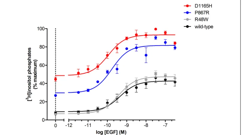
PLCg1(R48W), PLCg1(P867R), and PLCg1(D1165H) were chosen as a representative sample of the varying
degrees of basal activity increase seen in mutant forms of PLCg1 and were challenged with concentrations of
EGF ranging between 10-11.5 and 10-6 M (Figure 5). Consistent with the single-dose experiments, the mutant forms of PLCg1 show phospholipase hyper-activity compared to wild-type PLCg1, reaffirming EGF dependent
changes in activity in the mutant forms of PLCg1. In addition, these data suggest that certain mutant forms of
PLCg1, e.g. PLCg1(D1165H), are actively signaling in most cellular contexts. In contrast, other mutant forms of
PLCg1, e.g. PLCg1(R48W), are only hyperactive under certain conditions, such as elevated levels of EGF.
We also quantified lipase activity in the context of receptor-dependent activation for PLCg2, the other PLCg
isozyme in humans, which is regulated similarly to PLCg1. Like PLCg1, dysregulated PLCg2 activity drives
several diseases, including autoimmune disorders and cancer.8,9 Accordingly, mutant forms of PLCg2 found in cancer were assayed to determine EGFR-dependent lipase activity (Figure 6). Similar to PLCg1, all mutant forms
of PLCg2 showed elevated phospholipase activity over the wild-type enzyme in the presence and absence of EGF,
suggesting that this upregulated PLCg2 activity could also contribute to disease phenotypes.
Future Directions
Our data indicate that the point mutations in PLCg1 found in ATL are all gain-of-function, resulting in
hyper-activity of the phospholipase, both basally and in the context of receptor-dependent activation. These results raise multiple questions. For example, are additional mechanisms necessary to drive the onset of ATL when PLCg1 is
mutated, or is dysregulated lipase activity sufficient? If the latter scenario is true, is there a threshold level of PLCg1 activity required to drive cellular transformation? To begin to address these questions, we are currently
generating NIH3T3 fibroblast cell lines that stably over-express wild-type PLCg1, PLCg1(R48W),
PLCg1(P867R), or PLCg1(D1165H). We will define the capacity of each cell line to drive colony formation in
possibility is that colony formation will be correlated with basal PLCg1 activity, that is, the number of colonies
will increase as basal PLCg1 activity increases. By finding the answers to questions such as these, the oncogenic
role of PLCg1 will be better defined and we can explore the possibility of PLCg1 as a potential drug target.
Acknowledgements
Figure 1. A simplified schematic of T-cell receptor signaling. Upon stimulation by a peptide-major histocompatibility complex (MHC), the phosphorylated T-cell receptor recruits the non-receptor tyrosine kinase Zap-70. Zap-70 phosphorylates linker for activation of cells (LAT), which recruits PLCg1 into the
T-cell receptor signaling complex. Upon phosphorylation by interleukin-2-inducible T-T-cell kinase (ITK), PLCg1
is activated resulting in the hydrolysis of PIP2 into DAG and IP3.
Figure 2. Position (n=26) and frequency of substitutions (red balls) in PLCg1 for a cohort of 370 patients with
Figure 3A. Accumulation of [3H]inositol phosphates after an ~16 hour incubation with 10 mM LiCl. Colors indicate the fold change in basal PLCg1 activity compared to wild-type PLCg1; grey, FC<50; blue,
50<FC<1000; red, FC>1000. A mutant form of PLCg1 (PLCg1(Y747E/R748E)) previously shown by us to be
highly active was used as a control (Ref. 5). Data are shown as the mean +/- SEM of triplicate determinations pooled from three independent experiments.
Figure 4A. Receptor dependent activation of PLCg1 and/or EGFR in the presence or absence of EGF. Data are
presented as the mean +/- SEM of triplicate determinations pooled from two independent experiments.
Figure 4B. Receptor dependent activation of PLCg1 and/or EGFR in the presence or absence of EGF. Data are
Figure 5. Dose-response of PLCg1 phospholipase activity as a function of EGF concentration. Data are
presented as the mean +/- SEM of single determinations pooled from three independent experiments.
References
1. Rhee SG (2001). Regulation of phosphoinositide-specific phospholipase C. Annu Rev Biochem 70, 281- 312.
2. Ji, QS, et al. (1997). Essential role of the tyrosine kinase substrate phospholipase C-g1 in mammalian growth and development. Proc Natl Acad Sci USA 94, 2999-3003.
3. Asokan, SB, et al. (2014). Mesenchymal chemotaxis requires selective inactivation of myosin II at the leading edge via a noncanonical PLCg/PKCa pathway. Dev Cell 31, 747-760.
4. Kataoka K, et al. (2015). Integrated molecular analysis of adult T cell leukemia/lymphoma. Nat Genet 47, 1304-1315.
5. Ishitsuka, K & Tamura, K (2014). Human T-cell leukaemia virus type I and adult T-cell leukaemia-lymphoma. Lancet Oncol.15, e517–e526.
6. Chang, JS, et. al. (1997). Overexpression of Phospholipase C-g1 in Rat 3Y1 Fibroblast Cells Leads to Malignant Transformation. Cancer Res 57, 5465-5468.
7. Hajicek N, et al. (2013). Autoinhibition and phosphorylation-induced activation of phospholipase C-g isozymes. Biochem 52, 4810-4819.
8. Ombrello, MJ, et al. (2012) Cold urticaria, immunodeficiency, and autoimmunity related to PLCG2 deletions. N Engl J Med 366, 330-338.
9. Woyach, JA, et al. (2014). Resistance mechanisms for the Bruton’s tyrosine kinase inhibitor ibrutinib. N Engl J Med 370, 2286-2294.

![Figure 3A. Accumulation of [ 3 H]inositol phosphates after an ~16 hour incubation with 10 mM LiCl](https://thumb-us.123doks.com/thumbv2/123dok_us/8331166.2210094/8.918.61.861.61.531/figure-a-accumulation-inositol-phosphates-hour-incubation-licl.webp)