Original Research Article
A prospective study of deviated nasal septum giving rise
to other ENT pathologies
S. Ranga Pradheep Kumar, Seema V. Patel*
INTRODUCTION
Septal deviations can be caused by genetic influences, developmental, mechanical injuries, sinonasal polyposis, neoplasia and rarely by congenital malformations. Thus, the septal deviation can arise in utero, during delivery, and all through the whole life-span.1 Deviated nasal
septum (DNS) can be asymptomatic in an individual throughout their life span or may cause nasal obstruction and symptoms of rhinosinusitis like nasal discharge, facial pain, headache, sneezing, epistaxis, mild to severe loss of smell.2,3
DNS not only causes symptoms pertaining to the nose but also leading to ear and throat pathology. This is primarily due to Eustachian tube dysfunction (ETD), altering middle ear pressure and mouth breathing due to nasal obstruction caused by DNS leads to drying effect in the throat causing irritation and ultimately infection.4
The main aim of this study is to obtain a more in-depth view of deviated nasal septum leading to other associated ears, nose and throat (ENT) pathology and also which types of deviated nasal septum causes more "symptomatology" and ENT pathology.
ABSTRACT
Background: The deviated nasal septum (DNS) is a commonly occurring clinical condition that often causes nasal obstruction. This study, included two groups of subjects 100 each. The group A consists of subjects having DNS with nasal symptoms. The group B consists of subjects having DNS without nasal symptoms but with ear and throat symptoms. The present study makes an effort to review the types and its associated pathology.
Methods: A prospective study was carried out in the Department of ENT, Government Medical College and Hospital, Nagpur from September 2016 to October 2018. Data was collected and analysed.
Results: The mean age 31.17 years in group A and 30.07 years in group B, with male to female ratio of 1.17:1 in group A and 1.04:1 in group B. Left-sided septal deviation was more common than right-sided deviation. The most common symptoms in group A was nasal obstruction (94%) and in group B was otorrhoea (80%)."C" shaped DNS was the most common type in both the groups. Out of total 200 subjects comprising both the groups, 70 (35%) subjects had significant sinonasal disease, 138 (69%) subjects had ear pathology, 69 (34.5%) had Eustachian tube dysfunction, 35 (17.5%) had throat pathology. In this study, “S” shaped deviation was more prone to be associated with ENT pathology.
Conclusions: “S” shaped DNS was maximally associated with sinonasal pathology and there was a high correlation between the side of septal deviation to the side of ear pathology, particularly in asymptomatic DNS.
Keywords: Deviated nasal septum, Diagnostic nasal endoscopy, Eustachian tube
Department of Otorhinolaryngology, Head and Neck Surgery, Indira Gandhi Government Medical College, Nagpur, Maharashtra, India
Received: 06 September 2019
Revised: 16 December 2019
Accepted: 17 December 2019
*Correspondence:
Dr. Seema V. Patel,
E-mail: [email protected]
Copyright: © the author(s), publisher and licensee Medip Academy. This is an open-access article distributed under the terms of the Creative Commons Attribution Non-Commercial License, which permits unrestricted non-commercial use, distribution, and reproduction in any medium, provided the original work is properly cited.
METHODS
Study design and setting
A prospective study was carried out in the Department of Otorhinolaryngology, head and neck surgery in Government Medical College and Hospital, Nagpur for a period of two years, from September 2016 to October 2018. The study population included 200 cases attending in the Department of Otorhinolaryngology OPD.
Inclusion criteria
All patients from 11 to 55 years of age; patients with symptomatic deviated nasal septum; patients with asymptomatic deviated nasal septum associated with other ear and throat symptoms like ear discharge, hard of hearing, ear fullness, throat pain/discomfort were included in this study.
Exclusion criteria
Patients of less than 11 years and greater than 55 years, patient with any associated sinonasal or nasopharyngeal malignancy and patient with granulomatous disease of nose were excluded.
Plan of study
This study includes two groups of 100 subjects each. The group A consists of subjects having septal deviations with nasal symptoms irrespective of ear/throat symptoms. The group B consists of subjects having septal deviation without nasal symptoms but with ear and throat symptoms. All DNS patients from 11 to 55 years of age were included. Patient with any associated sinonasal or nasopharyngeal malignancy, granulomatous disease of the nose were excluded. A thorough clinical examination and diagnostic nasal endoscopy (DNE) were done to evaluate the nasal cavity and nasal septum in all the patients. All the subjects were advised X-ray PNS and CT-PNS if required.
Based on thorough DNE, we have classified the observed
deviation into following types like anterior deviation, posterior deviation, caudal dislocation, “C” shaped, “S” shaped deviation (either in cephalo-caudal or antero-posterior direction), spur, "C" shaped with the spur.
Statistical analysis
All statistical calculations were analysed by p value using
x2 tests with 2×2 contingency table by using Epi Info™
version 7.2.2.6.
RESULTS
In this study maximum subjects belong to age group of 21 to 30 years in group A 30 (30%) subjects and in group B 34 (34%) subjects with a mean age 31.17 years in group A and 30.07 years in group B (Table 1).
DNS was little more in males than females (Table 2) with male to female ratio of 1.17:1 in group A and 1.04:1 in group B.
Table 1: Age distribution in both groups (n=200).
Age group (in years)
Group A Group B
No. of
subjects (%)
No. of
subjects (%)
11-20 17 17 20 20
21-30 30 30 34 34
31-40 26 26 20 20
41-50 17 17 21 21
51-55 10 10 5 5
Total 100 100 100 100
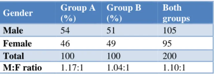
Table 2: Gender distribution in both groups (n=200).
Gender Group A (%)
Group B (%)
Both groups
Male 54 51 105
Female 46 49 95
Total 100 100 200
M:F ratio 1.17:1 1.04:1 1.10:1
Table 3: Side of septal deviation in both groups (n=200).
Deviation laterality
Group A Group B Both groups
No. of subjects % No. of subjects % Total no. of subjects %
Right 32 32 39 39 71 35.5
Left 40 40 44 44 84 42
Bilateral 28 28 17 17 45 22.5
Total 100 100 100 100 200 100
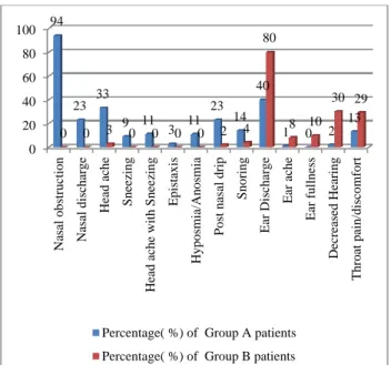
Septal deviation was more towards left side 84 (42%) than right side 71 (35.5%). The most common symptom in group A was nasal obstruction in 94 (94%) subjects, followed by otorrhoea in 40 (40%) subjects. Headache was seen in 33 (33%) subjects. Excessive sneezing was
Figure 1: Symptomatology in both the groups (n=200).
Maximum subjects in group A had "C" shaped DNS 44 (44%) followed by "S" shaped DNS 23 (23%). Maximum subjects in group B had "C" shaped DNS 41 (41%) followed by Spur in 26 (26%). In both the groups most common type of septal deviation was "C" shaped in 85 (42.5%) subjects (Table 4).
Table 4: Types of deviated nasal septum on nasal endoscopy (n=200). S. no. Types of deviation Group A Group B Both group %
1 Anterior
deviation 5 9 14 7
2 Posterior
deviation 7 4 11 5.5
3 Caudal
dislocation 1 3 4 2
4 C-shaped 44 41 85 42.5
5 S-shaped 23 13 36 18
6 Spur 14 26 40 20
7 C -shaped
with Spur 6 4 10 5
8 Total 100 100 200 100
In group A out of 100 (100%) subjects, 52 (52%) subjects had significant sinonasal pathology. In "S" shaped DNS 22 out of 23 (95.6%) subjects, followed by "C" shaped DNS 19 out of 44 (43.1%) subjects had sinonasal pathology. In group A, 58 (58%) subjects had ear pathology. In "C" shaped with spur 5 out of 6 (83.3%) subjects had ear pathology. In "S" shaped DNS 15 out of 23 (65.2%) subjects had ear pathology. In "C" shaped DNS 25 out of 44 (56.8%) subjects had ear pathology.
Table 5: Sinonasal, ear and throat pathology in different types of DNS in both groups (n=200).
Types of deviation No. of subjects Sinonasal pathology Ear pathology Throat pathology
Group A Group B Group A Group B Group A Group B
Anterior deviation 14 3 2 2 7 0 2
Posterior deviation 11 3 1 3 4 2 1
Caudal dislocation 4 0 1 1 3 0 0
C- shaped 85 19 3 25 33 3 7
S- shaped 36 22 9 15 12 6 4
Spur 40 2 1 7 17 2 7
C- shaped with Spur 10 3 1 5 4 1 0
There was more than one pathology seen in same subjects.
Table 5 presents sinonasal, ear and throat pathology in different types of DNS in both groups. In group A, 14 (14%) subjects had throat pathology. In “C” shaped DNS 3 out of 44 (6%) subjects had throat pathology. In “S” shaped DNS 6 out of 23 (26%) subjects had throat pathology.
In group B out of 100 subjects, 18 (18%) subjects had sinonasal pathology. Maximum cases of "S" shaped DNS 9 out of 13 subjects (69.2%) had sinonasal pathology. 80 (80%) subjects had ear pathology and 21 (21%) subjects had Throat pathology.
Out of total 200 subjects comprising both the groups, 70 (35%) subjects had significant sinonasal disease. Among these maximum association were observed in "S" shaped DNS 31 out of 36 (86.1%) subjects.
Based on the available statistical data in present study for occurrence of sinonasal pathology with “S” shaped DNS the calculated p-value by chi-square test with 2×2 contingency table is less than 0.00001 which is highly significant. The association between sinus disease and presence of other types of DNS (other than “S” shaped) is not statistically significant.
In group A, the associated anatomical variants on lateral nasal wall was concha bullosa (CB) in 27 (27%) subjects and paradoxical middle turbinate (PMT) in 20 (20%) subjects. The inferior turbinate was hypertrophied in 25 (25%) subjects. In 33 (33%) subjects polypi or osteomeatal complex (OMC) block was seen. CT Scan showed involvement of sinuses with homogenous opacity due to mucosal disease in 40 (40%) subjects.
0 20 40 60 80 100 Nasa l obstr u c tio n Nasa l dis ch a rge Hea d ache Sn eezing Hea d ache wit h Sn eezin g Epis tax is Hy p o sm ia/A n o sm ia Po st n asal d rip Sn o ring Ea r D ischa rge Ea r ach e Ea r fu lln ess Dec reased H eari n g Thr o at p ain /d isco m fort 94 23 33 9 11 3 11 23 14 40
1 0 2
13
0 0 3 0 0 0 0 2 4
80
8 10 30 29
Table 6: Sinonasal pathology in different types of DNS in both groups (n=200).
Types of deviation
No. of subjects with DNS
Sinonasal pathology in both groups
Polyp/ OMC
block Sinusitis Anatomical variation
Group A
Group B
Group A
Group B
PMT CB
Group A
Group B
Group A
Group B
Anterior deviation 14 5 1 2 2 2 1 1 2 1
Posterior deviation 11 4 2 1 1 0 1 1 3 0
Caudal dislocation 4 1 0 1 0 0 0 0 0 1
C-shaped 85 22 5 3 16 1 7 1 11 1
S-shaped 36 31 22 5 16 4 7 1 7 2
Spur 40 3 1 1 2 0 1 0 3 0
C shaped with spur 10 4 2 0 3 1 3 0 1 0
There was more than one pathology seen in some subjects.
In group B patients, sinus involvement was noted on CT scan in 21 (21%) subjects of whom 9 (9%) subjects had “S” shaped DNS. 4 (4%) had “C” shaped DNS, 4 (4%) had anterior deviation of septum. Interestingly none of these 21 subjects were complaining of any nasal symptoms.
Associated ear pathology
In group A, 58 subjects had ear pathology. In 44 subjects of “C” shaped DNS 25 (56.8%) had ear pathology and in 23 subjects of “S” shaped DNS 15 (65.2%) had ear
pathology. In group B, 80 subjects were associated with ear pathology. In 41 subjects of “C” shaped DNS 33 subjects (80.4%) had ear pathology and in 13 subjects of “S” shaped DNS 12 (92.3%) had ear pathology.
Out of 200 subjects, 138 (69%) subjects had ear pathology. In 85 (42.5%) subjects of "C" shaped DNS 58 (68.2%) had ear pathology, whereas in 36 (18%) subjects of "S" shaped DNS 27 (75%) subjects had ear pathology.
It was found that "S" shaped DNS were highly associated with ear pathology when compared with other types of septal deviation in both the groups.
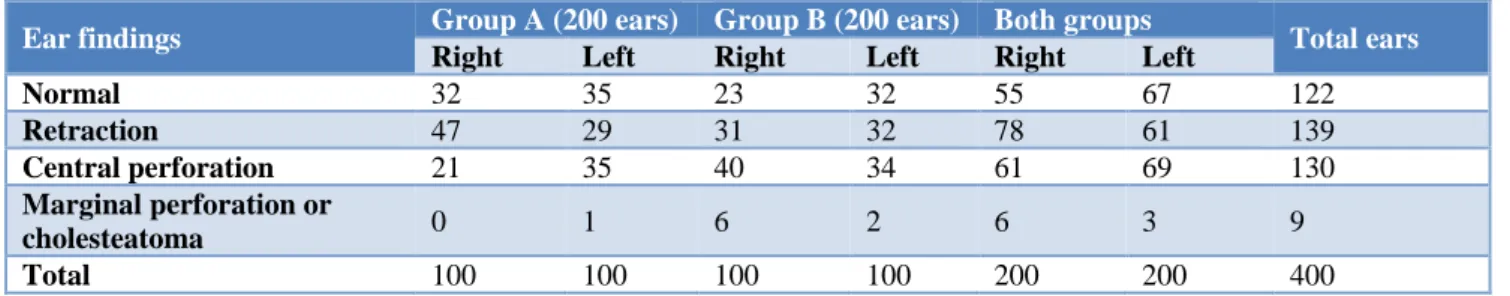
Table 7: Details of ear pathology in both groups (n=200).
Ear findings Group A (200 ears) Group B (200 ears) Both groups Total ears
Right Left Right Left Right Left
Normal 32 35 23 32 55 67 122
Retraction 47 29 31 32 78 61 139
Central perforation 21 35 40 34 61 69 130
Marginal perforation or
cholesteatoma 0 1 6 2 6 3 9
Total 100 100 100 100 200 200 400
Table 8: Side of septal deviation in relation to side of ear pathology in group A (n=100).
Side of deviation
No. of subjects (%) with septal deviation
Right ear
pathology Left ear pathology
Both ear
pathology Total
No. of
subjects %
No. of
subjects %
No. of
subjects %
No. of
subjects %
Right 32 6 18.75 6 18.75 5 15.6 17 53.1
Left 40 5 12.5 12 30 5 12.5 22 55
Bilateral 28 7 25 9 32.1 3 10.7 19 67.8
Total 100 18 18 27 27 13 13 58 58
In 200 subjects, 71 (35.5%) subjects had right sided septal deviation out of which the same side that is right side ear pathology was seen in 19 subjects showing 26.7% association whereas in opposite side that is left side ear pathology in 13 subjects showing 18.3% association.
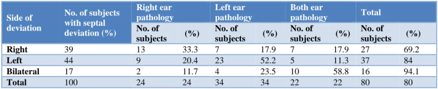
Table 9: Side of septal deviation in relation to side of ear pathology in group B (n=100).
Side of deviation
No. of subjects with septal deviation (%)
Right ear pathology
Left ear pathology
Both ear
pathology Total
No. of
subjects (%)
No. of
subjects (%)
No. of
subjects (%)
No. of
subjects (%)
Right 39 13 33.3 7 17.9 7 17.9 27 69.2
Left 44 9 20.4 23 52.2 5 11.3 37 84
Bilateral 17 2 11.7 4 23.5 10 58.8 16 94.1
Total 100 24 24 34 34 22 22 80 80
Table 10: DNS associated with ear and Eustachian tube pathology.
Study subjects DNS Ear pathology ETO pathology
No. of subjects % No. of subjects %
Group A 100 58 58 26 26
Group B 100 80 80 43 43
Both Groups 200 138 69 69 34.5
Table 11: Types of ETO in both groups (n=200).
Type of ETO on DNE
Group A Group B Both groups
No. of
subjects %
No. of
subjects %
No. of
subjects %
Type 1 (normal ostium) 74 74 57 57 131 65.5
Type 2 (inflammatory) 20 20 37 37 57 28.5
Type 3 (peritubal lymphoid tissue) 4 4 4 4 8 4
Type 4 (hypoplastic) 2 2 2 2 4 2
Type 5 (cicatrial) 0 0 0 0 0 0
Total 100 100 100 100 200 100
Associated ETD
In group A, 58 subjects had ear pathology and 26 subjects had Eustachian tube opening (ETO) pathology on endoscopy. The association between ETO pathology and ear pathology was 44.8% (26 out of 58) which is statistically highly significant. (corrected chi-square=15.1266, df=1, p=0.0001).
In group B, totally 80 subjects had ear pathology and 43 subjects had ETO pathology. The association between ETO pathology and ear pathology was 53.75% (43/80) which is statistically significant. (corrected chi-square =6.6325, df=1, p=0.01). Thus, the DNS causes ETD thereby causing ear pathology (Table 10).
Associated throat pathology
In group A, 14% of subjects had throat pathology. In 44 subjects of "C" shaped DNS 3 (6%) subjects had throat pathology and in 23 subjects of “S” shaped DNS 6 (26%) had throat pathology. Rest 5 subjects had different types of DNS. In group B, 21% of subjects had throat pathology. In 41 subjects of "C" shaped DNS 7 (17%) subjects had throat pathology and in 13 subjects of “S”
shaped DNS 4 (30.7%) had throat pathology. Rest of 10 subjects had different types of DNS (Table 5).
Treatment underwent
In Group A 99 (99%) subjects underwent septal correction after medical treatment, 50 subjects FESS were performed, 14 (14%) subjects underwent ear surgery for CSOM. Tympanoplasty was done in 13 (13%) subjects and MRM was done in 1 (1%) subject. In 13 cases of safe CSOM, the septal correction was done prior to ear surgery. In group B, 53 (53%) subjects underwent septal correction in 17 (17%) subjects FESS were performed, 9 (9%) subjects undergone tympanoplasty, 9 (9%) subjects tonsilloadenoid resection after septal correction. 14 (14%) subjects underwent modified radical mastoidectomy, 2 (2%) underwent cortical mastoidectomy followed by septal correction in 3 subjects. Rest patients of CSOM will undergo ear surgery, 3 months after septal correction.
DISCUSSION
Age and gender
Subjects above the age of 11 years were included in the study because children don't express accurate symptoms and may not co-operate for DNE. In this study maximum subjects belongs to the age group of 21 to 30 years which is similar to the study conducted by Mladina et al who found less incidence in primary school group which rises with age, suggest that these types could be influenced by the growth of the splanchnic cranial bones and the final angulation of the skull base during puberty and adolescence.5 There was Male dominance in cases of spur
in both groups, group A 1.8:1 and group B 1.6:1, which was similar to study done by Moorthy et al, Nayak et al and Mladina et al.5,6,9 Spur was common in male than
females which is typically a trauma-caused deformity, and it is generally accepted that males are more exposed to nasal trauma than females. In present study in which, M:F ratio for "C" shaped deviation was 0.86:1. In a study of children and adolescents in Zagreb, Croatia, "C" shaped was strikingly more frequent in females (76.9%) than in males (23.1%) and declared that "C" shaped DNS is a "female type" deformity.7
The side of the deformity
In present study left-sided septal deviation was more common than right-sided deviation in both the groups which is similar to the study by Moorthy et al and Mladina et al.5,7 A similar result was observed in the
Korean study (56.0% for left-sided and 39% for right-sided deformities) explained that during stages of labour while delivering of fetus, most common position is left occipito anterior and during extraction internal rotation happens towards midline at the true pelvis which is the narrowest passage in birth canal where nasal bones rotates under stress which leads to nasal deformity on left sides.5 Perhaps the finding of Quante et al, on Caucasian
newborns could explain this prevalence: a certain degree of overlapping between right and left parietal bones can occur during change in position of the baby’s head in the delivery canal.8 They found that the right parietal bone
was higher in about 50% of newborns, whereas the left was higher in only 20%; an equal level was found in about 30% of the newborns. Furthermore, they found that the incidence of nasal obstruction was about 20% in adults whose parietal bones had an unequal level, whereas it was remarkably lower (up to 6%) in those with an equal level.
Presenting complaints
In Group A 94% of subjects complained of Nasal obstruction found to be the most common symptom. This is similar to the study conducted by Moorthy et al.6
In a study done by Nayak et al, nasal obstruction (83%) was the most common complaint followed by headache (73%).9
Present study second most common symptom was otorrhoea in 40% of the subjects. Headache was seen in 33% subjects which may be because of sinusitis or mucosal contact points. Headache with sneezing was in 11% subjects of allergic rhinitis in group A. The study made by Kubba et al had sneezing in 12%.10
Associated sinonasal pathology
In Group A subjects, the associated anatomical variants on the lateral nasal wall were CB, and PMT which were observed in 27% and 20% subjects respectively which are similar to Moorthy et al showing CB 23% and PMT 15%.6 In a study conducted by Poorey et al showing CB
22%.11 In the present study CT scan showed polypi in
33% cases in Group A and 32% in the study conducted by Moorthy.6Among 44 subjects of "C" shaped DNS 16
(36.3%) had sinusitis and in 23 subjects of "S" shaped DNS 16 (69.5%) subjects had sinusitis. In group A out of 100 subjects, 52 subjects had significant sinonasal pathology. Among 44 subjects of "C" shaped DNS 19 subjects had sinonasal pathology with 43.1% association and in 23 subjects of "S" shaped DNS, 22 subjects had sinonasal pathology with 95.6% association present.
Out of total 200 subjects comprising both the groups 70 (35%) subjects had the significant sinonasal disease. In 36 subjects of “S” shaped DNS, 31 subjects had the sinonasal disease with 86.1% association. Based on the available statistical data from the present study for an occurrence of sinonasal pathology with “S” shaped DNS showing p<0.00001 which was highly significant. The association between sinonasal disease and presence of other types of DNS (other than “S” shaped) was not statistically significant (Table 5). This was similar to study conducted by Moorthy et al.6 Yousem et al
evaluated the morphologic features that predispose to sinusitis and concluded that patients with evidence of sinusitis on CT scanning had a higher degree of septal deviation than did those without.12 They further showed
that rates of sinusitis were not significantly different to the side of the septal deviation which was similar to the present study.
Similarly, Calhoun et al examined the paranasal sinus CT images of both asymptomatic and symptomatic patients.13
They clearly showed a strong correlation between septal deviation and sinus disease, although the degree of septal deviation was never qualified. They further documented a significant association with OMC block and ethmoid sinus disease only on the side to which the septum deviated.
As per the study conducted by Ozkurt et al, it was found that the presence of DNS based upon a degree of DNS (by calculating nasoseptal angle) created a risk factor for chronic rhinosinusitis.14 They declared that there are three
The first is the mechanical theory of Stammberger.15
Secretion accumulates in the sinus as a result of the narrowing of the osteomeatal complex and causes chronic rhinosinusitis by being infected later on.
The second theory is the aerodynamic theory.16
According to this theory, the mucociliary activity decreases following the nasal flow rate increase and mucosal dryness occur in relation to the nasal septal deviation and consequently, chronic rhinosinusitis develops.
The third theory is Bachert’s pressure theory.17
According to this theory, deviation or the posterior nasal septum causes chronic rhinosinusitis by creating pressure and air flow changes within the maxillary sinuses.
In the study conducted by Elahi et al showed ipsilaterally, that is, in the direction of septal deviation, OMC and sinus disease is directly attributable to the septal deviation in the absence of any other discernable factor.18
While on the side opposite the direction of septal deviation, a prominent bulla ethmoidalis and various middle turbinate abnormalities have been shown to be the cause of OMC obstruction. They also showed increasing OMC disease bilaterally with increasing septal deviation.
In a study conducted by Kennedy et al, the anterior ethmoid region were the most frequently involved area of the paranasal sinuses disease in relation to DNS.19
Associated ETD
The types of pharyngeal end of ETO was described based upon the study conducted by Prudente de et al.24 ETO
changes (Table 11) in the present study is similar to study conducted by Kaya et al and Akyildiz, Metin Yuksel et al who found that inflammatory, obstructive, and infective pathologies of the nose and paranasal sinuses were reported to have negative impacts on eustachian tube (ET) function.20,21 Among these factors, chronic nasal
obstruction due to septal deviation was suggested to cause chronic suppurative otitis media via ET dysfunction, deteriorated middle ear ventilation. The mechanisms of inflammation of nasopharyngeal opening of ET as a result of turbulent air streams, sniffing or insufflations of infected material into middle ear by Toynbee phenomenon.
In a study conducted by Gencer et al found that the development of paranasal sinuses and mastoid air cells during postnatal development process can be supported by the positive pressure of airflow on the nasal cavity and nasopharyngeal mucosa.22 They have found that nasal
pathologies like DNS decrease mastoid air volume and maxillary sinus volume by decreasing nasal airflow and so mastoid pneumatization and in this way have otitis media and other otological problems.
In a study conducted by Yeolekar et al they found that in patients of otitis media, there was associated DNS (80%), allergic rhinitis (14.5%), sinusitis (20%), polyp (2.5%) and adenoid in 11.5%.23
CONCLUSION
Deviated nasal septum was associated with significant sinonasal pathology. “S” shaped DNS was maximally associated with sinonasal pathology which was statistically significant. There is a high correlation between the side of septal deviation to the side of ear pathology, particularly in asymptomatic DNS. So when DNS is associated with any sinonasal or ear or throat pathology, one should consider its detail examination and if needed septal correction on before undertaking any definitive sinus or ear or adenotonsillar surgery. Especially "S" shaped DNS should be considered for septal correction in all cases.
ACKNOWLEDGEMENTS
Authors like to thank our Medical College and Hospital for its support and the patients for cooperating with the study protocol.
Funding: No funding sources Conflict of interest: None declared
Ethical approval: The study was approved by the Institutional Ethics Committee
REFERENCES
1. Pirsig W. Growth of the Deviated Septum and Its Influence on Midfacial Development.Facial Plast Surg. 1992;8(04):224-32.
2. Robinson, Jennifer. What Is a Deviated Septum. WebMD. 2016;12:11.
3. Alt J. "Disorders of Smell & Taste." American Rhinologic Society. 2015. Available at|: http://care.american-rhinologic.org/disorders_ of_smell_taste. Accessed on 3 November 2019. 4. Nanda MS, Kaur M, Bhatia S. Impact of
Septoplasty on hearing and middle ear function. Int J Res Med Sci. 2017;6(1):135.
5. Mladina R, Čujić E, Šubarić M, Vuković K. Nasal septal deformities in ear, nose, and throat patients. Am J Otolaryngol. 2008;29 (2):75- 82.
6. Moorthy PNS, Kolloju S, Madhira S, Jowkar AB. Clinical Study on Deviated Nasal Septum and Its Associated Pathology. Int J Otolaryngol Head & Neck Surg. 2014;3(2):75-81.
7. Šubarić M, Mladina R. Nasal septum deformities in children and adolescents: a cross sectional study of children from Zagreb, Croatia. Int J Pediatr Otorhinolaryngol. 2002;63(1):41-8.
9. Nayak DR, Balakrishnan R, Murty KD, Hazarika P. Endoscopic septoturbinoplasty: Our update series. Indian J Otolaryngol Head Neck Surg. 2002;54(1):20-4.
10. Kubba H, Bingham BJ. Endoscopy in the assessment of children with nasal obstruction. J Laryngol Otol. 2001;115(5):380-4.
11. Poorey VK, Gupta N. Endoscopic and Computed Tomographic Evaluation of Influence of Nasal Septal Deviation on Lateral Wall of Nose and Its Relation to Sinus Diseases. Indian J Otolaryngol Head Neck Surg. 2014;66(3):330-5.
12. Yousem DM, Kennedy DW, Rosenberg S. Ostiomeatal complex risk factors for sinusitis: CT evaluation. J Otolaryngol. 1991;20(6):419-24. 13. Calhoun KH, Waggenspack GA, Simpson CB,
Hokanson JA, Bailey BJ. CT Evaluation of the Paranasal Sinuses in Symptomatic and Asymptomatic Populations. Otolaryngol Neck Surg. 1991;104(4):480-3.
14. Ozkurt FE, Akdag M, Keskin I, Iskenderoglu AY, Orhan T. Relation Between the Nasal Septal Deviation and Chronic Rhinosinusitis. Int J Basic Clin Stud. 2014;3(1):25-30.
15. Stammberger H, Posawetz W. Functional endoscopic sinus surgery. Concept, indications and results of the Messerklinger technique. Eur Arch Otorhinolaryngol. 1990;247(2):63-76.
16. Blaugrund SM. Nasal obstruction. The nasal septum and concha bullosa. Otolaryngol Clin North Am. 1989;22(2):291-306.
17. Kumar H, Jain R, Douglas RG, Tawhai MH. Airflow in the Human Nasal Passage and Sinuses of Chronic Rhinosinusitis Subjects. 2016.
18. Elahi MM, Frenkiel S. Septal deviation and chronic sinus disease. Am J Rhinol. 2000;14(3):175-9. 19. Kennedy DW, Zinreich SJ, Rosenbaum AE, Johns
ME. Functional endoscopic sinus surgery. Theory and diagnostic evaluation. Arch Otolaryngol. 1985;111(9):576-82.
20. Kaya M, Dağlı E, Kırat S. Does Nasal Septal Deviation Affect the Eustachian Tube Function and Middle Ear Ventilation? Turkish Arch Otorhinolaryngol. 2018;56(2):102-5.
21. Akyildiz MY, Özmen ÖA, Demir UL, Kasapoğlu F, Coşkun HH, Basut OI, et al. Impact of Septoplasty on Eustachian Tube Functions. J Craniofac Surg. 2017;28(8):1929-32.
22. Gencer ZK, Özkiriş M, Okur A, Karaçavus S, Saydam L. The Possible Associations of Septal Deviation on Mastoid Pneumatization and Chronic Otitis. Otol Neurotol. 2013;34 (6):1052-7.
23. Yeolekar A, Dasgupta K. Otitis media: Does the onus lie on sinonasal pathology? Indian J Otol. 2011;17(1):8.
24. Prudente de JEA, Aquino JEAP, Zavarezzi DE, Carvalho MRMS, Aquino JNP. Endoscopic Avaliation of Pharyngeal Orifice of Eustachian Tube in Patients with Chronic Otitis. Int Arch. Otorhinolaryngol. 2007;11(2):106-8.