Original Research Article
An approach towards metastatic neck nodes and most common
site of primaries: a retrospective study
Rudra Prakash
1*, Smrity R. B. Dutta
2INTRODUCTION
Head and neck cancer (HNC) is a disease of the upper aero digestive tract and is one of the most frequently diagnosed cancers worldwide.1 A high rate of cancers involving the head and neck are reported across the Asian region, with notable variations between countries.2 Head and neck cancers account for 30 percent of the total cancer burden in India in which cancer of the tongue, mouth, pharynx account for more than 80 percent of the case.3 Disease prognosis is largely dependent on tumour stage and site. Patients with early stage disease have a 60–95% chance of cure with local therapy.4 One of the features of head and neck cancers is that it involves organs whose dysfunction is easily noticeable. Spread of the malignant tumours originating in this region to
regional lymph nodes is quite common. The actual incidence of spread to lymph nodes may vary slightly with different region or sub region affected. The prognosis of a given stage of primary tumour at any particular site in head and neck cancer becomes almost 50% worse in the presence of lymph nodes metastasis.5 Thus understanding the pattern of metastatic spread in head and neck cancers can help in management and prevention of future recurrences and reduce the risk of developing metastatic neck nodes.
Aims and objective
To find the most commonly involved neck nodes in metastatic head and neck cancer patients.
ABSTRACT
Background: Head and neck cancer is one of the most common cancers in India and worldwide. It accounts for 30 percent of the total cancer burden. Head and neck cancers are notorious for loco regional spread presenting late with metastastasis to cervical lymph nodes.
Methods: Our study was carried out on patients presenting with metastatic neck nodes at Silchar Medical College and Hospital, Assam from 1st April 2013 to 30th March 2015.
Results: In our study 167 patients presented with head and neck cancer with metastatic neck nodes. Metastatic nodes involving level II accounted for 51.4% of cases with primaries in oral cavity, base of tongue. Level III lymph nodes were involved in 48.6% of cases with primaries in the hypopharynx and larynx.
Conclusions: In our study it was found that hypopharynx is the most common primary head and neck cancer with metastatic neck node. The most commonly involved lymph nodes are level II and level III. This helps in understanding the pattern of micrometastases in head and neck cancer patients with N0 neck which makes way for the role of selective neck dissection in these groups of patients.
Keywords: Metastatic neck nodes, Primaries, Management of N0 neck
Department of ENT and Head Neck Surgery, 1AIIMS, Patna, Bihar, 2SMCH, Silchar, Assam, India
Received: 10 March 2019
Revised: 07 July 2019
Accepted: 11 July 2019
*Correspondence:
Dr. Rudra Prakash,
E-mail: [email protected]
Copyright: © the author(s), publisher and licensee Medip Academy. This is an open-access article distributed under the terms of the Creative Commons Attribution Non-Commercial License, which permits unrestricted non-commercial use, distribution, and reproduction in any medium, provided the original work is properly cited.
To find the common sites of primary of metastatic neck nodes in head and neck cancer patients.
METHODS
In our retrospective study carried out over a period of two years from 1st April 2013 to 30th March 2015 we evaluated head and neck cancer patients presenting with metastatic neck nodes attending the Department of ENT and Department of Radiotherapy at SMCH, Assam. Routine clinical ENT examination and panendoscopies were used to identify the primaries and HPE was done to confirm the diagnosis. Fine needle aspiration cytology (FNAC) was used to confirm the metastatic nature of neck nodes as it is an effective, safe diagnostic tool, reliable in the diagnosis of neck masses, relatively easy to perform and with low associated costs.6,7 Patient presenting with N0 neck and occult primaries were excluded from the study.
RESULTS
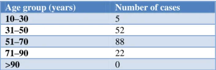
In our retrospective study from 1st April 2013 to 30th March 2015 167 patients presented with head and neck cancer with metastatic neck nodes at SMCH, Assam. The most common lymph node levels involved were level II and III. Metastatic nodes involving level II accounted for 51.4% of cases with their primaries situated in oral cavity, base of tongue, tonsil and parotid. Level III group of lymph nodes were involved in 48.6% of cases with primaries in the hypopharynx and larynx. The most common primaries of metastatic neck nodes were hypopharynx 25.6%, larynx 23%, base of tongue 20%, oral cavity 14.3% and tonsil 9%. Of all the cases males were 143 in number accounting for majority of cases and 85.6%, females were 24 in number accounting for a mere 14.7%. The male to female ratio was 6:1. Majority of cases belonged to 50 years to 70 years of age group being a total of 88 in number and accounting for 52.7%. Youngest patient with neck node was 18 years of age and oldest was 82 years of age.
Table 1: Distribution of most common sites of primaries with metastatic neck nodes.
Primary site No. of cases with neck nodes
Percentage (%)
Base of tongue 34 20
Tonsil 15 9
Larynx 38 23
Parotid 4 2.3
Oesophagus 1 0.7
Oral cavity 23 13.8
Hypopharynx 43 25.7
Temporal bone 1 0.7
Mandible 2 1.20
Maxilla 1 0.6
Nasopharynx 5 3
Total 167 100
Table 2: Distribution of number of affected males and females.
Sex Number of cases
Male 143
Female 24
Table 3: Distribution of cases in various age groups.
Age group (years) Number of cases
10–30 5
31–50 52
51–70 88
71–90 22
>90 0
DISCUSSION
Head and neck cancers account for 30 percent of the total cancer burden in India in which cancer of the tongue, mouth, pharynx account for more than 80 percent of the cases.3 In India the overall leading cancer in order of occurrence is oral cavity, lung, pharynx, oesophagus followed by stomach, making oral cancer the overall most common cancer.8 As is known, head and neck squamous cell carcinoma is a loco regional disease
notorious for regional and distant metastases,
representing the leading cause of death in head and neck squamous cell carcinoma patients.
While tobacco and alcohol abuse are the major risk factors for these tumours worldwide, chewed tobacco, betel nut, beedi smoking and Epstein Barr virus (EBV) are aetiological agents that are specific to Asia that result in an increased incidence of head neck cancers in this region of the world.9,10 Patients with head and neck cancer might present with, mass in the neck, neck pain, bleeding from the mouth, paranasal sinus congestion, especially with nasopharyngeal carcinoma, bad breath, sore tongue, painless ulcer or sores in the mouth that do not heal, white, red or dark patches in the mouth that will not go away, earache, unusual bleeding or numbness in the mouth, lump in the lip, mouth or gums, enlarged lymph nodes in the neck, slurring of speech (if the cancer is affecting the tongue), hoarseness of voice which persists for more than six weeks, sore throat which persists for more than six weeks, difficulty swallowing food, change in diet or weight loss.11
is the most common histological variant. Isolated supraclavicular nodal involvement is almost invariably related to malignant disease arising below the clavicle.
To understand the pattern of nodal metastases from head and neck primaries we need to understand the loco regional anatomy of head and neck lymphatic system. In 1981 the Memorial Sloan – Kettering Hospital published a number of levels within the neck which contain group of lymph nodes representing the first echelon sites for metastases from head and neck primary sites.14,15 These include Level Ia (sub mental), level Ib (submandibular), level II (upper jugular), level III (middle jugular), level IV (lower jugular), level V (posterior triangle), level VI (pre/para tracheal and laryngeal nodes), level VII (superior mediastinal nodes). Level Ia group of lymph nodes drain floor of mouth, anterior nasal cavity, anterior 2/3 of tongue, anterior mandibular alveolar ridge and lower lip. Level Ib drains oral cavity, nasal cavity, mid face soft tissue and submandibular gland. Level II group of lymph nodes drains oral cavity, nasal cavity, nasopharynx, oropharynx, hypopharynx, larynx and parotid. Level III drains oral cavity, naso/oro/hypo pharynx and larynx. Level IV drains hypopharynx, thyroid, cervical oesophagus and larynx. Level V drains naso/oropharynx, skin of posterior scalp and neck. Level VI drains thyroid, glottis, subglottis, PFS and cervical oesophagus.
Factors implicated in metastatic nodal disease include site of the tumour, size of tumour, tumour thickness, previous
treatment and tumour recurrence.16 Metastatic
involvement of various lymph nodes usually progresses from superior to inferior in an orderly fashion. The risk of nodal metastasis increases from anterior to posterior aspect of the upper aero digestive tract, that is, lips, oral cavity, oropharynx and hypopharynx. For tumours of the larynx and pharynx, the risk of nodal metastasis increases with progression from centre of the upper aero digestive tract to the periphery.17 However discontinuous or “skip metastases” were described in 3–10 percent of cases below dissected areas that were pathologically node negative.18 The tongue especially is known to cause “skip metastasis” to level IV. Contra lateral neck spread may occur early in those tumours situated in or near midline. Vocal cords are extremely avascular and have sparse lymphatic drainage hence metastases from them is uncommon. The T stage usually reflects tumour burden and is correlated with risk of nodal metastasis for any given primary site. Overall, the risk of metastasis is less than 15% for T1, 15% to 30% for T2, 30% to 50% for T3 and up to 75% for T4 head and neck primary squamous cell carcinomas.17
The region specific drainage translates well into clinical practice and it is possible to predict the site of a primary depending upon the level of lymph node involved. In a landmark study of 1155 patients with previously untreated head and neck squamous cell carcinoma
published by Lindberg in 1972.9 The topographical
distribution of clinically evident cervical metastases was set out. This identified distinct patterns of spread to the neck nodes based on the primary site. Histological proof of this was produced by Shah in 1990 in a series of 1119 neck dissections.19
As mentioned previously tongue, mouth, pharynx account for more than 80 percent of total cases of head and neck cancers. According to the NCRP’S consolidated report of the HBCR’S 2007-2011 the number of site specific head and neck cancer cases registered in region of Dibrugarh and Guwahati in decreasing order is other pharynx 653 (5309%), mouth 257 (21.2%), tongue 164 (13.5%), larynx 106 (8.8%) in Dibrugarh and other pharynx 1356 (47.9%), mouth 545 (19.3%), tongue 472 (16.7%), larynx 382 (13.5%) in Guwahati.8
The data is suggestive of the fact that in this part of India the most common site of primaries is pharynx followed by mouth and tongue and larynx being the least common of all.
The most common site of primaries of neck nodes usually are tonsil 41%, hypopharynx 21%, base of tongue 15%, oral cavity 10% and larynx 10%.19,20 In our study conducted at SMCH, Assam a tertiary care hospital which caters to the population of Barak valley we found that the most common primaries of metastatic neck nodes were hypopharynx 25.6%, larynx 23%, base of tongue 20%, oral cavity 14.3% and tonsil 9%. Our data is supported by the pattern and risk of metastatic disease as mentioned before. The most common lymph nodes involved were level II and level III. Metastatic nodes involving level II accounted for 51.4% of cases with their primaries situated in oral cavity, base of tongue, tonsil and parotid mainly. Level III group of lymph nodes were involved in 48.6% of cases with primaries mainly in the hypopharynx and larynx.
The significance of our study lies in the fact that around 15 to 20% of patients presenting with N0 neck clinically will have occult or micro metastases in the neck.21 Micrometastases are defined as deposits of cancer cells between 0.2 and 2 mm in size making them clinically undetectable but increasing the chances of recurrences in future if not dealt with.22 Current methods can’t identify microscopic nodal metastases.21
This gave the concept of elective/selective neck dissection in head and neck cancer patients presenting
with N0.23,24 Selective neck dissection includes 1) supraomohyoid neck dissection involving removal of
level I, II and III for primary tumour of oral cavity, 2) jugular neck dissection which involves removal of
of 39 months. There were 81 recurrences and 50 deaths in the elective-surgery group and 146 recurrences and 79 deaths in the therapeutic-surgery group. At 3 years, elective node dissection resulted in an improved rate of overall survival (80.0%; 95% confidence interval [CI], 74.1 to 85.8), as compared with therapeutic dissection (67.5%; 95% CI, 61.0 to 73.9), for a hazard ratio for death of 0.64 in the elective-surgery group (95% CI, 0.45 to 0.92; p=0.01 by the log-rank test). At that time, patients in the elective-surgery group also had a higher rate of disease-free survival than those in the therapeutic-surgery group (69.5% vs. 45.9%, p<0.001). Elective node dissection was superior in most subgroups without significant interactions.2
CONCLUSION
In our study it was found that hypopharynx is the most common primary head and neck cancer with metastatic neck node followed by larynx, base of tongue, oral cavity and tonsil. The most commonly involved lymph nodes are those in level II and level III. This helps us in understanding the pattern of micrometastases in head and neck cancer patients with N0 neck which makes way for the role of selective neck dissection in these groups of patients to prevent recurrences in the form of neck metastases in future and thus improve the quality of life of head and neck cancer patients.
Funding: No funding sources Conflict of interest: None declared
Ethical approval: The study was approved by the Institutional Ethics Committee
REFERENCES
1. Gregoire V, Lefebvre J-L, Licitra L, Felip E. On behalf of the EHNS-ESMO-ESTRO Guidelines Working Group. Squamous cell carcinoma of the head and neck:EHNS-ESMO-ESTRO Clinical Practice Guidelines for diagnosis, treatment and follow-up. Ann Oncol. 2010;21(Suppl. 5):v184–6. 2. D’Cruz A, Vaish R, Kapre N, Dandekar M, Gupta
S, Hawaldar R, et al. Elective versus Therapeutic Neck Dissection in Node-Negative Oral Cancer. N Engl J Med. 2015;373(6):521-9.
3. Cancers, N. & Sites, A. Chapter 9 Head and Neck Cancers. 2011.
4. D'cruz A, Lin T, Anand AK, Atmakusuma D,
Calaguas MJ, Chitapanarux I, et al. Consensus recommendations for management of head and neck cancer in Asian countries: A review of international guidelines. Oral Oncol. 2013;49(9):872–7.
5. Shah JP, Patel SG, Singh B. Jatin Shah’s Head and
Neck Surgery and Oncology, 4th ed. Philadelphia: Elsevier; 2012: 426.
6. el Hag IA, Chiedozi LC, al Reyees FA, Kollur SM.
Fine needle aspiration cytology of head and neck masses. Seven years’ experience in a secondary care hospital. Acta Cytol. 2003;47(3):387-92.
7. Ellison E, LaPuerta P, Martin SE. Supraclavicular masses: results of a series of 309 cases biopsied by fine needle aspiration. Head Neck. 1999;21(3):239-46.
8. Kumar V, Ramesh C, Reddy R. Leading sites of
cancer in India. Consolidated report of the HBCR, 2007-2011. 2010;1:1–16.
9. Alvi A, Myers EN, Johnson JT. Cancer of the Oral
Cavity. In: Myers EN, Suen JY, editors. Cancer of the head and neck. Philadelphia: W.B. Saunders Company; 1996: 321–361.
10. Mayne ST, Morse DE, Winn DM. Cancers of the
oral cavity and pharynx. In: Schottenfeld D, Fraumeni FJ, editors. Cancer epidemiology and prevention. Oxford Univ Press; 2006.
11. Scottish Executive Health Department. Scottish
Referral Guidelines for Suspected Cancer.
Edinburgh: Scottish Executive Health Department; 2002. Available at: http://www.show.scot.nhs. uk/sehd/mels/hdl2002_45.pdf. Accessed on 3 June 2019.
12. Calabrese L, Jereczek-fossa BA, Jassem J.
Diagnosis and management of neck metastases from an unknown primary. Acta Otorhinolaryngol Ital. 2005;5:2-12.
13. Ansari J, Glaholm J. Management of an unknown primary carcinoma. In: Watkinson John C. Stell & Maran’s Textbook of Head ND Neck Surgery and Oncology, 5th ed. Boca Raton: CRC Press; 2012: 690.
14. Paleri V, Watkinson John C. Metastatic neck
disease. In: Watkinson John C. Stell & Maran’s Textbook of Head ND Neck Surgery and Oncology, 5th ed. Boca Raton: CRC Press; 2012: 663.
15. Chao CKS. Practical essentials of intensity
modulated radiation therapy. 2nd ed. Philadelphia: Lippincott Williams & Wilkins; 2005.
16. Paleri V, Watkinson John C. Metastatic neck
disease. In: Watkinson John C. Stell & Maran’s Textbook of Head ND Neck Surgery and Oncology, 5th ed. Boca Raton: CRC Press; 2012: 669.
17. Shah JP, Patel SG, Singh B. Cervical Lymph Nodes.
In: Thomas Stefanie J, editor. Jatin Shah’s Head and Neck Surgery and Oncology, 4th ed. Philadelphia: Elsevier; 2012: 430.
18. Cummings B, Kim J, O’Sullivan B. Radiation
therapy and management of the cervical lymph nodes. In: Gaertner Schmidt R, editor. Cummings Otolaryngology Head and Neck Surgery, 4th ed. Philadelphia: Elsevier; 2005: 2592.
19. Paleri V, Watkinson John C. Metastatic neck
disease. In: Watkinson John C. Stell & Maran’s Textbook of Head ND Neck Surgery and Oncology,5th ed. Boca Raton: CRC Press; 2012: 666.
21. Shah JP, Patel Snehal G, Singh B. Cervical Lymph Nodes. In: Thomas Stefanie J, editor. Jatin Shah’s Head and Neck Surgery and Oncology, 4th ed. Philadelphia: Elsevier; 2012: 432.
22. Paleri V, Watkinson John C. Metastatic neck
disease. In: Watkinson John C.Stell & Maran’s Textbook of Head ND Neck Surgery and Oncology, 5th ed. Boca Raton: CRC Press; 2012: 667.
23. Royal College of Pathologists. Standards and
Datasets for Reporting Cancers: Datasets for histopathology reports on head and neck carcinomas and salivary neoplasms. 2nd Edition. London: The Royal College of Pathologists; 2005. Available at: http://www.rcpath.org/resources/pdf/HeadNeck DatasetJun05.pdf. Accessed on 3 June 2019.
24. Robbins KT, Clayman G, Levine PA, Medina J,
Sessions R, Shaha A, et al. Neck dissection classification update: revisions proposed by the American Head and Neck Society and the American Academy of Otolaryngology-Head and Neck Surgery. Arch Otolaryngol Head Neck Surg. 2002;128(7):751-8.