http://www.cmbl.org.pl DOI: 10.2478/s11658-006-0044-0 Received: 20 March 2006
Revised form accepted: 28 June 2006
© 2006 by the University of Wrocław, Poland
* E-mail: [email protected]
Abbreviations used: DHNs – dehydrins; Kn-type – dehydrins containing n-copies of K-segments; KnS-type – dehydrins containing n-copies of K-segments followed a single copy of S-segment; LEA – late embryogenesis abundant; SKn-type – dehydrins containing a single copy of S-segment followed by n-copies of K-segments; YnKn-type – dehydrins containing n-copies of Y-segments followed by n-copies of K-segments; YnSKn-type – dehydrins containing n-copies of Y-segments followed a single copy of S-segment and n-copies of K-S-segments
PLANT DEHYDRINS – TISSUE LOCATION, STRUCTURE AND FUNCTION
TADEUSZ RORAT*
Institute of Plant Genetics, PAS, Strzeszyńska 34, 60-479 Poznań, Poland
involved in cold acclimation processes. The main question arising from the in vitro findings is whether each DHN structural type could possess a specific function and tissue distribution. Much recent in vitro data clearly indicates that dehydrins belonging to different subclasses exhibit distinct functions.
Key words: Dehydration stress, Drought, Cold acclimation, Freezing tolerance, LEA proteins, Dehydrin
INTRODUCTION
Under environmental conditions generating reduced water potential, plants produce an array of proteins as a part of a global stress response to protect the cell metabolism [1, 2]. The synthesis of hydrophilic proteins is a major part of the response to water-deficit conditions [2, 3]. These proteins were first characterized in cotton during the late stages of embryogenesis, and called LEA proteins (Late Embryogenesis Abundant). Thereafter, proteins homologous to the cotton LEAs were identified in the seeds of many higher plants [4]. All of the LEA proteins are characterized by a high glycine content, high hydrophilicity and a low secondary structure [3]. Three major groups of LEA proteins have been defined on the basis of sequence similarity and structural characteristics: group 1, group 2 and group 3 [5-7].
Group 1 LEA proteins, of which the wheat Em protein is the type sequence, are only found in plants. They are unstructured in solution [8] and contain a 20-residue amino acid motif family [5]. The group 3 LEA proteins are characterized by a repeated 11-mer amino acid motif whose consensus sequence has been broadly defined as ΦΦE/QXΦKE/QKΦXE/D/Q, where Φ represents a hydrophobic residue [9]. Homologous proteins to the group 3 LEAs have been discovered in organisms other than plants, including nematodes and prokaryotes [9-11]. The group 3 LEA proteins, as with the classic examples from the anhydrobiotic nematode Aphelenchusavenae [12], and from the bullrush Typha latifolia [13], are natively unfolded in solution, but seem to be more structural on drying. Group 2 LEA proteins, called dehydrins, are mainly found in plants and are typically accumulated in dehydrating plant tissues such as developing seeds during maturation, or in vegetative tissues subjected to environmental stress such as drought, low temperature and salinity [2, 14]. The accumulation of dehydrins is one of the prominent components of plant adaptation to severe environmental conditions [14]. Dehydrins are distributed in a wide range of organisms including higher plants, algae, yeast and cyanobacteria [14-17].
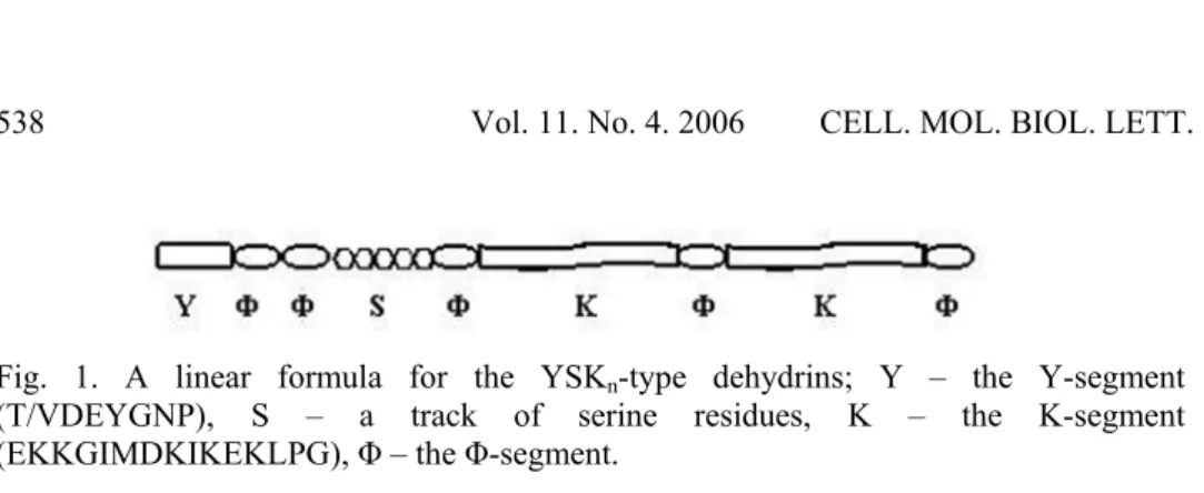
Fig. 1. A linear formula for the YSKn-type dehydrins; Y – the Y-segment (T/VDEYGNP), S – a track of serine residues, K – the K-segment (EKKGIMDKIKEKLPG), Φ – the Φ-segment.
The Φ-segments are not preserved, and show considerable variation between different DHNs. The K-segment is the only segment present in all dehydrins. It resembles a lipid-binding class A2 amphipathic α-helical segment found in apoliporoteins and α-synucleins [14, 18]. Class A amphiphatic helices have well-demarcated polar and non-polar faces with negatively charged residues opposite the hydrophobic face and positively charged residues at the polar/non-polar interface [19]. Some other notable characteristics of DHNs are a lack of cysteine and tryptophan, a high percentage of charged and polar amino acids (hydrophilic) and the ability to remain in solution after boiling [14, 19, 20]. The number and order of the Y-, S- and K-segments define different DHN sub-classes: YnSKn, YnKn, SKn, Kn and KnS. Structural analyses shown that the DHNs appear to lack a defined structure in their pure form; however, in the presence of a structural cosolvent such as trifluoro-ethanol (TFE), a DSP16 dehydrin from Craterostigma plantagineum was found to display α-helical structures [21]. A 35-kD DHN-related protein from cowpea (Vignaunguiculata) was also shown to adopt α-helical structure in the presence of SDS; this is postulated to be mediated by amphipathic α-helical structures formed by K-segments [22].
The genes encoding DHNs are a redundant family. For example, six Dhn genes have been characterized in Arabidopsis [23], thirteen in Hordeum vulgare [24, 25], and at least two in many other plant species [26, UniProt Knowledgebase, http://www.expasy.org/cgi-bin/].
Immunohistochemical and fractionation studies revealed that dehydrins are not only accumulated under the conditions of water deficit that occur during the late stages of embryogenesis [2, 27-31] and in vegetative tissues in response to stress leading to cellular dehydration such as drought, low temperature and salinity [1, 2, 14, 21, 22, 32-36], but they are also present in nearly all vegetative tissues during normal growth conditions [25, 32, 34, 37, 38]. Such a wide distribution of dehydrins in various tissues during plant growth and in response to stress leading to cellular dehydration suggests that these proteins may play an essential role in plant growth and in stress tolerance. A major question is how different dehydrin types function in different tissues during plant growth and under cell dehydration conditions.
Though the precise function of dehydrins has not been established yet, much recent in vitro data clearly indicates that dehydrins belonging to different subclasses exhibit distinct functions. In vitro experiments revealed that YSKn -type DHNs bind to lipid vesicles that contain acidic phospholipids [39], while others (KnS-type) were shown to bind metals [40, 41], scavenge hydroxyl radicals [42], protect lipid membranes against peroxidation [43], or display cryoprotective activity towards freezing-sensitive enzymes [44-46]. Two other types, the SKn- and K-type, seem to be mainly involved in cold acclimatization and drought resistance.
LOCATION OF DEHYDRINS IN PLANT TISSUES
Many studies demonstrated that DHNs are distributed in different tissues during plant growth and development. Some dehydrins are mainly found in mature seeds. Two examples of such distribution are RAB18 (Y2SK2-type) from
Arabidopsis and RAB17 (DHN1; YSK2) from Zea mays; they exhibit
localization in all the parts of the embryo and in the endosperm of mature seeds [27, 47]. Another is pea (Pisum sativum) DHN-COG (SK2) dehydrin, which is accumulated in mid- to late embryogenesis in developing cotyledons and in dehydration-stressed seedlings [28]. In mature cotyledons, the DHN-COG protein comprises about 2% of the total seed proteins. Carrot ECP40 (YSK2) was found to localize in the endosperm and zygotic embryos in mature seeds [29]. Other dehydrins, such as a 24-kD (MAT1; YK) and a 26/27-kD (MAT9; YK) were purified from mature soybean (Glycine max) seeds [30, 31]; a 35-kDa dehydrin (Y2K) was purified from dry seeds of cowpea (Vigna unguiculata) [22]. Most of the known dehydrins are localized in vegetative tissues under normal growth conditions, and in floral organs. For example, Arabidopsis
ERD14 (SK2) and ERD10 (LTI29, SK3-type) have been shown to localize in root tips, and in the vascular tissues of roots, stems, leaves and flowers [32]. Accumulation in the cells of all tissues of the shoots including the epidermal, cortical, phloem and xylem tissues was demonstrated for peach PCA60 DHN (YK-type) [44]. Other dehydrins, e.g. wheat WCOR410 (SK3) were found to accumulate preferentially in the vascular transition area of roots, leaves and crowns [35]. Some dehydrins exhibit a localization specific to cell types, e.g. pollen sacs, guard cells, root meristematic cells [32] or plasmodesmata [48]. RAB18 (Y2SK2) from Arabidopsis showed accumulation specific to the stomatal guard cells of stems, leaves and flowers [32]. From all these studies, it can be concluded that there are different types of DHNs that can localize in the same tissues during normal growth and that most of them are to be found in vascular tissues and surrounding cells. Under dehydration conditions, which naturally occur in the late stages of seed development, and in mature seeds, the DHNs are located in different tissues.
dehydrins, and they also appeared in other tissues than under normal growth conditions. For example, the Arabidopsis ERD14 and ERD10 (LTI29) dehydrins, which were primarily localized in root tips and in the vascular tissues of roots, stems, leaves and flowers in plants grown under normal growth conditions, were detected in cells of all the tissues in cold-stressed plants [32]. Similar data was obtained for the distribution of DHN24 from Solanum
sogarandinum [38] and P-80 from Hordeum vulgare [33] in cold-acclimated
plants. Other dehydrins, such as LTI30 (K6) from Arabidopsis, were not detected in plants grown in control conditions, but did accumulate in all the tissues of the roots, in the vascular tissues of stems, leaves and flowers, and in the pollen sacs under cold conditions [32]. Similarly, wheat WCS120 DHN (K6) family proteins are mainly localized in the vascular transition zone of wheat crown tissues in cold-acclimated plants, but have no detectable level in the mature xylem and shoot apical meristem or lateral root primordial [34]. Another wheat DHN (WCOR410; SK3) was found to accumulate preferentially during cold acclimation in the vascular transition area of the roots, leaves and crown [35]. Unlike the dehydrins mentioned above, the Arabidopsis stomatal guard cell RAB18 dehydrin (Y2SK2) was not induced by low temperature, but it was strongly induced by exogenous ABA. A high elevation was also observed in the ERD14 content in plants subjected to ABA and NaCl treatments [32]. The DSP14 (SK) and DSP16 DHN-like (YSK2) proteins from Craterostigma
plantagineum were detected in seeds, roots and leaves under normal growth
conditions. In drought-dehydrated plants, they were present in all types of cells, but preferentially in phloem sieve tube elements in leaves and in embryonic cells in seeds [49]. Another dehydrin, TAS14 (YSK2) from Lycopersiconesculentum, was barely detected in control plants but strongly accumulated in developing adventious root primordia, the vascular tissue of the shoots and in the differentiated cortical cells of stems and leaves in salt-stressed plants [36]. This data clearly shows that dehydrins belonging to different sub-classes may accumulate in the same organs or tissues in plants grown under normal conditions, and that their amounts substantially increase under the cell dehydration conditions that occur during seed maturation or in vegetative tissues subjected to environmental stress such as drought, low temperature and elevated salinity.
SUBCELLULAR LOCALIZATION
a YSK2-type, localizes to the nucleus and cytoplasm [27]. The nuclear localization of RAB17 (DHN1) was dependent on the phosphorylation of the S-segment [27, 55], but wheat WCS120 (K6) protein, which does not possess an S-segment, was also found to localize in the cytoplasm and nucleus [34]. Those authors suggest that the numerous lysine-rich repeats present in the WCS120 proteins may play a role in their nuclear import. Peach PCA60 (YK) was found in the cytosol and nucleus, and also found to be associated with the chloroplasts [44]. Some other DHNs containing the S-segment, such as DSP16 (YSK2) from the resurrection plant Cratereostigma plantagineum [17], RAB21 (YSK2) from
Oryza sativa [56], RAB16-like dehydrin from birch (Betula pubescens Ehrh.) [45], and DHN10 (KS) and DHN24 (SK3) from Solanum sogarandinum [37, 38], have been found to localize only in the cytosol. The wheat DHN WCOR410 (SK3) was found to accumulate in the vicinity of the plasma membrane of cells in the vascular transition area [35]. Recent fractionation studies of winter wheat, winter rye and maize showed the presence of two DHN-like proteins associated with the mitochondria [49]. Another fractionation study showed the presence of spinach CAP85 predominately in the cytosol but also associated with the endoplasmic reticulum [57].
POST-TRANSLATIONAL MODIFICATIONS
Much recent data indicated that dehydrin proteins are post-translationally modified. Phosphorylation of DHNs seems to be a major post-translational modification. It was first reported for maize DHN1 (Rab17; YSK2-type) [58]. As shown, the phosphorylated protein fragment was found to be the S-segment [59]. The region between amino acid positions 66 and 96 was shown to be necessary for the targeting of RAB17 (DHN1) to the nucleus. It contains the serine cluster followed by three acidic amino acids (EEE) as a putative consensus site for protein kinase2 (CK2) phosphorylation [55] and a stretch of basic amino acids (RRKK) resembling a nuclear localization signal-binding domain found in mammalian nucleolar phosphoprotein Nopp140, yeast NSR1 and simian virus 40 large T antigen-type nuclear localization signal (NLS). The binding of DHN1 (Rab17) to peptides containing the nuclear localization signal of the simian virus 40 antigen was shown to be dependent on the phosphorylation of DHN1 [27]. Mutation in the consensus site for CK2 recognition resulted in the in vitro
absence of the phosphorylated form of DHN1 and in a strong decrease in the content of the protein in isolated nuclei of transgenic Arabidopsis. These results suggest that the phosphorylation of DHN1 by protein kinase CK2 is the relevant step for its nuclear location, either by facilitating binding to specific proteins or as a direct part of the nuclear targeting apparatus [55]. Phosphorylation was also reported for other DHNs, such as tomato TAS14 (YSK2) [36], DSP16 from
smpp.) and Pistaciavera L. can be glycosylated [61, 62]. One of the blueberry DHNs has been cloned, and the gene was found to encode a K5 DHN [62].
Features specifying the localization of DHNs to the nucleus
FUNCTIONAL STUDIES OF DEHYDRINS
As dehydrins are among several ubiquitous dehydration-stress responsive protein types in plants, it was speculated that they protect cells against damage caused by cellular dehydration [1, 14]. The wide distribution of dehydrins in the vegetative tissues of plants grown under normal conditions suggests that these proteins may also play an essential role during plant growth. A major question is how each structural dehydrin type functions in different tissues during plant growth and during cellular dehydration. The precise function each of dehydrin type inplanta has not been established. To get more insight in the potential roles DHNs play in plants, their cryoprotective properties, ability to bind lipids and metals, and antioxidative activity have been thoroughly analyzed in in vitro
studies.
THE FUNCTIONS OF DEHYDRINS FROM IN VITRO FINDINGS
The ability of dehydrins to bind proteins and lipids
In pure form, DHNs are thought to be intrinsically unstructured [21, 22], but they may form intrinsic structures when bound to target molecules [3]. The K-segment present in all DHNs resembles a lipid-binding class A2 amphipathic α-helical segment found in apolipoproteins and α-synucleins [18]. The α-synuclein protein binds to acidic phospholipids and vesicles with small diameters, and this is accompanied by pronounced α-helicity [18]. The presence of the K-segment raises the question of whether DHNs bind lipids, bilayers or phospholipid vesicles. If the K-segment forms an α-helical structure similar to that of the A2 amphipathic segment, one of its roles would be hydrophobic interactions with membranes and denatured proteins [14]. It has been hypothesized that dehydrins function as surfactant molecules, acting synergistically with compatible solutes to prevent coagulation of colloids and a range of macromolecules [14]. However, direct evidence for such function in planta has not been established. There is evidence from in vitro experiments that DHNs have a propensity to engage in invitro hydrophobic interactions that may involve the formation of amphipathic α-helices by the K-segment. Circular dichroism spectrum analysis of the approximately 35-kD cowpea (Vigna unguiculata) dehydrin (Y2K) revealed that in the presence of 10 mM sodium dodecyl sulfate, the protein formed amphipathic α-helices [22]. In the absence of SDS, the CD spectrum of the 35-kD dehydrin showed a strong negative band near 197 nm and a weak band at approximately 222 nm, which is characteristic of polypeptides that lack a well-defined secondary structure; only random coil conformation was found in the native state. The α-helical structure was also found to be formed in the presence of SDS in CuCOR19 (K3S), a dehydrin from
band at 197 nm and promoted a stronger negative band in the range of 205 to 235 nm, meaning that CuCOR19 formed α-helices in the presence of SDS, just as the cowpea dehydrin does [46]. Similar data, with no defined secondary structure, was obtained for a recombinant C. plantagineum DSP16 (YSK2) dehydrin purified from bacteria. Dilute aqueous buffer solutions of the DSP16 do not display a well-defined three-dimensional structure in terms of the canonical secondary structural elements [21]. It was suggested that the random coil structure might form a layer cohesive to other structures and have the ability to bind water [1]. From these findings, DHNs seem to protect plant cells against dehydration by means of their random coil structure, which maintains protein structure and binds water. On the other hand, the apparent structure-promoting effect of 10 mm SDS on the 35-kD cowpea dehydrin suggests that dehydrins in vivo may contain α-helical structures capable of lipid binding. The 35-kD dehydrin is of particular interest because it cosegregates with chilling tolerance in cowpea; this has been demonstrated during seedling emergence under chilling conditions [64]. Cowpea varieties expressing the 35-kDa DHN showed an enhanced chilling tolerance during seedling emergence; this was not seen in varieties lacking this protein, although no difference in electrolyte leakage was observed either, indicating that this difference is not due to specific plasma membrane protection [22].
More direct evidence for the capability of dehydrins to bind lipids was provided by studies on maize (Zea mays) DHN1 dehydrin (RAB17; YSK2-type), isolated from mature seeds [39]. As shown, DHN1 binds in vitro to lipid vesicles that contain acidic phospholipids, and this binding was found to be more favorable to vesicles of smaller diameter (SUV) prepared from negatively charged phospholipids containing phosphatidic acid (PA), phosphatidyl-Ser (PS) and phosphatidyglycerol (PG) [39]. The CD spectrum analysis of structural changes in DHN1 upon binding to PA-derived vesicles revealed a significant shift of the spectrum at 208 to 222 nm, suggesting that the protein adopted an α-helical structure after binding to PA-derived lipid vesicles. Note that no changes in the CD spectrum were found when the DHN1 was incubated with phospholipids derived from phosphatidyl-choline (PC) [39]. The association of DHN1 with PA-derived vesicles results in an apparent increase in the α-helicity of the protein by 9%, similar to that observed in the presence of 10 mM SDS. The increase in the α-helicity of maize DHN1 when bound to phospholipid vesicles
Radical-scavenging ability
As recently reported, other functions have been proposed for dehydrin proteins. It was demonstrated that CuCOR19 dehydrin from Citrus unshiu, a K3S-type, enhanced cold tolerance in transgenic tobacco plants and prevented in vitro
peroxidation of liposomes; this inhibitory activity against liposome oxidation was higher than that of albumin, glutathione, proline and sucrose [43]. Lipid peroxidation is a free radical-mediated degradative process that involves polyunsaturated fatty acids and results in the formation of lipid radicals. Those authors suggest that this dehydrin facilitates plant cold acclimation by acting as radical-scavenging protein to protect membrane systems under cold stress [43]. In addition to activity against lipid peroxidation, it was demonstrated that CuCOR19 had the ability to scavenge hydroxyl and peroxyl radicals [42]. It was revealed that the glycine, histidine and lysine residues in the amino acid sequence of CuCOR19 are the major residues that were targeted by hydroxyl radicals, and suggested that CuCOR19 is a hydroxyl radical-scavenging protein that may reduce oxidative damage induced by water stress in plants. Hydroxyl radicals are generated by a metal/H2O2 system during cellular dehydration, and they are postulated to be extremely cytotoxic.
Metal-binding activity
Another citrus dehydrin, CuCOR15, a KS-type, was shown to have metal-binding activity, and the specific metal-metal-binding domain in the protein sequence was identified [41]. The metal-binding property of the CuCOR15 dehydrin was tested using immobilized metal ion affinity chromatography (IMAC). Fe3+, Co2+, Ni2+, Cu2+ and Zn2+ bound to CuCOR15, but Mg2+, Ca2+ and Mn2+ did not. The highest affinity was detected for Cu2+. The dehydrin was able to bind up to 16 Cu2+ ions. IMAC indicated that His residues contributed to Cu2+-dehydrin binding. Those authors also identified a core sequence for Cu2+ binding, HKGEHHSGDHH, which was located near the N-terminal end [41]. From these results, those authors concluded that CuCOR15 functions as radical-scavenger, reducing metal toxicity in plant cells under water-stressed conditions. This antioxidative activity may be a crucial function of KnS-type dehydrins in conditions leading to the generation of hydroxyl radicals in plants. It was also reported that ITP dehydrin (iron transport protein; KS) from castor bean (Ricinus communis) could participate in iron transport [40], and that Arabidopsis ERD14 and celery VBA45 dehydrin are involved in calcium binding, which is dependent on phosphorylation of the proteins [52, 60]. Though the ITP dehydrin is similar to CuCOR15, it was shown to have the highest affinity for Fe2+ [40].
Cryoprotective activity
glycine betaine and proline, or BSA [46]. Analysis of the circular dichroism spectrum of CuCOR19 showed that the major secondary structure of CuCOR19 in the solution is a random coil. It is likely that the random coil structure of dehydrins may play an important role in the cryoprotection of freezing-sensitive enzymes. Low temperature reduces the activity of oligomeric proteins by the dissociation of subunits. Randomly coiled moieties of CuCOR19 could make cohesive layers with the surface of the oligomers, and prevent the disassociation of the active forms. Functional analyses of cryoprotective and antifreeze activity have been also reported for Prunus persica PCA60 dehydrin (YK) [44] and the
Betula pubescens one [45]. PCA60 preserved the in vitro enzymatic activity of lactate dehydrogenase after several freeze-thaw cycles in liquid nitrogen. PCA also exhibited distinct antifreeze activity as evidenced by ice crystal morphology and thermal hysteresis [44]. Dehydrins from Betula pubescens were shown to enhance α-amylase activity in the presence of polyethylene glycol [45]. These in
vitro tests suggest that some dehydrins may protect enzymes under low
temperature stress in vivo.
Contribution to stress tolerance
Many studies reported a positive correlation between the accumulation of dehydrin transcripts or proteins and the tolerance to freezing, drought, and salinity [25, 32, 34, 35, 38, 47, 63-69]. Transgenic plants and heterologous expression in yeast overexpressing Dhn genes have also been used to elucidate the potential role of DHN proteins in stress tolerance. Transgenic tobacco plants overexpressing spinach CAP85 (K11) dehydrin did not show a significant improvement in freezing tolerance [70]. Similarly, overexpression or antisense inhibition of the RAB18 (Y2SK2) gene had no effect on freezing tolerance in
Arabidopsis [47]. However, overexpression of the citrus CuCOR19 (K3S) gene in tobacco results in a slight decrease in ion leakage during chilling and freezing [43]. Recently, Puhakainen et al. [23] provided the data that overexpression of multiple Arabidopsis Dhn genes such as LTI29 (ERD10, SK3-type) and LTI30 (K6) resulted in increased freezing tolerance and improved survival under exposure to low temperatures, demonstrating that dehydrins do contribute to freezing tolerance. The data presented above suggests that some dehydrin types are involved in plant tolerance to freezing.
EACH DHN STRUCTURAL TYPE EXHIBITS A SPECIFIC FUNCTION
As mentioned above, the number and order of Y-, S- and K-segments define different DHN sub-classes designated YnSKn, YnKn, SKn, Kn and KnS. The main question arising from in vitro findings is whether each DHN structural type could possess a specific function and tissue distribution. Much recent in vitro
YSK2-type dehydrins bind lipids in vitro
It was recently demonstrated that the maize YSK2 DHN1 displayed in vitro binding activity to phospholipid vesicles and that the binding was more favorable to vesicles of smaller diameter (SUV) prepared from negatively charged phospholipids. Upon binding to PA-derived lipid vesicles, the DHN1 adopted an α-helical structure. The two K-segments present in the protein might be involved in membrane binding [39, 51]. The increase in α-helicity of the DHN1 when bound to phospholipid vesicles invitro may suggest that the DHN1 also takes on α-helical structures when associated with vesicles invivo [39]. The preference of DHN1 to bind PA-derived phospolipids is intriguing, as PA is a minor lipid fraction in plant cells (1 to 2% of the total lipids), but its levels typically increase with activation of phospholipase D activity in response to abiotic stress, including drought [71, 72]. PA-derived vesicles undergo bilayer-to-hexagonal phase transitions at acidic pH and in the presence of a high concentration of Ca2+ [73]. It may be speculated that the YSK2-type DHN1 may stabilize membranes by an inhibitory effect on the transition of the PA-derived vesicle to the hexagonal phase, or by altering membrane interfacial charge density to decrease the facilitated fusion of negatively charged vesicles. Localization of the DHN1 in the nucleus and cytoplasm raises the question which cellular compartment is the target for binding lipid vesicles. The DHN1 is mainly localized and phosphorylated in the embryo and endosperm tissues of mature seeds [27]. Other YSK2-type dehydrins, e.g. DSP16 from
C. plantagineum, localizes in leaf tissues during drought. This data suggests that YSKn-type DHNs might protect membranes in conditions that induce cellular dehydration. Another YSK2 dehydrin, TAS14 from Lycopersicon esculentum, was found to localize in different vegetative tissues during normal growth. Considering all of the above, the assumption can be made that YSKn-type dehydrins bind in vivo to lipid vesicles and stabilize their structure in conditions inducing cellular dehydration by an inhibitory effect on the transition of PA-derived vesicles to the hexagonal phase, and under normal growth conditions, they may contribute to maintaining the functional structure of the membranes. The interaction of DHN1 with membranes is regulated in a phosphorylation-dependent manner.
KnS-type dehydrins display an in vitro radical-scavenging activity
Much recent data indicated that dehydrins of the KnS-type exhibit radical-scavenging activity. As mentioned above, K3S-type CuCOR19 dehydrin from
Citrusunshiu exhibited hydroxyl and peroxyl radical-scavenging activity invitro
proline, glycine betaine and sucrose. A potential function of CuCOR19 in membrane protection was further evidenced by the expression of citrus CuCOR19 in transgenic tobacco plants (Nicotiana tobacum L.). Malondialdehyde content enhanced by chilling stress in control plants was lower in tobacco plants expressing citrus dehydrin; note that the transgenics displayed a lower level of electrolyte leakage than the control [42]. This antyoxidative activity may be a crucial function of KnS dehydrins in conditions leading to generate hydroxyl radicals in a metal/H2O2 system in plants during cellular dehydration. Cu2+ and Fe2+ ions are the most efficient at generating hydroxyl radicals. Lipid peroxidation is induced by free radicals (autooxidation), photooxidation in the presence of singlet oxygen 10
2, or enzyme reaction (lipoxygenase, cyclooxygenase). In membrane unsaturated fatty acids, hydroxyl radicals induce a process leading to the formation of lipid radicals. Peroxidation causes a loss of unsaturated fatty acids and membrane disfunction through modifications in fluidity, thus affecting ion transport, selective permeability, enzyme activity and receptor availability. By binding metals, the CuCOR19 reduces the potential to form hydroxyl radicals under water-stressed conditions. The activity of KnS-type dehydrins to scavenge hydroxyl radicals makes these proteins an important antioxidative factor in cells under cellular dehydration stress.
The metal-binding activity of KnS-type dehydrins
Another property of KnS-type dehydrins was found to be their metal-binding ability. Citrus unshiu dehydrin, CuCOR15, which is a KS-type, was shown to bind metals in vitro; the highest affinity was detected for Cu2+ binding [41]. A histidine-rich domain in the protein, the consensus HKGEHHSGDHH, was the core sequence for Cu2+-dehydrin binding [41]. It was also reported that another KS-type protein, ITP from castor bean (Ricinus communis) binds iron ions and participates in iron transport [40]. Cu2+ and Fe2+ ions are the most efficient at generating hydroxyl radicals. By binding metals, CuCOR15 and ITP reduce the potential to form hydroxyl radicals in plant cells under water-stressed conditions. This data suggests that KnS-type dehydrins reduce metal toxicity to form hydroxyl radicals, and protect membranes against lipid peroxidation and the destabilization of their function. On the other hand, it has been demonstrated that metal-binding is also exhibited by the SKn-type dehydrins. Arabidopsis ERD14 and celery VBA45 dehydrins have been shown to bind calcium; however, this was dependent on the phosphorylation of the proteins [52, 60].
SKn- and Kn-type DHNs may participate in plant acclimation to low
temperature
By contrast to the situation with YnSKn- and KnS-type DHNs, there is no direct
vascular tissues of transporting organs (roots, stems), the vascular tissues of leaves, the roots tips and the apical part of the shoots [32, 35, 38, 49]. Upon cold stress, the proteins are accumulated in cells of all the tissues, but the most substantial accumulation was found in the vascular tissues and surrounding cells. Furthermore, the data for the wheat WCOR410 [35] and potato DHN24 [38] revealed that their accumulation in response to low temperature correlated well with the capacity of the plants to cold acclimate and develop freezing tolerance. The vascular system and the apical meristems are crucial for plant growth and survival, and it therefore seems likely that the localization of the SKn DHNs in these parts of unstressed plants is required for the protection of mechanisms for water and molecule transport to the rapidly dividing and growing cells of the apical part and leaf tissues. The presence of the DHNs in the roots tips might promote water influx into the actively dividing parenchymal cells of the root meristem. Similarly, in cells surrounding the xylem vessels, SKn DHNs might function as water attractants during the transport of water from the xylem vessels to sink tissue. Accumulation of the SKn DHNs in vascular tissues in cold stressed plants might promote protection of mechanisms of water and molecule transport to the apical parts and leaves for their proper acclimation. During freezing, intercellular ice crystal formation is initiated in both the subepidermal and perivascular tissues [74, 75]. Understandably, cells bordering these regions are more likely to be affected by dehydration and higher ionic stress, which result from water migration to the growing extracellular ice crystal. Thus, it seems reasonable to assume that plant tolerance to abiotic stresses relies primarily on the ability to protect the vascular area against dehydration. As also reported, the accumulation of SKn-type dehydrins was not only associated with cold acclimation. Some of the SKn dehydrins, such as DHN-COG from Pisum, were found to accumulate in cotyledons in mid- to late embryogenesis, where severe dehydration conditions occur, and then in dehydration-stressed seedlings [28]. Others, like DSP14 from Craterostigma plantagineum were detected in seeds, roots and leaves. In drought-dehydrated plants, DSP14 was present in all types of cells, but preferentially in phloem sieve tube elements in leaves and in embryonic cells in the seeds [48]. The tissue-specific localization and accumulation of SKn-type dehydrins in the vascular area in the cold may suggest that this type of dehydrin is involved in some unknown mechanisms that protect water and molecule transport to growing young tissues for their proper acclimation. In cell dehydration conditions during seed maturation, they may protect molecules against the loss of water.
tolerance of the plants [63]. The meristematic crown, the most freezing-tolerant tissue, accumulates more of the WCS proteins than the basal region of the crown, shoot and roots. These observations are consistent with the fact that winter wheat survival is determined by the capacity of the crown meristematic tissues to survive the winter [77]. The WCS proteins are localized mainly in the vascular transition zone with no detectable level in the mature xylem and shoot apical meristem or lateral root primordia [34]. Also, LTI30 was shown to localize in vascular tissues during low temperature treatment [32]. Although there is no direct evidence from in vitro experiments on a potential role of the K-type dehydrins, their localization in the vascular transition zone, like the SKn-type dehydrins, suggests that both the SKn- and K-type dehydrins may participate in similar protection mechanisms against low temperature stress or are involved in acclimation processes that protect transport systems upon stress for a correct acclimation of meristematic apical tissues and leaves.
CONCLUSION
To summarize the data presented above, the following conclusions may be drawn.
- Proteins belonging to group 2 of the LEA, the so-called dehydrins, are widely distributed in the plant kingdom.
- On the basis of amino acid sequence similarity and structural characteristics, dehydrins can be divided into different types.
- DHNs can be localized in different vegetative tissues during growth under normal conditions, and are substantially accumulated in the cells of all tissues under conditions leading to cell dehydration, such as drought, low temperature and salinity, and during natural dehydration processes that occur during seed maturation and in dry seeds.
- There is no correlation between dehydrin type and tissue localization. Several DHN types may be localized in the same tissue.
- The precise function of dehydrins in planta has not been established but in
vitro findings revealed that each dehydrin type could have a specific
REFERENCES
1. Ingram, J. and Bartels, D. The molecular basis of dehydration tolerance in plants. Annu. Rev. Plant Physiol. Plant Mol. Biol. 47 (1996) 377-403. 2. Allagulova, Ch.R., Gilamov, F.R., Shakirova, F.M. and Vakhitov, V.A. The
plant dehydrins: structure and functions. Biochemistry (Moscow) 68 (2003) 945-951.
3. Garay-Arroyo A., Colmenoro-Florest J.M., Garciarrubio A. and Covarrubias A.A. Highly hydrophilic proteins in prokaryotes and eucaryotes are common during conditions of water deficit. J. Biol. Chem. 275 (2000) 5668-5674. 4. Dure, L., Crouch, M., Harada, J., Ho, T.-H.D., Mundy, J., Quatrano, R.,
Thomas, T. and Sung, Z.R. Common amino acid sequence domains among the LEA proteins of higher plants. Plant Mol. Biol. 12 (1989) 475-486. 5. Cuming, A. C. LEA proteins. In Seed Proteins (Shewry, P. R. and Casey, R.,
Eds.), (1999) pp. 753-780, Kluwer Academic Publishers, Dordrecht.
6. Bray, E. A. Molecular responses to water deficit. Plant Physiol. 103 (1993) 1035-1040
7. Wise, M.J. LEAping to conclusions: a computational reanalysis of late embryogenesis abundant proteins and their possible roles. BMC Bioinform. 4 (2003) 52.
8. McCubbin, W.D., Kay, C.M. and Lane, B.G. Hydrodynamic and optical properties of the wheat germ Em protein. Can. J. Biochem. Cell Biol. 63 (1985) 803-811.
9. Dure III, L. Occurrence of a repeating 11-mer amino acid sequence motif in diverse organisms. Protein Pept. Lett. 8 (2001) 115-122.
10. Solomon, A., Salomon, R., Paperna, I. and Glazer, I. Desiccation stress of entomopathogenic nematodes induces the accumulation of a novel heat stable product. Parasitology 121 (2000) 409-416.
11. Browne, J., Tunnacliffe, A. and Burnell, A. Plant desiccation gene found in a nematode. Nature (London) 416 (2002) 38.
12. Goyal, K., Tisi, L., Basran, A., Browne, J., Burnell, A., Zurdo, J. and Tunnacliffe, A. Transition from natively unfolded to folded state induced by desiccation in an anhydrobiotic nematode protein. J. Biol. Chem. 278 (2003) 12977-12984.
13. Wolkers, W.F., McCready, S., Brandt, W.F., Lindsey, G.G. and Hoekstra, F.A. Isolation and characterization of a D-7 LEA protein that stabilizes glasses in vitro. Biochim. Biophys. Acta 1544 (2001) 196-206.
14. Close, T.J. Dehydrins: A commonality in the response of plants to dehydration and low temperature. Physiol. Plant 100 (1997) 291-296.
15. Campbell, S.A. and Close, T.J. Dehydrins: genes, proteins, and association with phenotypic traits. New Phytol. 137 (1997) 61-74.
17. Mitwisha, L., Brandt, W., McCread, L. and Lindsey, G.G. HSP12 is a LEA-like protein in Saccharomyces cerevisiae. Plant Mol. Biol. 37 (1998) 513-521.
18. Davidson, W.S., Jonas, A., Clayton, D.F. and George, J.M. “Stabilization of alpha-synuclein secondary structure upon binding to synthetic membranes.” J. Biol. Chem. 273 (1998) 9443-9449.
19. Segrest, J.P., Deloof, H., Dohlman, J.G., Brouilette C.G. and Anantharamaiah, G.M. Amphipathic helix motif: classes and properties. Proteins Struct. Funct. Genet. 8 (1990) 103-117.
20. Close, T.J., Kortt, A.A. and Chandler, P.M. A cDNA-Based Comparison of Dehydration-Induced Proteins (Dehydrins) in Barley and Corn. Plant Mol. Biol. 13 (1989) 95-108.
21. Lisse, T., Bartels, D., Kalbitzer, H.R. and Jaenicke, R. The recombinant dehydrinlike desiccation stress protein from the resurrection plant
Craterostigmaplantagineum displays no defined three-dimensional structure in its native state. Biol. Chem. 377 (1996) 555-561.
22. Ismail, A.M., Hall, A.E. and Close, T.J. Purification and partial characterization of a dehydrin involved in chilling tolerance during seedling emergence of cowpea. Plant Physiol. 120 (1999a) 237-244.
23. Puhakainen, T., Hess, M.V., Mäkela, P., Svenson, J., Heino, P. and Palva, E.T. Overexpression of multiple dehydrin genes enhances tolerance to freezing stress in Arabidopsis. Plant Mol. Biol. 54 (2004) 743-753.
24. Choi, D.W., Zhu, B. and Close, T.J. The barley (Hordeum vulgare L.) dehydrin multigene family: sequences, allele types, chromosome assignments, and expression characteristics of 11 Dhn genes of cv Dicktoo. Theor. Appl. Genet. 98 (1999) 1234-1247.
25. Rodriguez, E.M., Svenson, J.T., Malatrasi, M., Choi, D.-W and Close, T.J. Barley Dhn13 encodes a KS-type dehydrin with constitutive and stress responsive expression. Theor. Appl. Genet. 110 (2005) 852-858.
26. Svenson, J., Ismail, A.M., Palva, E.T and Close, T.J. Dehydrins. In: Sensing, Signalling and Cell Adaptation (Storey, K.B. and Storey, J.M. Eds.), Elsevier Science B.V. (2002) 155-171.
27. Goday, A., Jensen, A.B., Culianezmacia, F.A.. Alba, M.M., Figueras, M., Serratosa, J., Torrent, M. and Pages, M. The maize abscisic acid-responsive protein RAB17 is located in the nucleus and interacts with nuclear-localization signals. Plant Cell 6 (1994) 351-360.
28. Robertson, M. and Chandler, P.M. A dehydrin cognate protein from pea (Pisumsativum L.) with an atypical pattern of expression. Plant Mol. Biol. 26 (1994) 805-816.
30. Momma, M., Haraguchi, K., Saito, M., Chikuni, K. and Harada. K. Purification and characterization of the acid soluble 26-kDa polypeptide from soybean seeds. Biosci. Biotechnol. Biochem. 61 (1997) 1286-1291. 31. Momma, M., Kaneko, S., Haraguchi, K. and Matsukura, U. Peptide mapping
and assessment of cryoprotective activity of 26/27-kDa dehydrin from soybean seeds. Biosci. Biotechnol. Biochem. 67 (2003) 1832-1835.
32. Nylander, M., Svensson, J., Palva, E.T. and Welin, B.V. Stress-induced accumulation and tissue-specific localisation of dehydrins in Arabidopsis thaliana. Plant Mol. Biol. 45 (2001) 263-279.
33. Bravo, L.A., Close, T.J., Corcuera, L.J. and Guy, C.L. Characterization of an 80- kDa dehydrin-like protein in barley responsive to cold acclimation. Physiol. Plant. 106 (1999) 177-183.
34. Houde, M., Daniel, C., Lachapelle, M., Allard, F., Laliberte, S. and Sarhan, F. Immunolocalization of freezing-tolerance-associated proteins in the cytoplasm and nucleoplasm of wheat crown tissues. Plant J. 8 (1995) 583-593.
35. Danyluk, J., Perron, A., Houde, M., Limin, A., Fowler, B., Benhamou, N. and Sarhan, F. Accumulation of an acidic dehydrin in the vicinity of the plasma membrane during cold acclimation of wheat. Plant Cell 10 (1998) 623-638.
36. Godoy, J.A., Lunar, R., Torresschumann, S., Moreno, J., Rodrigo, R.M. and Pintortoro, J.A. Expression, tissue distribution and subcellular-localization of dehydrin Tas14 in salt-stressed tomato plants. Plant Mol. Biol. 26 (1994) 1921-1934.
37. Rorat, T., Grygorowicz, W.J., Irzykowski, W. and Rey, P. Expression of KS-type dehydrins is primarily regulated by factors related to organ type and leaf developmental stage under vegetative growth. Planta 218 (2004) 878-885.
38. Rorat,T., Szabala, B.M., Grygorowicz, W.J., Wojtowicz, B., Yin, Z. and Rey, P. Expression of SK3-type dehydrin in transporting organs is associated with cold acclimation in Solanum species. Planta 224 (2006) 205-221. 39. Koag, M-C., Fenton, R.D., Wilken, S. and Close, T.J. The binding of maize
DHN1 to lipid vesicles. Gain of structure and lipid specificity. Plant. Physiol. 131 (2003) 309-316.
40. Krüger, C., Berkowith, O., Stephan, U.W. and Hell, R. A metal-binding member of the late embryogenesis abundant protein family transports iron in the phloem of Ricuinus communis L. J. Biol. Chem. 277 (2002) 25062-25062.
41. Hara, M., Fujinaga, M. and Kuboi, T. Metal binding by citrus dehydrin with histidine-rich domains. J. Exp. Bot. 56 (2005) 2695-2703.
43. Hara, M., Terashima. S, Fukaya, T. and Kuboi, T. Enhancement of cold tolerance and inhibition of lipid peroxidation by citrus dehydrin in transgenic tobacco. Planta 217 (2003) 290-298.
44. Wisniewski, M., Webb, R., Balsamo, R., Close, T.J., Yu, X.M. and Griffith, M. Purification, immunolocalization, cryoprotective, and antifreeze activity of PCA60: A dehydrin from peach (Prunus persica). Physiol. Plant. 105 (1999) 600-608.
45. Rinne, P.L.H., Kaikuranta, P.L.M., van der Plas, L.H.W. and van der Schoot, C. Dehydrins in cold-acclimated apices of birch (Betula pubescens
Ehrh.): production, localization and potential role in rescuing enzyme function during dehydration. Planta 209 (1999) 377-388.
46. Hara, M., Terashima, S. and Kuboi, T. Characterization and cryoprotective activity of cold-responsive dehydrin from Citrus unshiu. J. Plant. Physiol. 158 (2001) 1333-1339.
47. Lang, V. and Palva, E.T. The expression of a RAB-related gene, RAB18, is induced by abscisic-acid during the cold-acclimation process of Arabidopsis thaliana (L) Heynh. Plant Mol. Biol. 20 (1992) 951-962.
48. Karlson, D.T., Fujino, T., Kimura, S., Baba, K., Itoh, T. and Ashworth, E.N. Novel plasmodesmata association of dehydrin-like proteins in cold acclimation red-osier dogwood (Cornus sericea). Tree Physiol. 23 (2003) 759-767.
49. Schneider, K., Wells, B., Schmelzer, E., Salamini, F. and Bartels, D. Desiccation leads to the rapid accumulation of both cytosolic and chloroplastic proteins in the resurrection plant Craterostigma plantagineum
Hochst. Planta 189 (1993) 120-131.
50. Egerton-Warburton, L.M., Balsamo, R.A. and Close, T.J. Temporal accumulation and ultrastructural localization of dehydrins in Zea mays. Physiol. Plant. 101 (1997) 545-555.
51. Borovskii, G.B., Stupnikova, I.V., Antipina, A.I. and Voinikov, V.K. Accumulation of protein, immunochemically related to dehydrins in the mitochondria of cold treated plants. Dokl. Akad. Nauk 371 (2000) 251-254. 52. Heyen, B.J., Alsheikh, M.K., Smith, E.A., Torvik, C.F., Seals, D.F. and
Randall, S.K. The calcium-binding activity of a vacuole-associated, dehydrin–like protein is regulated by phosphorylation. Plant Physiol. 130 (2002) 675-687.
53. Asghar, R., Fenton, R.D., Demason, D.A. and Close, T.J. Nuclear and cytoplasmic localization of maize embryo and aleurone dehydrin. Protoplasma 177 (1994) 87-94.
54. Bracale, M., Levi, M., Savini, C., Dicorato, W. and Galli, M.G. Water deficit in pea root tips: Effects on the cell cycle and on the production of dehydrin-like proteins. Ann. Bot. 79 (1997) 593-600.
56. Mundy, J. and Chua, N.H. Abscisic acid and water-stress induce the expression of a novel rice gene. Embo J. 7 (1988) 2279-2286.
57. Neven, L., Haskell, G.,D.W., Hofig, A., Li, Q.B. and Guy, C.L. Characterization of a spinach gene responsive to low- temperature and water-stress. Plant Mol. Biol. 21 (1993) 291-305.
58. Vilardell, J., Goday, A., Freire, M.A., Torrent, M., Martinez, M.C., Torne, J. M. and Pages, M. Gene, sequence, developmental regulation and protein phosphorylation of RAB17 in maize. Plant Mol. Biol. 14 (1990) 423-432. 59. Plana, M., Itarte, E., Eritja, R., Goday, A., Pages, M. and Martinez, M.C.
Phosphorylation of maize RAB-17 protein by casein kinase-2. J. Biol. Chem. 266 (1991) 22510-22514.
60. Alsheikh, M.K., Heyen, B.J., Randall, S.K. Ion binding properties of the dehydrin ERD14 are dependent upon phosphorylation. J. Biol. Chem. 278 (2003) 40882-40889.
61. Golan-Goldhirsh, A., Peri, I., Birk, Y. and Smirnoff, P. Inflorescence bud proteins of Pistaciavera. Trees-Struct. Funct. 12 (1998) 415-419.
62. Levi, A., Panta, G.R., Parmentier, C.M., Muthalif, M.M. Arora, R., Shanker, S. and Rowland, L.J. Complementary DNA cloning, sequencing and expression of an unusual dehydrin from blueberry floral buds. Physiol. Plant. 107 (1999) 98-109.
63. Sarhan, F., Oullet, F. and Vazquez-Tello, A. The wheat wcs120 gene family: a useful model to understand the molecular genetics of freezing tolerance in cereals. Physiol. Plant. 101 (1997) 439-445.
64. Ismail, A.M., Hall, A.E. and Close, T.J. Allelic variation of a dehydrin gene cosegregates with chilling tolerance during seedling emergence. Proc. Natl. Acad. Sci. U. S. A. 96 (1999b) 13566-13570.
65. Whitsitt, M.S., Collins, R.G. and Mullet, J.E. Modulation of dehydration tolerance in soybean seedlings. Plant Physiol. 114 (1997) 917-925.
66. Cellier, F., Conéjéro, G., Breitler, J-C. and Casse, F. Molecular and physiological responses to water deficit in tolerant and drought-sensitive lines of sunflower. Plant Physiol. 116 (1998) 319-328.
67. Ismail, A.M., Hall, A.E. and Close, T.J. Chilling tolerance during emergence of cowpea associate with a dehydrin and slow electrolyte leakage. Crop Sci. 37 (1997) 1270-1277.
68. Tabaei-Aghdaei, S.R., Harrison, P. and Pearce, R.S. Expression of dehydratio-stress-related genes in the crowns of wheatgresses species [Lophopyrum elongatum (Host) A. Love and Agropyrondesertorum (Fisch. Ex Link.) Schult. having contrasting acclimation to salt, cold and drought. Plant Cell Environ. 23 (2000) 561-571.
69. Zhu, B., Choi, D.W., Fenton, R. and Close, T.J. Expression of the barley dehydrin multigene family and the development of freezing tolerance. Mol. Gen. Genet. 264 (2000) 145-153.
spinach cold-acclimation proteins in tobacco. Plant Physiol. 116 (1998) 1367-1377.
71. Frank, W., Munnik, T., Kerkmann K., Salamini F. and Bartels D. Water deficit triggers phospholipase D activity in the resurrection plant
Craterostigmaplantagineum. Plant Cell 12 (2000) 111-123.
72. Munnik, T. Phosphatidic acid: an emerging plant lipid second messenger. Trends Plant Sci. 6 (2001) 227-233.
73. Cullis, P.R., Hope, M.J. and Tilcock C.P.S. Lipid polymorphism and the roles of lipids in membranes. Chem. Phys. Lipids 40 (1986) 127-144
74. Pearce, R.S. Extracellular ice and cell shape in frost-stressed cereals leaves: a low temperature scanning-electron microscopy study. Planta 175 (1988) 313-324.
75. Pearce, R.S. and Ashworth E.N. Cell shape and localization of ice in leaves of overwintering wheat during frost stress in the field. Planta 188 (1992) 324-331.
76. Welin, B.V., Olson, A., Nylander, M. and Palva, E.T. characterization and differential expression of DHN/LEA/RAB- like genes during cold-acclimation and drought stress in Arabidopsis thaliana. Plant Mol. Biol. 26 (1994) 131-144.