R E S E A R C H
Open Access
DNA-nanoparticle assemblies go organic:
Macroscopic polymeric materials with nanosized
features
Elad D Mentovich
1, Konstantin Livanov
1, Deepak K Prusty
2, Mukules Sowwan
3and Shachar Richter
1*Abstract
Background:One of the goals in the field of structural DNA nanotechnology is the use of DNA to build up 2- and 3-D nanostructures. The research in this field is motivated by the remarkable structural features of DNA as well as by its unique and reversible recognition properties. Nucleic acids can be used alone as the skeleton of a broad range of periodic nanopatterns and nanoobjects and in addition, DNA can serve as a linker or template to form DNA-hybrid structures with other materials. This approach can be used for the development of new detection strategies as well as nanoelectronic structures and devices.
Method:Here we present a new method for the generation of unprecedentedall-organicconjugated-polymer nanoparticle networks guided by DNA, based on a hierarchical self-assembly process. First, microphase separation of amphiphilic block copolymers induced the formation of spherical nanoobjects. As a second ordering concept, DNA base pairing has been employed for the controlled spatial definition of the conjugated-polymer particles within the bulk material. These networks offer the flexibility and the diversity of soft polymeric materials. Thus, simple chemical methodologies could be applied in order to tune the network's electrical, optical and mechanical properties.
Results and conclusions:One- two- and three-dimensional networks have been successfully formed. Common to all morphologies is the integrity of the micelles consisting of DNA block copolymer (DBC), which creates an all-organic engineered network.
Keywords:DNA block copolymer, DNA-Nanoparticle Assemblies, Self- Organization, Micelles
Background
The use of DNA to build 2- and 3-D nanostructures is based on its remarkable structural features as well as on its unique and reversible recognition properties. In this newly-established field, called structural DNA nanotech-nology, nucleic acids can be used alone as the skeleton of a broad range of periodic nanopatterns and nanoob-jects [1-5]. In addition, DNA can serve as a linker or template to form DNA-hybrid structures with other materials [6-9]. DNA utilized in this context leads to new detection strategies [10,11] as well as nanoelectro-nic structures [12] and devices [13].
While many examples in the field of structural DNA nanotechnology, like the ones presented above, deal with discrete objects or 2-D periodic structures not exceeding the size of one micron, much less attention has been devoted to bulk-DNA materials of macroscopic dimen-sions exhibiting nano-structured features. Again, one can distinguish between structures composed exclusively of nucleic acids like DNA hydrogels, and networks con-sisting of both synthetic polymers and nucleic acids. The building blocks of pristine DNA networks are branched, double-stranded (ds) DNA crossovers, which are cova-lently connected by DNA ligase. They are biocompatible, biodegradable, inexpensive to fabricate and easily molded into desired shapes and sizes. The salient fea-tures of such hydrogels are that the gelling process is achieved under physiological conditions and the encap-sulation of drugs, including proteins and even live * Correspondence:[email protected]
1
Faculty of Exact Sciences and Center for Nanoscience and Nanotechnology, Tel-Aviv University, Ramat Aviv, Tel-Aviv 69978, Israel
Full list of author information is available at the end of the article
mammalian cells, are easily accomplished [14]. In the al-ternative hybrid approach, networks are generated from synthetic polymers with oligonucleotides as cross-linking units. Such materials are sensitive to temperature [15] and DNA host molecules [16] and change their mechan-ical properties reversibly upon the addition of DNA sequences [17]. The latter trigger can even be employed to release incorporated semiconductor quantum dots on demand [18]. More-regular structures with high micro-scopic order are obtained by cross-linking gold nanopar-ticles with oligonucleotides into crystalline states. By choosing the sequence it is even possible to generate dif-ferent crystal structures [19].
In this contribution we present a new method for the generation of unprecedented all-organic conjugated-polymer nanoparticle networks guided by DNA, which are based on a hierarchical self-assembly process. First, microphase separation of amphiphilic block copolymers induced the formation of spherical nanoobjects. As a second ordering principle, DNA base pairing was employed for controlled spatial definition of the conjugated-polymer particles within the bulk material. These networks offer the flexibility and the diversity of soft polymeric materials. As such, simple chemical methodologies could be applied in order to tune the net-work's electrical, optical and mechanical properties.
In recent years, several methods have been developed to generate linear DNA block-copolymer (DBC) struc-tures by "grafting–onto" strategies connecting the ter-mini of the biological and the organic polymer segments [20-22]. For water-soluble polymers, efficient attachment to the nucleic-acid component was achieved in water or buffer solutions, while hydrophobic polymers were coupled in high yields on the solid phase.
Results and discussion
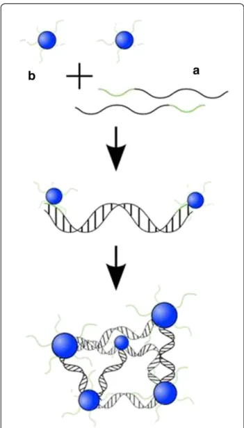
Since our aim was to produce electronically-active, nanostructured bulk materials with the help of DNA, we used a DNA block copolymer with the hydrophobic seg-ment, a polyfluorene derivative covalently linked to sin-gle stranded DNA (22mer). DNA-b-PF micelles were generated simply through dissolving the polymer [23] in a buffer solution followed by heating to 95°C and cooling to room temperature overnight. The resulting spherical aggregates with cores consisting of polyfluorene and shells composed of single-stranded (ss) DNA were visua-lized on a mica surface by atomic-force microscopy (AFM), showed an average height of 8 ± 3 nm (see Add-itional file 1).
On the basis of this self-assembled building block, higher-ordered structures were fabricated by hybridization (Figure 1). A rigid duplex was designed consisting of 120 base pairs (bp) and two terminal 24mer overhangs that were complementary to the DNA present in the corona of
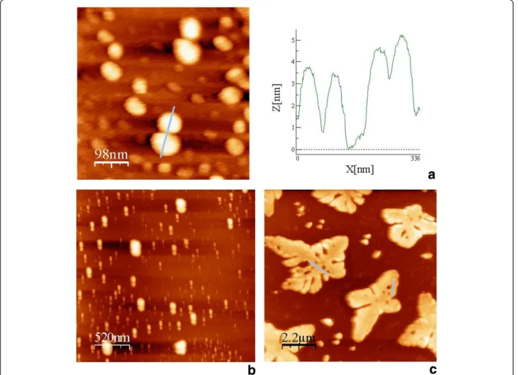
DBC micelles . After mixing micelles and linker strands the resulting structures were analyzed by AFM and Trans-mission Electron Microscopy (TEM). As revealed by the former technique, linear assemblies, two-dimensional sheets and three-dimensional fractals were formed and are shown in Figures 2a, 2b and 2c, respectively.
For the one- and two-dimensional structures, either in tapping mode or in phase mode, micelle cores were identified as bright spots. Moreover, both types of struc-tures showed characteristic spacing of the microphase-separated conjugated polymers within the DNA matrix. The duplex of 166 bp bridging the core of the micelles exhibits a length of 56.4 nm on the assumption of 0.34 nm per nucleotide. Statistical analysis of the
characteristic spacing of conjugated polymer cores gave distances of 50 ± 7 and 66 ± 7 nm for one- and two-dimensional assemblies, respectively, which is in good agreement with the theoretical value (see Additional 1 Figure S1 and Fig. S2). For the 3-D fractals, microdo-mains within the material could be measured only at the edges of the sample, exhibiting average distances of 80 ± 5 nm (Figure 2c, inset).
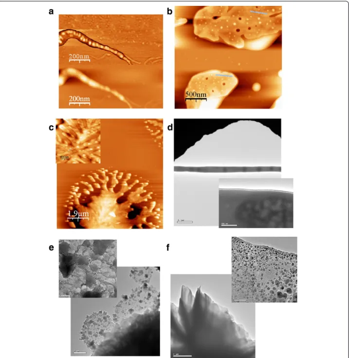
While AFM gives information about the ordering on the surface of the material, TEM allowed investigation of the bulk structure. For the TEM study, DNA comple-mentary to that of the corona of the DBC micelles was immobilized on a gold-coated grid via an Au-S-bond, with the use of a previously published procedure [24,25]. Interestingly, high contrast was observed between mi-celle cores and DNA within the sample. This might be attributed to the semiconducting properties of polyfluor-ene of the DBCs and is in sharp contrast to other block-copolymer systems investigated by TEM [26]. As for AFM, one-dimensional structures could be well visua-lized by TEM. The micelle cores that were separated through linker DNA, appeared as dark spots (Figure 2d). On average, the PFO domains were 800 nm apart,, indi-cating the formation of aggregates.
Following this, the two-dimensional structures were investigated by TEM at lower and higher magnifications. The low-magnification images of the DNA micelle net-works show films of varying thickness appearing as shades of grey (Figure 2e). While these images did not allow the identification of individual DBC particles, such structures were visible at higher magnification (Figure 2f). The
micelle cores appeared as dark spots scattered within a matrix of brighter contrast. Different film thicknesses cor-respond to areas with different grey scale. It should be noted that quantitative distance analysis cannot be per-formed for imperfect crystals as exist in our sample. How-ever, TEM undoubtedly proves microstructure formation of DBC micelles induced by linker DNA (Figure 2e, inset). The 3-D fractals appeared as darker areas in the TEM picture at lower magnification (Figure 2f ). At higher magnification, individual particles could be identified at the edges of the structures (Figure 2f inset), confirming the observation of the AFM measurements. The pres-ence of 1-D chains, 2-D sheets and 3-D fractals have been reported for inorganic nanoparticles equipped with various covalently attached surface functionalities in-cluding DNA [27]. Our structural analysis reveals similar behavior; however, the materials presented here consist exclusively of organic building blocks and, even more important, rely on two self-assembly processes, namely, Watson-Crick base-pairing of DNA and microphase sep-aration of the organic polymer segment in an aqueous environment.
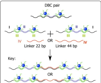
To demonstrate the flexibility of our approach, we fab-ricated a second type of DNA hybrid material with dif-ferent structural features. The system contained four building blocks (Figure 3). The sequence of the 115mer template I was chosen in such a way that hybridization with sequence II results in a central double-stranded DNA part, which is flanked by two annealing sites for the DBC micelles III (2x22 bp). The remaining 44mer overhangs of the template allow formation of extended structures by hybridization with sequence IV, a 24mer.
After annealing of all components in a single
hybridization reaction, the resulting structures were ana-lyzed by AFM on a mica surface. The AFM images sug-gest the formation of one-dimensional extended DNA chains decorated with pairs of DBC micelles (Figure 4a and 4b).
Statistical analysis of the separation of particles revealed two characteristic distances. The first was 7 ± 2 nm, which can be ascribed to the spacing of two DBC micelles by sequence II. This separation is in good agreement with the calculated value of 7.7 nm. The sec-ond characteristic distance was found to be 13 ± 2 nm. This can be explained by linking DBC micelle pairs by sequence IV. Again good agreement with the calculated value (14 nm) was observed.
Finally, we varied the size of sequence IV, which con-nects template-strand I. When the length of IV was increased from 24 to 44 bp, completely different struc-tures were detected by AFM, namely, dendritic architec-tures. Such surface topologies have been found for other polymers as well [28]. This great structural difference can be attributed to the changed connection between
template-strands I. When sequence IV is composed of 22 bp, exclusively ds DNA is present, whereas when IV consists of 44 bp, ds stretches alternate with ss stretches of 22 bp. We ascribe the different structures to the dif-ference in persistence length of the ds DNA (50 nm) versus that of ss DNA (2-3 nm). In similar experiments with polymers, the rigidity of the backbone was found to be an important parameter determining the structural features of large assemblies. In a separate study, we will
report on the detailed growth mechanism of the den-dritic structures.
Conclusions
In summary, we have shown the fabrication of 1-, 2- and 3-D structures composed of DNA and DBCs. Common to all morphologies is the integrity of the micelles con-sisting of DBCs. Moreover, the micelles represent not only structural features but act as cross-linking units as
well. Most noteworthy is the structural diversity of the morphologies controlled by variation of the DNA sequences. One cannot expect to realize the same degree of order as that obtained for pristine DNA assemblies based on the tile and origami approaches [29]. The same holds true for networks generated by oligonucleotide functiona-lized gold nanoparticle networks. There is a striking differ-ence between the bioorganic structures presented here and the all DNA and bioinorganic assemblies. The DNA conjugated-polymer superstructures rely on two self-assembly processes, while in pristine DNA and the Au nanoparticle-DNA assemblies only Watson-Crick base pairing is responsible for superstructure formation. In this respect, the materials presented here should be compared to block-copolymers which have also been reported con-taining microphase-separated semiconducting domains [30,31]. We have demonstrated that typical morphologies of conventional block copolymer systems can be extended by the utilization of DNA block copolymers allowing add-itional structure control by the DNA segment.
Methods
DBC micelles
poly(9,9-dioctylfluorene) was functionalized with one terminal hydroxyl group as described previously[23]. After conversion of 1 to the phosphoramidite polymer (2), the desired diblock-copolymer architecture, DNA-b -PF (3) was obtained by grafting 2 onto the immobilized DNA and incubating with concentrated ammonia to achieve liberation from the solid support and de-protection of the nucleobases. The nucleic acid segment was composed of the 22mer sequence 5’-CCTCGCT CTGCTAATCCTGTTA-3’ with a molecular weight of 6670 g/mol while the organic polymer consisted of poly-fluorene with a total number average molecular weight of 5000 g/mol.
Pairs
0.1 nmol of sequence 1 was immersed in 500μL PBS buffer solution (pH = 7.4) and added to 0.2 nmol F8PB1147 block copolymer micelles solution immersed
in 100μL PBS buffer and left for 24 h at room temperature for hybridization. After hybridization took place, 50μL of the medium were deposited on a mica surface, left for 4 h and dried with nitrogen gas.
3D network
For the preparation of the double-strand DNA con-nector, 71.4μLof a solution containing 0.1 nmol of se-quence 3 was mixed with 38.46μL of 0.1 nmol of sequence 4. The block-copolymer solution (0.1 nmol in 10μL) was diluted to a volume of 0.5 mL with PBS buf-fer. The two solutions were then mixed and deposited on a mica surface.
HR-TEM imaging
TEM measurements were performed on gold-coated TEM grids that were immersed in 100μLof a solution of the network of interest of complementary thiolated ssDNA strands (0.5 nmol in 100μL). These grids were kept in solution for 24 h and then dried with nitrogen gas before imaging.
DNA sequences
Sequence 1 5' AATCATACGTACTCAACTGCTG GG AGCGAGACGATTAGGACAAT AACTTGGGTATGCT
GTCAGATGGCTCG GGAGCGAGACGATTAGGACA
AT AATCATACGTACTCAACTGCTG 3'
Sequence 2 5' CGA GCC ATC TGA CAG CAT ACC CAA GTT3'
Sequence 3 5'TGG CCA CGA ACA AAC AAG ACA AGA GAG TAA GTC TGA TCT GGA GAG GTC GGA AAT CAT AGA AAC CAC ACG AAT GAT AAG GCA TGG AGG TAA AAG GCA TCA ATA ACA GGA TTA GCA GAG CGA GG 3'
Sequence 4 5' TTG ATG CGT TTT ACC TCC ATG CCT TAT CAT TCG TGT GGT TTC TAT GAT TCT CGA CCT CTC CAG ATC AGA CTT ACT CTC TTG TCT TGT TTG TTC GTG GCC ATA ACA GGA TTA GCA GAG CGA GG3'
Thiolated DNA5' (Thiol C6) ATA CCC ACG CCG AAA CAA GC
Additional file
Additional file 1:Atomic-Force-Microscopy analysis and statistical data of the spacing lengths of the network are presented.
Abbreviations
DBC: DNA block copolymer; TEM: Transmission Electron Microscopy.
Competing interests
The authors declare no competing interests.
Acknowledgements
We thank Prof Andreas Herrmann (RUG, Netherlands) for supplying the micelles and fruitful discussions. SR thanks the Bikura Fund for financial support
Author details
1
Faculty of Exact Sciences and Center for Nanoscience and Nanotechnology, Tel-Aviv University, Ramat Aviv, Tel-Aviv 69978, Israel.2University of
Groningen, Zernike Institute for Advanced Materials, Nijenborgh 4, 9747AG, Groningen, The Netherlands.3Nanotechnology Research Laboratory,
Materials Engineering Department, Al-Quds University, East Jerusalem, Palestinian Authority.
Authors’contributions
ED and KL carried out the AFM studies and the DNA hybridization. DP synthesized the micelles. All participated in the design of the experiments, coordination and helped to draft the manuscript. All authors read and approved the final manuscript.”
Received: 13 February 2012 Accepted: 30 May 2012 Published: 30 May 2012
References
1. Fu TJ, Seeman NC:Biochemistry (Mosc)1993,32:3211. 2. Li X, Yang X, Qi J, Seeman NCJ:Am Chem Soc1996,118:6131. 3. Rothemund PWK, Papadakis N, Winfree E:PLoS Biol2004,2:e424. 4. Rothemund PWK:Nature2006,440:297.
5. He Y, Ye T, Su M, Zhang C, Ribbe AE, Jiang W, Mao CD:Nature2008,
452:198.
6. Mirkin CA, Letsinger RL, Mucic RC, Storhoff JJ:Nature1996,382:607. 7. Alivisatos AP, Johnsson KP, Peng X, Wilson TE, Loweth CJ, Bruchez MP,
Schultz PG:Nature1996,382:609.
8. Storhoff JJ, Mirkin CA:Chem Rev1999,99:1849.
9. Niemeyer CM, Adler M, Gao S, Chi LF:Angew Chem Int Ed2000,39:3055. 10. Taton TA, Mirkin CA, Letsinger RL:Science2000,289:1757.
11. Ke YG, Lindsay S, Chang Y, Liu Y, Yan H:Science2008,319:180. 12. Braun E, Eichen Y, Sivan U, Ben-Yoseph G:Nature1998,391:775. 13. Keren K, Berman RS, Buchstab E, Sivan U, Braun E:Science2003,302:1380. 14. Um SH, Lee JB, Park N, Kwon SY, Umbach CC, Luo D:Nat Mat2006,5:797. 15. Nagahara S, Matsuda T:Polym Gels Net1996,4:111.
16. Murakami Y, Maeda M:Biomacromolecules2005,6:2927. 17. Lin DC, Yurke B, Langrana NAJ:Mat Res2005,20:1456. 18. Liedl T, Dietz H, Yurke B, Simmel F:Small2007,3:1688.
19. Park SY, Lytton-Jean AKR, Lee B, Weigand S, Schatz GC, Mirkin CA:Nature
2008,451:553.
20. Alemdaroglu FE, Ding K, Berger R, Herrmann A:Angew Chem Int Ed2006,
45:4206.
21. Li Z, Zhang Y, Fullhart P, Mirkin CA:Nano Lett2004,4:1055.
22. Safak M, Alemdaroglu FE, Li Y, Ergen E, Herrmann A:Adv Mat2007,19:1499. 23. Kwak M, Gao J, Prusty DK, Musser AJ, Markov VA, Tombros N, Stuart MCA,
Browne WR, Boekema EJ, ten Brinke G, Jonkman HT, van Wees BJ, Loi MA, Herrmann A:Angew Chem Int Ed2011,50:3206.
24. Ghabboun J, Sowwan M, Cohen H, Molotsky T, Borovok N, Dwir B, Kapon E, Kotlyar A, Porath D:Appl Phys Lett2007,91:173101.
25. Porath D, Bezryadin A, de Vries S, Dekker C:Nature2000,403:635. 26. Chiu JJ, Kim BJ, Kramer EJ, Pine DJJ:Am Chem Soc2005,127:5036. 27. Nie ZH, Petukhova A, Kumacheva E:Nat Nanotech2010,5:15.
28. Granasy L, Pusztai T, Borzsosnyi T, Toth GI, Tegze G, Warren JA, Douglas JF:
PMag2006,86:3757.
29. Bordier B, Peralaheape M, Degols G, Lebleu B, Litvak S, Sarihcottin L, Helene C:Proc Natl Acad Sci U S A1995,92:9383.
30. Marsitzky D, Klapper M, Müllen K:Macromolecules1999,32:8685. 31. Sommer M, Lang AS, Thelakkat M:Angew Chem Int Ed2008,47:7901.
doi:10.1186/1477-3155-10-21
Cite this article as:Mentovichet al.:DNA-nanoparticle assemblies go organic: Macroscopic polymeric materials with nanosized features.