ORIGINAL ARTICLE
The influence of in vitro pectin
fermentation on the human fecal microbiome
So‑Jung Bang
1†, Gayoung Kim
1†, Mi Young Lim
2, Eun‑Ji Song
2,3, Dong‑Hyun Jung
1, Jun‑Seok Kum
4,
Young‑Do Nam
2,3, Cheon‑Seok Park
1*and Dong‑Ho Seo
2*Abstract
Pectin is a complex dietary fiber and a prebiotic. To investigate pectin‑induced changes in the gut microbiome and their effects on the short chain fatty acids (SCFAs) production, we performed in vitro pectin fermentation using the feces of three Korean donors. The pectin degradations in all three donors were observed. While the donors displayed differences in baseline gut microbiota composition, commonly increased bacteria after pectin fermentation included Lachnospira, Dorea, Clostridium, and Sutterella. Regarding SCFAs, acetate levels rapidly increased with incubation with pectin, and butyrate levels also increased after 6 h of incubation. The results suggest that pectin fermentation increases bacterial species belonging to Clostridium cluster XIV (Lachnospira, Dorea, and Clostridium), with Lachnospira displaying the greatest increase. The results also confirm that pectin fermentation leads to the production of acetate and butyrate.
Keywords: Pectin, Fecal microbiota, Fermentation, Short chain fatty acids, Prebiotic
© The Author(s) 2018. This article is distributed under the terms of the Creative Commons Attribution 4.0 International License (http://creat iveco mmons .org/licen ses/by/4.0/), which permits unrestricted use, distribution, and reproduction in any medium, provided you give appropriate credit to the original author(s) and the source, provide a link to the Creative Commons license, and indicate if changes were made.
Introduction
Recent studies have demonstrated that the gut micro-biota plays important roles in human health, and are associated with diseases. The pathogenic mechanisms of various diseases and disorders, such as irritable bowel syndrome (Hyland et al. 2014), Crohn’s disease (Gevers et al. 2014), and non-alcoholic steatohepatitis/non-alco-holic fatty liver disease (Icaza-Chávez 2013; Sánchez et al.
2017) are associated with the composition and diver-sity of the gut microbiota (Laparra and Sanz 2010). The constitution of the gut microbiota can be influenced by endogenous and environmental factors, such as one’s die-tary, antibiotic, xenobiotic, and probiotic intakes (Falony et al. 2016).
Prebiotic food ingredients feed the intestinal microbi-ota, and can be used to selectively promote the growth of specific microbiota (Laparra and Sanz 2010). Most
prebiotics are carbohydrates, such as inulin, fructooligo-saccharide, and human milk oligofructooligo-saccharide, which are not digested by human digestive enzymes (Bindels et al.
2015; Coppa et al. 2006). Microorganisms in the intes-tines produce energy and short-chain fatty acids (SCFAs) including acetate, propionate, and butyrate, affecting the host though prebiotic fermentation (Flint et al. 2012). The SCFAs produced by the gut microbiota have positive effects on immune function and ameliorate metabolic diseases such as obesity and type 2 diabetes (Clemente et al. 2012; Gerritsen et al. 2011).
Differences in intestinal microbiota composition can exist depending on diet (Conlon and Bird 2014; David et al. 2014), and in particular, on the types of nutrients ingested in various countries, environments, and cul-tures (Marques et al. 2010). Asian diets tend to be high in carbohydrates, while western country diets are relatively high in fat (LeCroy and Stevens 2017; Li et al. 2017). The Korean diet, one of the Asian diets, tends to be a high-vegetable diet compared with the typical western diet because of traditional foods including Kimchi (Kim et al.
2016; Song and Joung 2012; Lee et al. 2002).
Pectin is a complex polysaccharide found in the cell walls of a variety of vegetables and fruits, which is mainly
Open Access
*Correspondence: [email protected]; [email protected]
†So‑Jung Bang and Gayoung Kim contributed equally to this work 1 Graduate School of Biotechnology and Institute of Life Science
and Resources, Kyung Hee University, Yongin 17104, Republic of Korea
2 Research Group of Healthcare, Korea Food Research Institute,
Wanju 55365, Republic of Korea
composed of d-galacturonic acid (GalA) with α-(1-4) gly-cosidic linkages (Sriamornsak 2003). Pectin is a candidate prebiotic because it is not well degraded by human diges-tive enzymes, but is by microorganisms (Holloway et al.
1983). Pectin is degraded by the gut microbiota, produc-ing SCFAs and changproduc-ing the composition of the intestinal microbiota (Chung et al. 2016; Marounek and Dušková
1999). Although many studies have confirmed the deg-radation of pectin and the production of SCFAs by gut microbiota species of the genera Bifidobacterium, Faecal-ibacterium, Anaerostipes, and Roseburia (Duncan et al.
2004), very few metagenomics studies have been per-formed on the human gut microbiota following in vitro fermentation of pectin. In addition, no studies have been performed examining pectin utilization and gut micro-biota changes in Koreans, which have high vegetable con-sumption. Thus, in this study, we explored the process of pectin degradation and the compositional changes in the gut microbiota of three Korean subjects after pectin fermentation.
Materials and methods
Donor information
All donors (males, ages 29, 30, and 30) were healthy and did not have any gastrointestinal disease. Donors had consumed a regular diet and had not received antibiotic treatment in the last 6 months. The study was approved by the Institutional Review Board of Kyung Hee Univer-sity (IRB file no. KHSIRB-17-004). All experiments were performed in accordance with relevant guidelines and regulations. Informed consent was obtained from all participants.
Fecal collection
Fecal samples (20 g) were collected from three volun-teers under anaerobic conditions and transported within 1 h after excretion. Fecal samples were diluted fivefold with sterile phosphate-buffered saline in an anaerobic chamber. After mixing, the resultant fecal slurries were homogenized and immediately inoculated in the pre-pared medium.
Growth media
Pectin from citrus peel was purchased from Sigma-Aldrich (St. Louis, MO, USA), and was composed of > 74% galacturonic acid. Because a basal medium is non-selective and supports the growth of several organ-isms, we selected a basal medium for this study; the basal medium used was the chopped meat (CM) broth containing 15% (v/v) of bovine rumen fluid (CMR). Each liter of CMR consisted of peptone (30.0 g), yeast extract (5.0 g), dipotassium phosphate (5.0 g), l-cysteine (0.5 g), and resazurin (0.001 g). Pectin was added to a
final concentration of 1%. CMR medium with 1% pectin (CMRP) was stirred on a hot plate to dissolve the pec-tin, transferred to serum vials, and capped. Sealed CMRP medium was flushed with 99.5% CO2 gas and sterilized by autoclave at 121 °C for 15 min.
Batch culture incubations
To investigate changes in microbial diversity and pectin degradability, 100 μL of prepared feces was inoculated into 20 mL of CMRP medium (baseline). Cultures were incubated at 37 °C at 150 rpm, with sampling at various incubation times (0, 6, 12, 18, 24, 36, and 48 h). All sam-ples were immediately frozen and stored at − 72 °C. Incu-bations were performed in CMRP medium in triplicate.
Determination of total sugar
The total sugar in culture was measured by the phenol– sulfuric acid method. A 5% phenol solution (200 μL) was added to 100 μL of culture supernatant from each time point. The reaction mixture was mixed with 800 μL of 99% sulfuric acid and vortexed. After cooling for 20 min at 25 °C, 250 μL of each mixture was added to a 96-well microplate. The absorbance of the phenol–sulfuric acid was measured at 550 nm using an iMark Microplate Absorbance Reader (BioRad Laboratories, Inc., Hercules, CA, USA).
Determination of reducing sugar
The reducing sugar in culture was measured using 3,5-dinitrosalicylic acid (DNS). Culture supernatants from each time point (20 μL) were diluted in 80 μL deionized water. Reducing sugar was detected by adding 300 μL of DNS solution and boiling for 5 min. After cool-ing on ice, the absorbance was measured at 555 nm uscool-ing an iMark Microplate Absorbance Reader.
Thin liquid chromatography (TLC) analysis
TLC analysis was performed with TLC Silica gel 60G F25425 Glass plates (Merk Millipore, Billerica, MA, USA) after activating at 110 °C for 5 min. Culture supernatants from each time point were loaded onto TLC plates and placed in a TLC chamber containing a 5:2:3 (v/v/v) sol-vent mixture of 1-butanol:acetic acid:water for degrada-tion product analysis. Plates were dried, rapidly soaked into 0.3% (w/v) 1-naphthol and 5% (v/v) sulfuric acid in methanol, dried again, and placed on a 110 °C oven for 10 min.
DNA extraction and 16S rRNA gene sequencing
bead-beating twice for 5 min. The V1–V2 regions of 16S rRNA genes were amplified by polymerase chain reaction (PCR) using universal primers (8F and 338R) with bar-code sequences for multiplexing sample reads. PCR was performed using a PCR Thermal Cycler Dice (Takara, Shuzu, Japan) and recombinant Taq DNA polymerase (Takara). The PCR conditions were as follows: 95 °C for 5 min; 30 cycles of 30 s at 95 °C, 1 min at 61 °C, and 40 s at 72 °C; and 5 min at 72 °C. The amplified 16S rRNA gene products were purified with an AccuPrep PCR Puri-fication Kit (BIONEER, Daejeon, Korea).
PCR product concentrations were measured on a Nan-oDrop ND-1000 (NanNan-oDrop Technologies Inc., Wilm-ington, DE, USA) and mixed to a constant concentration such that the total concentration was 1 mg. The Ion Xpress Plus Fragment Library Kit (Thermo Scientific, Wilmington, DE, USA) was used to form the amplicon library according to the manufacturer’s instructions, and quantification of the amplicon library was per-formed using a Bioanalyzer 2100 (Agilent Technologies, Inc., Santa Clara, CA, USA). The amplicon library was sequenced on an Ion Torrent PGM system (Thermo Sci-entific, Wilmington, DE, USA).
Bioinformatic analysis
The quantitative insights into microbial ecology (QIIME) pipeline (Caporaso et al. 2010) was used to analyze the sequences. After quality checking the FASTQ file, the barcode sequences of the amplicons were removed. Sequences of 300–440 bp size were filtered and dimers and chimeras were removed, based on Ribosomal Data-base Project (RDB) dataData-base, using ultra-fast sequence analysis (USEARCH 8.1 to ensure high quality. Opera-tional taxonomic units were analyzed based on the Greengenes 13_8 database (McDonald et al. 2012) iden-tified with 97% similarity. Principal coordinates analy-sis (PCoA; based on the Bray–Curtis distance) was performed using QIIME 1.9.1. The identification of microbial taxa that were significantly associated with the incubation time was conducted using multivariate asso-ciation with a linear model (Morgan et al. 2012). Asso-ciations with a Benjamini–Hochberg false discovery rate-corrected p value (q value) of < 0.1 were considered significant. The raw data was uploaded to NCBI sequence read archive database (accession number: DRA006695).
SCFA concentration analysis
CMRP medium supernatants containing the fecal inocu-lum of the three donors at each incubation time (0, 6, 12, 18, 24, 36, and 48 h) were transferred to new tubes, and aliquots were frozen at − 20 °C for SCFA analysis. Before analysis, samples were filtered through membrane fil-ters (pore size: 0.25 μm). SCFA analysis was performed
by ion chromatography (IC) using a 940 Professional IC Vario (Methrohm, Herisau, Switzerland) composed of a two-channel peristaltic pump and a 945 Professional Detector Vario conductivity detector, with an 889 IC Sample Center (Methrohm). IC Net 3.1 software was used to record the data. Ion exclusion was performed on a Metrosep Organic Acid 250/7.8 column (Methrohm) and 0.1% sulfuric acid was used as the mobile phase, at a flow rate of 0.5 mL/min and pressure of 6.99 MPa. Sam-ples (20 μL) were injected into columns maintained at 30 °C. The peak height, peak area, and retention time of recorded samples and acetic acid, propionic acid, butyric acid, valeric acid, and isovaleric acid standards were used to measure concentrations.
Results
Pectin degradation by human feces according to incubation time
early stages. Galacturonic acid, the final degrada-tion product of pectin, was produced after 6–12 h in all samples and was completely consumed at later time points. In the donor 3 sample, the production of galacturonic acid was faster than in the other sam-ples, which confirmed that more pectin degradation products were produced. These results are consistent with the total carbohydrate and reducing sugar analy-ses described above, and indicates that the intestinal microbiota differed in each donor.
Fecal microbial composition of each donor at baseline
To investigate the in vitro fermentation of pectin by the human fecal microbiota, we anaerobically incubated the feces of the three donors with 1% pectin for 18 h. The compositions of the fecal microbiota samples were observed at baseline (in raw fecal samples which were not mixed with medium or pectin) and after every 6 h of incubation time using 16S rRNA gene sequencing.
We first examined the initial fecal microbiota compo-sitions of each donor. At the phylum level, Firmicutes
(57.39% of average relative abundance) and Bacteroi-detes (42.11%) were dominant in all three donors, and
Proteobacteria (0.26%) and Actinobacteria (0.02%) were also present in small proportions (Fig. 2a). The
Firmicutes/Bacteroidetes ratios were higher in donor 1 (2.21 ratio) and donor 3 (1.52 ratio) compared to donor 2 (0.78 ratio). We next examined the top 20 most abundant genera in each baseline sample (Fig. 2b). Donors 1, 2, and 3 had 18, 12, and 11 genera comprising > 1% relative abundance, respectively. In all three donors, Bacteroides
and Ruminococcaceae were the most abundant. The relative abundance of Bacteroides in donor 2 was much higher (46.34%) than that in donors 1 and 3 (19.20 and 37.39%, respectively). In addition, each donor displayed differences in bacterial abundance. For example, donor 3 harbored more Lachnospiraceae and Faecalibacterium
than the others, while donor 1 and donor 2 harbored more Streptococcus, unclassified Rikenellaceae, Lactoba-cillus, Lachnospiraceae, and Prevotella.
Changes in fecal microbial composition during pectin fermentation
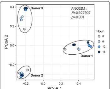
To evaluate the influence of pectin fermentation on the overall structure of the fecal microbiota, we conducted PCoA based on Bray–Curtis distances. Samples from each donor clustered separately from those of the other donors, showing that the donor had the greatest effect on microbiome composition, even after incubation with pectin for 18 h (Fig. 3). However, among samples from each donor, the microbial composition changed with increased incubation time. For donor 3, for example, the
baseline sample was most similar to the sample after 6 h pectin incubation, followed by those taken at 12 and 18 h.
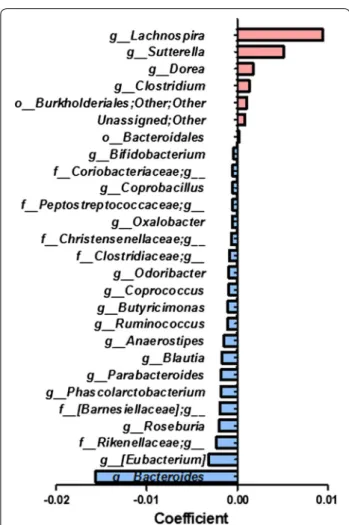
To identify microbes whose abundances significantly changed with increased incubation time, we explored the associations between bacterial abundance and incubation time by linear regression analysis. The levels of several specific bacteria were associated with time. For exam-ple, Lachnospira, Sutterella, Dorea, and Clostridium were significantly increased over time, while Bacteroides and
Roseburia were significantly decreased (Fig. 4 and Addi-tional file 1: Figure S1).
SCFA formation during pectin fermentation
When we analyzed the SCFA changes in each sample every 6 h, we observed rapid increases in acetate after 6 h, which continuously increased up to 18 h, then rap-idly decreased by 36 h. Propionate increased after 48 h, and while butyrate did not rapidly increase like acetate, it increased by approximately 28% by 48 h (Fig. 5).
Discussion
The contributions of the gut microbiome to health and nutrition depend on its composition, which is affected by different factors, including lifestyle and diet (Conlon and Bird 2014). Gut microbiota composition can be changed by including indigestible carbohydrates (prebiotics) in one’s diet (Flint et al. 2012). Pectin is a prebiotic dietary fiber that affects the gut microbiota (Woods and Gor-bach 2001). In this study, we investigated the utilization of pectin by the gut microbiota and analyzed microbiota composition changes with in vitro pectin fermentation through metagenomics analysis.
Pectin was mainly degraded between 0 and 18 h in all three donors, but the samples showed differences in their pectin degradation ability. These differences in substrate utility depend on the composition of the microbiome (Flint et al. 2008). When pectin was digested, galactu-ronic acid is produced (De Vries et al. 1982). Monosac-charide such as galacturonic acid is used as an energy source by bacteria and contributes to the development and maintenance of the gut microbiota (Zoetendal et al.
2012). In this study, the microbiota of donor 3 showed relatively high pectin utilization, and as expected, this sample contained higher baseline Lachnospiraceae and
Faecalibacterium levels than in the other samples. Lach-nospiraceae has been demonstrated to express carbohy-drate-active and pectin-degrading enzymes (Biddle et al.
2013), and Faecalibacterium also has reported pectin degradation ability (Lopez-Siles et al. 2012). In addi-tion, the sample from donor 2 consumed more galactu-ronic acid than the others once the reducing sugar level increased after 6 h. These results indicate that differences in the baseline levels of different microbiome compo-nents result in differences in pectin degradation and utili-zation in each donor.
We observed comprehensive overall gut microbiota changes after pectin fermentation in all three donors compared to their baseline compositions. We not only observed differences between each donor’s gut micro-biota, but also changes with increased incubation time by PCoA. We assumed that specific gut microbiota were utilizing pectin as a substrate; for example, Prevo-tella and Butyrivibrio spp., which express pectinolytic enzymes (Marounek and Dušková 1999). With increased incubation time with pectin, we observed increases in
Lachnospira, Sutterella, Dorea, and Clostridium. The Fig. 4 Significantly changed taxa according to pectin incubation
time (q value < 0.1)
Clostridium coccoides group (cluster XIVa) includes
Lachnospira, Dorea, and Clostridium (Lopetuso et al.
2013). Lachnospira was the most increased and has been reported to use pectin as a substrate (Wojciechowicz et al. 1980) and Lachnospira plays a role that produced SCFA (Duvallet et al. 2017; Jones et al. 2014). Pectin degradation by Lachnospira affects the growth of other bacteria, including other saccharolytic bacteria, via cross-feeding (Salyers and Leedle 1983). Increases in Dorea and
Clostridium may be due to Lachnospira cross-feeding. These results indicate that pectin promotes the presence of species in Clostridium cluster XIV, including Lach-nospira, Dorea, and Clostridium. We also observed that some bacteria, including Bacteroides, which can utilize pectin (Gibson and Roberfroid 1995), decreased with increasing pectin incubation time, possibly due to envi-ronmental nutritional limitations caused by selective cul-ture with pectin and carbohydrate consumption.
The production of SCFAs, including acetate, propi-onate, and butyrate, is affected by the composition of the gut microbiota, the utilization of carbohydrate sub-strates, and the gut environment, including the pH and other nutritional factors (Cummings and Englyst 1987; Yuan et al. 2006). Walker et al. (2005) reported that the production acetate and propionate increased at pH 6.5.
Lachnospira produces the most pectin lyase at pH 6.1– 6.3 (Silley 1986), and in pectin culture, mainly produces acetate and lactate (Dušková and Marounek 2001). The
Clostridium genus is known to mainly produce butyrate (Rajilić-Stojanović and de Vos 2014). Based on this, we suggest that Lachnospira produced galacturonic acid through the digestion of pectin and acetate, and that this environment restrained the growth of Bacteroides by causing a mildly acidic pH. Produced galacturonic acid affected the composition of the gut microbiota, enhanc-ing cluster XIVa species belongenhanc-ing to Dorea and Clostrid-ium, which then produced butyrate (Duncan et al. 2007; Walker et al. 2005). We observed decreased acetate after 18 h of pectin incubation, indicating that not only galac-turonic acid but also acetate was utilized by bacteria like
Faecalibacterium to produce butyrate (Duncan et al.
2004; Ramirez-Farias et al. 2008).
This result suggests that acetate-producing bacteria like Lachnospira and Faecalibacterium caused increased butyrate levels via butyrate synthesis using acetate as a substrate (Khan et al. 2012; Rios-Covian et al. 2015). Based on this, it appears that pectin degradation results in a gut microbiota growth environment associated with the development of acetate and butyrate.
In conclusion, we demonstrate that in Korean indi-viduals, pectin can change the gut microbiota by meas-uring total sugar levels and microbial composition
over time. Pectin was completely degraded by the gut microbiota at 6, 12, and 18 h, and Lachnospira and Fae-calibacterium, which can utilize pectin, were increased. Pectin-induced changes in the gut microbiota increased the formation of associated SCFAs from 6 h on, when pectin was decomposed. Therefore, we confirmed that pectin fermentation in gut microbiota samples from Korean individuals induced microbiota compositional changes. Increased pectin utilization and correspond-ing changes to gut microbiome composition may be beneficial to human health. Further analysis of the gut microbiomes of larger numbers of donors, in addition to experiments regarding cross-feeding and in vivo gut microbiota changes, would provide more accurate results.
Abbreviations
SCFAs: short chain fatty acids; CMR: bovine rumen fluid; CMRP: bovine rumen fluid pectin; DNS: 3,5‑dinitrosalicylic acid; TLC: thin liquid chromatography; PCR: polymerase chain reaction; QIIME: the quantitative insights into microbial ecology; PCoA: principal coordinates analysis; IC: ion chromatography; USE‑ ARCH: ultra‑fast sequence analysis; RDP: Ribosomal Database Project.
Authors’ contributions
C‑SP and D‑HS designed and coordinated all the experiments; S‑JB performed the experiments and analyzed the gut microbiota data; GK conducted experi‑ ments on pectin degradation of human fecal microbiome; MYL, E‑JS, and Y‑DN performed statistical analysis and metagenome analysis of human fecal microbial change; D‑HJ performed anaerobic cultivation of the human fecal microbiome; J‑SK confirmed the SCFA production by ion chromatography analysis. S‑JB, GK, C‑SP, and D‑HS prepared the manuscript, evaluated the results and revised the manuscript. All authors read and approved the final manuscript.
Author details
1 Graduate School of Biotechnology and Institute of Life Science
and Resources, Kyung Hee University, Yongin 17104, Republic of Korea.
2 Research Group of Healthcare, Korea Food Research Institute, Wanju 55365,
Republic of Korea. 3 Department of Food Biotechnology, Korea University
of Science and Technology, Daejeon 34113, Republic of Korea. 4 Research
Group of Food Processing, Korea Food Research Institute, Wanju 55365, Republic of Korea.
Competing interests
The authors declare that they have no competing interests.
Availability of data and materials
Not applicable.
Consent for publication
Not applicable.
Ethics approval and consent to participate
All procedures performed in studies involving human participants were in accordance with the ethical standards of the institutional and/or national research committee and with the 1964 Helsinki declaration and its later amendments or comparable ethical standards.
Additional file
Funding
This research was supported by the Main Research Program (E0170602‑02) of the Korea Food Research Institute funded by the Ministry of Science and ICT for Dr. Y‑D Nam and was supported by a National Research Founda‑ tion of Korea (NRF) Grant funded by the Korean government (MEST) (No. 2017R1A2B4004218) for Dr. C‑S Park.
Publisher’s Note
Springer Nature remains neutral with regard to jurisdictional claims in pub‑ lished maps and institutional affiliations.
Received: 30 May 2018 Accepted: 12 June 2018
References
Biddle A, Stewart L, Blanchard J, Leschine S (2013) Untangling the genetic basis of fibrolytic specialization by Lachnospiraceae and Ruminococ-caceae in diverse gut communities. Diversity 5(4):627–640. https ://doi. org/10.3390/d5030 627
Bindels LB, Delzenne NM, Cani PD, Walter J (2015) Towards a more comprehen‑ sive concept for prebiotics. Nat Rev Gastroenterol Hepatol 12(5):303–310. https ://doi.org/10.1038/nrgas tro.2015.47
Caporaso JG, Kuczynski J, Stombaugh J, Bittinger K, Bushman FD, Costello EK, Fierer N, Pena AG, Goodrich JK, Gordon JI (2010) QIIME allows analysis of high‑throughput community sequencing data. Nat Methods 7(5):335 Chung WS, Walker AW, Louis P, Parkhill J, Vermeiren J, Bosscher D, Duncan SH,
Flint HJ (2016) Modulation of the human gut microbiota by dietary fibres occurs at the species level. BMC Biol 14:3. https ://doi.org/10.1186/s1291 5‑015‑0224‑3
Clemente JC, Ursell LK, Parfrey LW, Knight R (2012) The impact of the gut microbiota on human health: an integrative view. Cell 148(6):1258–1270. https ://doi.org/10.1016/j.cell.2012.01.035
Conlon MA, Bird AR (2014) The impact of diet and lifestyle on gut microbiota and human health. Nutrients 7(1):17–44. https ://doi.org/10.3390/nu701 0017
Coppa GV, Zampini L, Galeazzi T, Gabrielli O (2006) Prebiotics in human milk: a review. Dig Liver Dis 38:S291–S294. https ://doi.org/10.1016/s1590 ‑8658(07)60013 ‑9
Cummings JH, Englyst HN (1987) Fermentation in the human large intestine and the available substrates. Am J Clin Nutr 45(5):1243–1255
David LA, Maurice CF, Carmody RN, Gootenberg DB, Button JE, Wolfe BE, Ling AV, Devlin AS, Varma Y, Fischbach MA, Biddinger SB, Dutton RJ, Turnbaugh PJ (2014) Diet rapidly and reproducibly alters the human gut microbiome. Nature 505(7484):559–563. https ://doi.org/10.1038/natur e1282 0 De Vries J, Rombouts F, Voragen A, Pilnik W (1982) Enzymic degradation of
apple pectins. Carbohydr Polym 2(1):25–33
Duncan SH, Holtrop G, Lobley GE, Calder AG, Stewart CS, Flint HJ (2004) Con‑ tribution of acetate to butyrate formation by human faecal bacteria. Br J Nutr 91(6):915–923. https ://doi.org/10.1079/BJN20 04115 0
Duncan SH, Belenguer A, Holtrop G, Johnstone AM, Flint HJ, Lobley GE (2007) Reduced dietary intake of carbohydrates by obese subjects results in decreased concentrations of butyrate and butyrate‑producing bacteria in feces. Appl Environ Microbiol 73(4):1073–1078. https ://doi.org/10.1128/ AEM.02340 ‑06
Dušková D, Marounek M (2001) Fermentation of pectin and glucose, and activ‑ ity of pectin‑degrading enzymes in the rumen bacterium Lachnospira multiparus. Lett Appl Microbiol 33(2):159–163
Duvallet C, Gibbons SM, Gurry T, Irizarry RA, Alm EJ (2017) Meta analysis of microbiome studies identifies shared and disease‑specific patterns. Nat Commun 8(1):1784. https ://doi.org/10.1038/s4146 7‑017‑01973 ‑8 Falony G, Joossens M, Vieira‑Silva S, Wang J, Darzi Y, Faust K, Kurilshikov A,
Bonder MJ, Valles‑Colomer M, Vandeputte D, Tito RY, Chaffron S, Ryme‑ nans L, Verspecht C, De Sutter L, Lima‑Mendez G, D’hoe K, Jonckheere K, Homola D, Garcia R, Tigchelaar EF, Eeckhaudt L, Fu J, Henckaerts L, Zhernakova A, Wijmenga C, Raes J (2016) Population‑level analysis of gut microbiome variation. Science 352(6285):560–564. https ://doi. org/10.1126/scien ce.aad35 03
Flint HJ, Bayer EA, Rincon MT, Lamed R, White BA (2008) Polysaccharide utiliza‑ tion by gut bacteria: potential for new insights from genomic analysis. Nat Rev Microbiol 6(2):121–131. https ://doi.org/10.1038/nrmic ro181 7 Flint HJ, Scott KP, Louis P, Duncan SH (2012) The role of the gut microbiota in
nutrition and health. Nat Rev Gastroenterol Hepatol 9(10):577–589. https ://doi.org/10.1038/nrgas tro.2012.156
Gerritsen J, Smidt H, Rijkers GT, de Vos WM (2011) Intestinal microbiota in human health and disease: the impact of probiotics. Genes Nutr 6(3):209–240. https ://doi.org/10.1007/s1226 3‑011‑0229‑7
Gevers D, Kugathasan S, Denson LA, Vazquez‑Baeza Y, Van Treuren W, Ren B, Schwager E, Knights D, Song SJ, Yassour M, Morgan XC, Kostic AD, Luo C, Gonzalez A, McDonald D, Haberman Y, Walters T, Baker S, Rosh J, Stephens M, Heyman M, Markowitz J, Baldassano R, Griffiths A, Sylvester F, Mack D, Kim S, Crandall W, Hyams J, Huttenhower C, Knight R, Xavier RJ (2014) The treatment‑naive microbiome in new‑onset Crohn’s disease. Cell Host Microbe 15(3):382–392. https ://doi.org/10.1016/j.chom.2014.02.005 Gibson GR, Roberfroid MB (1995) Dietary modulation of the human
colonic microbiota: introducing the concept of prebiotics. J Nutr 125(6):1401–1412
Holloway WD, Tasman‑Jones C, Maher K (1983) Pectin digestion in humans. Am J Clin Nutr 37(2):253–255
Hyland NP, Quigley EM, Brint E (2014) Microbiota‑host interactions in irritable bowel syndrome: epithelial barrier, immune regulation and brain‑gut interactions. World J Gastroenterol 20(27):8859–8866. https ://doi. org/10.3748/wjg.v20.i27.8859
Icaza‑Chávez M (2013) Gut microbiota in health and disease. Rev Gastroenterol Mex 78(4):240–248
Jones ML, Ganopolsky JG, Martoni CJ, Labbé A, Prakash S (2014) Emerging science of the human microbiome. Gut Microbes 5(4):446–457. https :// doi.org/10.4161/gmic.29810
Khan MT, Duncan SH, Stams AJ, van Dijl JM, Flint HJ, Harmsen HJ (2012) The gut anaerobe Faecalibacterium prausnitzii uses an extracellular electron shuttle to grow at oxic‑anoxic interphases. ISME J 6(8):1578–1585. https :// doi.org/10.1038/ismej .2012.5
Kim SH, Kim MS, Lee MS, Park YS, Lee HJ, SA Kang, Lee HS, Lee KE, Yang HJ, Kim MJ, Lee YE, Kwon DY (2016) Korean diet: characteristics and histori‑ cal background. J Ethnic Foods 3(1):26–31. https ://doi.org/10.1016/j. jef.2016.03.002
Laparra JM, Sanz Y (2010) Interactions of gut microbiota with functional food components and nutraceuticals. Pharmacol Res 61(3):219–225. https :// doi.org/10.1016/j.phrs.2009.11.001
LeCroy MN, Stevens J (2017) Dietary intake and habits of South Asian immi‑ grants living in western countries. Nutr Rev 75(6):391–404. https ://doi. org/10.1093/nutri t/nux02 3
Lee MJ, Popkin BM, Kim S (2002) The unique aspects of the nutrition transition in South Korea: the retention of healthful elements in their traditional diet. Public Health Nutr 5(1A):197–203. https ://doi.org/10.1079/PHN20 01294
Li J, Hou Q, Zhang J, Xu H, Sun Z, Menghe B, Zhang H (2017) Carbohydrate staple food modulates gut microbiota of mongolians in China. Front Microbiol 8:484. https ://doi.org/10.3389/fmicb .2017.00484
Lopetuso LR, Scaldaferri F, Petito V, Gasbarrini A (2013) Commensal Clostridia: leading players in the maintenance of gut homeostasis. Gut Pathog 5(1):23
Lopez‑Siles M, Khan TM, Duncan SH, Harmsen HJ, Garcia‑Gil LJ, Flint HJ (2012) Cultured representatives of two major phylogroups of human colonic Faecalibacterium prausnitzii can utilize pectin, uronic acids, and host‑ derived substrates for growth. Appl Environ Microbiol 78(2):420–428. https ://doi.org/10.1128/AEM.06858 ‑11
Marounek M, Dušková D (1999) Metabolism of pectin in rumen bacteria Butyrivibrio fibrisolvens and Prevotella ruminicola. Lett Appl Microbiol 29(6):429–433. https ://doi.org/10.1046/j.1472‑765X.1999.00671 .x Marques TM, Wall R, Ross RP, Fitzgerald GF, Ryan CA, Stanton C (2010) Program‑
ming infant gut microbiota: influence of dietary and environmental factors. Curr Opin Biotechnol 21(2):149–156. https ://doi.org/10.1016/j. copbi o.2010.03.020
Morgan XC, Tickle TL, Sokol H, Gevers D, Devaney KL, Ward DV, Reyes JA, Shah SA, LeLeiko N, Snapper SB, Bousvaros A, Korzenik J, Sands BE, Xavier RJ, Huttenhower C (2012) Dysfunction of the intestinal microbiome in inflammatory bowel disease and treatment. Genome Biol 13(9):R79. https ://doi.org/10.1186/gb‑2012‑13‑9‑r79
Rajilić‑Stojanović M, de Vos WM (2014) The first 1000 cultured species of the human gastrointestinal microbiota. FEMS Microbiol Rev 38(5):996–1047. https ://doi.org/10.1111/1574‑6976.12075
Ramirez‑Farias C, Slezak K, Fuller Z, Duncan A, Holtrop G, Louis P (2008) Effect of inulin on the human gut microbiota: stimulation of Bifidobacterium adolescentis and Faecalibacterium prausnitzii. Br J Nutr 101(4):541–550. https ://doi.org/10.1017/S0007 11450 80198 80
Rios‑Covian D, Gueimonde M, Duncan SH, Flint HJ, de los Reyes‑Gavilan CG (2015) Enhanced butyrate formation by cross‑feeding between Faecali-bacterium prausnitzii and Bifidobacterium adolescentis. FEMS Microbiol Lett. https ://doi.org/10.1093/femsl e/fnv17 6
Salyers AA, Leedle JAZ (1983) Carbohydrate metabolism in the human colon. In: Hentges DJ (ed) Human intestinal microflora in health and disease. Academic Press, London, pp 129–146
Sánchez B, Delgado S, Blanco‑Miguez A, Lourenço A, Gueimonde M, Margolles A (2017) Probiotics, gut microbiota, and their influence on host health and disease. Mol Nutr Food Res. https ://doi.org/10.1002/mnfr.20160 0240 Silley P (1986) The production and properties of a crude pectin lyase from
Lachnospira multiparas. Lett Appl Microbiol 2(2):29–31. https ://doi. org/10.1111/j.1472‑765X.1986.tb015 09.x
Song Y, Joung H (2012) A traditional Korean dietary pattern and metabolic syndrome abnormalities. Nutr Metab Cardiovasc Dis 22(5):456–462. https ://doi.org/10.1016/j.numec d.2010.09.002
Sriamornsak P (2003) Chemistry of pectin and its pharmaceutical uses: a review. Silpakorn Univ Int J 3(1–2):206–228
Walker AW, Duncan SH, McWilliam Leitch EC, Child MW, Flint HJ (2005) pH and peptide supply can radically alter bacterial populations and short‑ chain fatty acid ratios within microbial communities from the human colon. Appl Environ Microbiol 71(7):3692–3700. https ://doi.org/10.1128/ AEM.71.7.3692‑3700.2005
Wojciechowicz M, Heinrichova K, Ziołecki A (1980) A polygalacturonate lyase produced by Lachnospira multiparus isolated from the bovine rumen. J Gen Microbiol 117(1):193–199. https ://doi.org/10.1099/00221 287‑117‑1‑193
Woods MN, Gorbach SL (2001) Influences of fibers on the ecology of the intestinal flora. In: Spiller GA (ed) Handbook of dietary fibre in human nutrition. CRC, New York, pp 257–270
Yuan H, Chen Y, Zhang H, Jiang S, Zhou Q, Gu G (2006) Improved bioproduc‑ tion of short‑chain fatty acids (SCFAs) from excess sludge under alkaline conditions. Environ Sci Technol 40(6):2025–2029