345
© 2017 by the Serbian Biological Society How to cite this article: Wang D, Ren H, Erihemu, Zheng Z. Isolation, identification and antagonistic activity evaluation of actinomycetes in barks of nine trees. Arch Biol Sci. 2017;69(2):345-51.
Isolation, identification and antagonistic activity evaluation of actinomycetes in barks of
nine trees
Dong-sheng Wang1,*, Hai-ke Ren1, Erihemu2 and Zhi-yi Zheng3
1 College of Life Science, Shanxi Normal University, Linfen, Shanxi, China 2 College of Food Science, Shanxi Normal University, Linfen, Shanxi, China 3 Huaihai Industrial Group Co. Ltd., Changzhi, Shanxi, China
*Corresponding authors: [email protected]
Received: April 29, 2016; Revised: May 30, 2016; ; Accepted: June 3, 2016; Published online: November 4, 2016
Abstract: Actinomycetes are important producers of novel bioactive compounds. New sources need to be explored for isolating previously unknown bioactive compound-producing actinomycetes. Here we evaluated the potential of bark as a natural source of novel bioactive actinomycete species. Bark samples were collected from nine tree species at different elevations (1600-3400 m a.s.l.) on Qin Mountain, Shaanxi Province, China. Actinomycetes were cultivated, enumerated and isolated using serial dilution and spread-plate techniques. The antimicrobial activity of actinomycete isolates was analyzed using an agar block method against 15 typical bacterial and fungal species and plant pathogens. The dominant isolates were identified by 16S rRNA-based sequence analysis. Results showed that actinomycete counts in bark samples of
Quercus liaotungensis Koidz. was the highest among all trees species tested. The numbers of actinomycete species in bark samples were highest in Q. aliena var. acutiserrata and Spiraea alpina Pall. Antagonistic activity was detected in approxi-mately 54% of the actinomycete isolates. Of these, 20 isolates (25%) showed broad-spectrum antagonistic activity against ≥5 of the microorganisms tested. In conclusion, the bark on coniferous and broadleaf trees possesses a high diversity of actinomycetes and serves as a natural source of bioactive compound-producing actinomycetes.
Key words: antagonistic potentiality; bioactive compound; Gause’s synthetic agar; modified humid acid agar; Qin Mountain
INTRODUCTION
Approximately 45% of bioactive microbial metabo-lites are produced by actinomycetes [1]. Isolation of novel bioactive compound-producing actinomycetes is fundamental to research and the development of new drugs urgently needed for human health and agriculture. Actinomycetes are widely distributed in natural ecosystems. Extensive studies have explored soils, sediments and water bodies from which nu-merous bioactive actinomycete species were isolated. Representatives are Streptomyces and Micromonos-pora [1]. In recent decades, it has become increasingly difficult to find novel bioactive actinomycetes in the above natural ecosystems. In this context, alternative ecosystems, especially the internal and external envi-ronments of plants, have attracted substantial atten-tion of researchers [2-7].
provide nutritive substances and irregular precipita-tion is the source of water for microbial activities. In this way, the bark of trees could satisfy the growth re-quirements of actinomycetes, providing conditions for the colonization with optimal numbers of endophytic actinomycetes. Recently, Kitouni et al. [13] isolated 10 actinomycete isolates from oak and cedar barks, of which 4 isolates were found to be antagonistic to typical bacteria. However, no research has specifically analyzed the characteristics of actinomycetes in bark of typical trees on Qin Mountain.
To this end, the present study investigated actino-mycete populations residing in the barks of nine spe-cies of coniferous and broadleaf trees on Qin Moun-tain. We evaluated the potential for isolating novel bioactive compound-producing actinomycetes from the special ecosystem of bark by a colony count of actinomycetes, determination of the total number of actinomycetes species and identification of the domi-nant actinomycete species, followed by an evaluation of their antagonistic activity. The results were analyzed to explore the distribution of antagonistic actinomy-cetes in the bark of woody trees so as to facilitate ac-tinomycete screening for novel bioactive-compound producers from natural resources.
MATERIALS AND METHODS
Study site and bark sampling
This study was carried out on Qin Mountain (33°57ʹ-34°58ʹN, 107°45ʹ-107°53ʹE), Shaanxi Province, China. Qin Mountain is a natural geographic and climatic
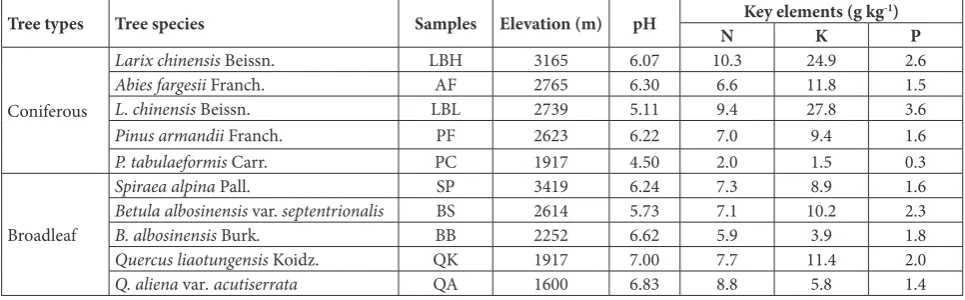
boundary between north and south China. There are five climatic belts, from warm temperate to alpine frigid zone, and seven altitudinal belts of vegetation on Qin Mountain. Nine typical species of trees, includ-ing four coniferous species and five broadleaf species, were selected from the seven vegetation belts. The co-niferous species included Larix chinensis Beissn. (LB), Abies fargesii Franch. (AF), Larix chinensis Beissn. (LB) and Abies fargesii Franch. (AF). The broadleaf species included Spiraea alpina Pall. (SP), Betula albosinensis var. septentrionalis (BS), B. albosinensis Burk. (BB), Quercus liaotungensis Koidz. (QK) and Q. aliena var. acutiserrata (QA). For LB, bark samples were collected at two elevations, 3165 m (LBH) and 2739 m (LBL). Elevations of other samples are shown in Table 1. The pieces of bark, 1.0 cm in thickness (0.3 cm for SP and BB), were cut off using a sterile blade at 1.0-1.5 m above the ground (0.5 m for SP). The samples were sealed in sterile polyethylene bags, transported to the laboratory, and stored in the dark at 4°C until use.
Actinomycete cultivation, enumeration and isolation
Serial dilution and spread-plate techniques [14] were used to isolate actinomycetes from bark samples. The samples were surface-sterilized by dipping in 70% ethanol and 0.1% mercuric chloride, each for 1 min. Serial dilutions were prepared by adding 5 g of grinded bark to 45 mL of sterile distilled water (10-1)
in a conical flask, followed by oscillation at 160 rpm for 10 min. and further dilution to 10-5. The dilutions
of 10-3 to 10-5 were used. Two agar media were tested:
Gause’s synthetic agar (20 g soluble starch, 1 g KNO3, 0.5 g K2HPO4, 0.5 g MgSO4·7H2O, 0.5 g NaCl, 0.01 g
Table 1. Sources and chemical properties of samples.
Tree types Tree species Samples Elevation (m) pH N Key elements (g kgK -1) P
Coniferous
Larix chinensis Beissn. LBH 3165 6.07 10.3 24.9 2.6
Abies fargesii Franch. AF 2765 6.30 6.6 11.8 1.5
L. chinensis Beissn. LBL 2739 5.11 9.4 27.8 3.6
Pinus armandii Franch. PF 2623 6.22 7.0 9.4 1.6
P. tabulaeformis Carr. PC 1917 4.50 2.0 1.5 0.3
Broadleaf
Spiraea alpina Pall. SP 3419 6.24 7.3 8.9 1.6
Betula albosinensis var. septentrionalis BS 2614 5.73 7.1 10.2 2.3
B. albosinensis Burk. BB 2252 6.62 5.9 3.9 1.8
Quercus liaotungensis Koidz. QK 1917 7.00 7.7 11.4 2.0
FeSO4, 10 g agar, 1000 mL distilled water) and modi-fied humic acid agar (10 g humic acid, 0.5 g Na2HPO4, 1 g KCl, 0.05 g; CaCl2 1 g MgSO4·7H2O, 10 g agar, 1000 mL distilled water). All media were supplemented with 80 mg L-1 potassium dichromate to inhibit the
growth of bacteria and fungi. After inoculation, all plates were incubated at 28°C for 15 days. Actinomy-cete colonies were identified by visual examination of the cultural and morphological characteristics; micro-scopic examination was performed if needed. Mor-phologically distinct colonies were transferred onto Gause’s synthetic agar slants separately, incubated at 28°C for 7 days, and then stored in the dark at 4°C. All experiments were performed in triplicate. The average number of actinomycete colonies on each plate was counted. Data are reported as colony-forming-unit (CFU) g-1 stove-dry bark. The colony numbers were
compared between different tree species by t-test in SAS 9.0 statistical software (SAS Institute Inc., Cary, NC, USA.).
Actinomycete identification
The dominant actinomycete isolates and isolates with broad-spectrum antagonistic activity were identified by 16S rRNA-based sequence analysis. Actinomy-cete DNA was extracted from pure isolates using the method described by Saito and Miura [15]. Partial 16S rRNA gene fragments were amplified by polymerase chain reaction (PCR) using the bacterial primers 27F: 5’-AGAGTTTGATCCTGGCTCAG-3’ and 1541R: 5’-AAGGAGGTGATCCAGCCGCA-3’. Amplification was carried out in a DNA Engine thermal cycler (BIO-RAD, USA), using a 50-µL reaction mixture contain-ing 4 µL Taq DNA polymerase (2.5 U µL-1, Genscript,
Nanjing), 5 µL 10× buffer (Transgene, Beijing), 1 µL 20 mM deoxynucleoside triphosphate (Transgene), 37µL of sterile distilled water, 1 µL of each primer (50 µM), and 1 µL of template. The PCR thermocy-cling conditions were as follows: initial denaturation at 94°C for 4 min; 30 cycles at 94°C for 1 min, 56°C for 1 min, 72°C for 2 min; and a final elongation at 72°C for 10 min. PCR reactions were purified and sequenced by Genscript Biotech (Nanjing) Co., Ltd, China. The obtained sequences were compared with available reference sequences in the EMBL/GenBank/ DDBJ databases and deposited in GeneBank under the accession Nos. KF447933–KF447962.
Antimicrobial activity assay
The antimicrobial activity of actinomycete isolates isolated on Gause’s synthetic agar was analyzed using an agar block method [16] against 4 bacterial species and 11 fungal species, which were provided by the Microbiology Laboratory in Shanxi Normal Univer-sity. The bacterial species included Escherichia coli E1, Staphylococcus aureus S4 and two pathogens of konjac soft rot, Serratia sp. H1 and Dickeya dadantii subsp. Dadantii D3. The fungal species included Penicillium sp. P1, Candida tropicalis C1 and nine plant patho-gens, Verticillium dahliae V2, Fusarium oxysporum FO1, F. solani (Mart.) Sacc FSS1, F. sulphureum FS1, F. oxysporum f. sp. cucumerinum FOC1, F. oxysporum f. sp. niveum FON1, F. solani FS3, F. oxysporum f. sp. vasinfectum FOV1, and Didymella bryoniae DB1.
Antagonistic potentiality assay of bark actinomycetes
The antagonistic potentiality of bark actinomycetes (APBA) was calculated by considering the number and antimicrobial spectrum of actinomycete isolates in the bark ecosystem using the following equation [17]:
where m and n are the numbers of tested bark samples and actinomycete isolates with antagonistic activity, respectively; Tn is the number of target microorgan-isms to which the actinomycete isolate is antagonistic.
RESULTS
Actinomycete colony counts and species number on Gause’s synthetic agar
The average number of actinomycete species of broadleaf trees was 72.4% higher than coniferous trees with no statistically significant difference (P>0.05). For broadleaf trees, species diversity was rich in SP, BS and QA, while it was poor in BB and QK. For co-niferous trees, species diversity was rich in AF and LBL, while it was poor in LBH, PF and PC. (Table 2).
Actinomycete colony counts and species number on modified humic acid agar
Humic acid is a macromolecular organic matter formed by the breakdown and resynthesis of dead plants. Actinomycetes recovered on modified humic acid agar could use humic acid as the sole carbon and energy source. On modified humic acid agar, acti-nomycete counts in bark samples of broadleaf trees (except for SP) were significantly more than those of coniferous trees (except for PC, P<0.05). Geographi-cally, the colony counts of actinomycetes correlated with elevations (r=-0.766, P<0.01).
Among all tree species, the total number of acti-nomycete species in bark samples was highest in AF and lowest in LBH. There was no significant
differ-ence in the average of the total number of actinomy-cete species between broadleaf and coniferous trees (P<0.05) (Table 2).
Dominant actinomycete species
A total of 142 actinomycete isolates were obtained from the bark samples on two media. The dominant isolates were classified into 19 species of five gen-era: Streptomyces spp. (73.7%), Micromonospora spp. (10.5%), Nocardiopsis spp. (5.3%), Actinoplanes spp. (5.3%) and Umezawaea spp. (5.3%) (Table 3).
Antagonistic activity of bark actinomycetes
A total of 79 actinomycete isolates were obtained from the bark samples on Gause’s synthetic agar. Of these, 43 isolates were found to be antagonistic to at least one of the 15 microorganisms tested. The average num-ber of actinomyceteisolates in broadleaf tree barks was 38.9% higher than that in coniferous tree barks, with no statistically significant difference (P>0.05). For broadleaf tree barks, with increasing elevation the numbers of antagonistic isolates showed significant decreases followed by significant increases. For
conif-Table 2. Colony counts and species number of actinomycetes (103 CFU g-1 dry bark).
Samples CountsGause’s synthetic agar1 Species numbers CountsModified humic acid agarSpecies numbers
Larix chinensis Beissn. 53.7±8.0c 3 0.3±0.1f 1
Abies fargesii Franch. 29.0±11.3cd 10 20.9±4.5f 10
L. chinensis Beissn. 1.3±0.6d 9 1.5±0.3f 6
Pinus armandii Franch. 3.4±0.3d 5 19.8±1.8f 6
P. tabulaeformis Carr. 0.2±0.1d 2 478.0±30.3c 8
Spiraea alpina Pall. 11.9±0.5d 14 8.2±3.1f 6
Betula albosinensis var. septentrionalis 3.9±0.8d 11 238.9±19.1e 7
B. albosinensis Burk. 181.3±14.4b 9 576.2±54.0b 5
Quercus liaotungensis Koidz. 637.8±143.6a 4 982.4±55.9a 6
Q. aliena var. acutiserrata 151.8±14.6b 12 415.8±31.5d 8
Different letters in the same column refer to significant difference at P<0.05 (t-test).
Table 3. Taxonomic distribution and origin of dominant actinomycete species.
Family Genera Isolates Total Coniferous treesSpecies Broadleaf trees
Streptomycetaceae Streptomyces 22 14 6 10
Pseudonocardiaceae Umezawaea 1 1 1 0
Nocardiopsaceae Nocardiopsis 3 1 1 1
Micromonosporaceae Micromonospora 2 2 1 1
Actinoplanes 2 1 1 0
erous tree barks, the numbers of antagonistic isolates in coniferous tree barks first increased and then de-creased with increasing elevation (Fig. 1).
The ratio of antagonistic isolates in broadleaf tree barks was 12.1% higher than in coniferous tree barks, with no statistically significant difference (P>0.05). With increasing elevation, the ratio of antagonistic isolates substantially decreased in both coniferous and broadleaf tree barks (Fig. 1).
Antagonistic potentiality
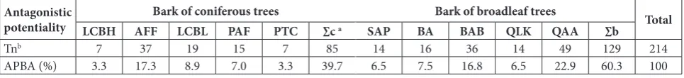
The APBA for coniferous tree barks was lower than for broadleaf tree barks. This indicated that the to-tal reserve of antagonistic actinomycetes was higher in broadleaf tree barks than in coniferous tree barks. APBA substantially varied in the barks of different tree species. For coniferous trees, the APBA in AFF was higher than that in other tree species. For broad-leaf trees, the APBA in QAA was higher than in other tree species (Table 4).
Distribution of antagonistic bark actinomycetes
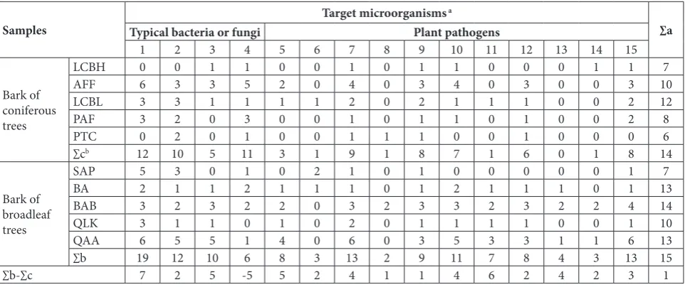
The number of isolates antagonistic to each target microorganism differed in bark samples between
broadleaf and coniferous trees. For 14 out of 15 target microorganisms, the number of antagonistic isolates was 1-7-fold higher in broadleaf trees than in conifer-ous trees. An exception was C. tropicalis, where the number of antagonistic isolates was 5-fold higher in coniferous trees than in broadleaf trees (Table 5).
The numbers of isolates antagonistic to the differ-ent target microorganisms varied in bark samples of single tree species. Moreover, the number of isolates antagonistic to each target microorganism varied in bark samples among different tree species. Additional-ly, the total antagonistic spectra of bark actinomycete isolates substantially varied in different tree species. (Table 5).
Identification of 5 broad-spectrum antagonistic isolates
The phylogeny and morphology of 5 broad-spectrum antagonistic isolates are shown in Figs. 2 and 3.
DISCUSSION
In this study, high actinomycete diversity was shown and a large number of actinomycetes with great ap-plication potential were residing in the barks of nine different tree species at different elevations on Qin Mountain. These results prove that living tree barks are natural resources with great antagonistic actino-mycete reserves, which should receive more attention in research and in the development of new antibiotics and antitumor agents. Additionally, the distribution of antagonistic actinomycetes in barks was dependent on tree species. Even for the same tree species, the distribution of antagonistic bark actinomycetes varied at different elevations. This indicated that regional environmental conditions, especially elevation, could affect the distribution of bioactive compound-pro-ducing bark actinomycetes.
Fig. 1. Numbers and relative abundances of antagonistic isolates. AN, AB − the averages of coniferous tree barks and broadleaf tree barks, respectively.
Table 4. Antagonistic potentiality of actinomycetes in tested barks.
Antagonistic
potentiality LCBH AFFBark of coniferous treesLCBL PAF PTC ∑c a SAP BABark of broadleaf treesBAB QLK QAA ∑b Total
Tnb 7 37 19 15 7 85 14 16 36 14 49 129 214
APBA (%) 3.3 17.3 8.9 7.0 3.3 39.7 6.5 7.5 16.8 6.5 22.9 60.3 100
a ∑c − the sum of An or APBA of the 5 coniferous tree barks; ∑b − the sum of An or APBA of the 5 broadleaf tree barks. b T
We obtained 5 broad-spectrum antagonistic ac-tinomycete isolates that inhibited the growth of ≥10 target microorganisms. These were identified as Strep-tomyces avidinii, S. malachitospinus, S. laculatispora, S. cyaneofuscatus and S. olivochromogenes, respectively. Previously, S. cyaneofuscatus [18] and S. olivochromo-genes [19] were found to produce valinomycin and phospholipase, respectively. To our knowledge, our work provides the first evidence regarding the antago-nistic activity of S. avidinii, S. malachitospinus and S. laculatispora. These potentially novel strains of strep-tomycetes provide alternative resources for research
and the development of new antibiotics. Further study needs to identify and characterize their bioactive compounds, especially those from the S. avidinii-, S. malachitospinus- and S. laculatispora-relatedisolates. Previous studies have proven that composted bark suppresses specific soil-borne diseases of plants, in-cluding root rot, collar rot and some other diseases caused by Phytophthora spp., Pythium spp. and Fu-sarium wilt [20-22]. In the present study, 54.4% of the
Fig. 2. Phylogenetic tree of the 5 broad-spectrum antagonistic isolates.
Samples Target microorganisms
a
∑a Typical bacteria or fungi Plant pathogens
1 2 3 4 5 6 7 8 9 10 11 12 13 14 15
Bark of coniferous trees
LCBH 0 0 1 1 0 0 1 0 1 1 0 0 0 1 1 7
AFF 6 3 3 5 2 0 4 0 3 4 0 3 0 0 3 10
LCBL 3 3 1 1 1 1 2 0 2 1 1 1 0 0 2 12
PAF 3 2 0 3 0 0 1 0 1 1 0 1 0 0 2 8
PTC 0 2 0 1 0 0 1 1 1 0 0 1 0 0 0 6
∑cb 12 10 5 11 3 1 9 1 8 7 1 6 0 1 8 14
Bark of broadleaf trees
SAP 5 3 0 1 0 2 1 0 1 0 0 0 0 0 1 7
BA 2 1 1 2 1 1 1 0 1 2 1 1 1 0 1 13
BAB 3 2 3 2 2 0 3 2 3 3 2 3 2 2 4 14
QLK 3 1 1 0 1 0 2 0 1 1 1 1 0 0 1 10
QAA 6 5 5 1 4 0 6 0 3 5 3 3 1 1 6 13
∑b 19 12 10 6 8 3 13 2 9 11 7 8 4 3 13 15
∑b-∑c 7 2 5 -5 5 2 4 1 1 4 6 2 4 2 3 1
a.Target microorganisms: 1 – Staphylococcus aureus,2 – Escherichia coli, 3 – Penicillium sp., 4 – Candida tropicalis, 5 – Serratia sp., 6 – Dickeya dadantii
subsp. dadantii, 7 – Verticillium dahliae, 8 – Fusarium oxysporum, 9 – Fusarium solani(Mart.)Sacc., 10 – Fusarium sulphureum, 11 – Fusarium oxysporum f. sp. cucumerinum, 12 – Fusarium oxysporum f. sp. niveum, 13 – Fusarium solani, 14 – Fusarium oxysporum f. sp. vasinfectum,15 –
Didymella bryoniae.
b∑c and ∑b − the sum of antagonistic isolates in coniferous and broadleaf trees, respectively; ∑a − the number of target microorganisms which all the
isolates in each bark sample inhibited.
actinomycete isolates showed antagonistic activity. Ac-cordingly, it is concluded that the antagonistic actino-mycetes in living tree barks propagated and produced bioactive compounds during the composting process. This may be one of the mechanisms of suppression activity of bark composts against plant pathogens.
In conclusion, a large number of bioactive com-pound-producing actinomycetes remain unexplored in the barks of living trees. These microbial resources should receive more attention in research and for the development for new antibiotics, antitumor and an-tiplant pathogen agents.
Acknowledgments:We are grateful for Dr. Chaofeng Lin for the English revision.
Conflict of interest disclosure: There is no conflict of interest.
REFERENCES
1. Bérdy J. Bioactive microbial metabolites. J Antibiot. 2005;58:1-26.
2. Watanabe Y, Shinzato N, Fukatsu T. Isolation of actino-mycetes from termites’ guts. Biosci Biotechnol Biochem. 2003;67:1797-801.
3. Okoro CK, Brown R, Jones AL, Andrews BA, Asenjo JA, Goodfellow M, Bull AT. Diversity of culturable actinomy-cetes in hyper-arid soils of the Atacama Desert, Chile. Anto-nie van Leeuwenhoek. 2009;95:121-33.
4. Promnuan Y, Kudo T, Chantawannakul P. Actinomycetes isolated from beehives in Thailand. World J Microbiol Bio-technol. 2009;25:1685-9.
5. Verma VC, Gond SK, Kumar A, Mishra A, Kharwar RN, Gange AC. Endophytic actinomycetes from Azadirachta indica A. Juss.: Isolation, diversity, and anti-microbial activ-ity. Microb Ecol. 2009;57:749-56.
6. Kumar V, Bharti A, Gupta VK, Gusain O, Bisht GRS. Actino-mycetes from solitary wasp mud nest and swallow bird mud nest: isolation and screening for their antibacterial activity. World J Microbiol Biotechnol. 2012;28:871-80.
7. Jiang Y, Han L, Chen X, Yin M, Zheng D, Wang Y, Qiu S, Huang X. Diversity and bioactivity of cultivable animal fecal actinobacteria. Adv Appl Microbiol. 2013;3:1-13.
8. Bascom-Slack CA, Ma C, Moore E, Babbs B, Fenn K, Greene JS, Hann BD, Keehner J, Kelley-Swift EG, Kembaiyan V, Lee SJ, Li P, Light DY, Lin EH, Schorn MA, Vekhter D, Bou-langer LA, Hess WM, Vargas PN, Strobel GA, Strobel SA. Multiple, novel biologically active endophytic actinomycetes isolated from upper Amazonian rainforests. Microb Ecol. 2009;58:374-83.
9. Cunha-Queda A, Ribeiro H, Ramos A, Cabral F. Study of biochemical and microbiological parameters during com-posting of pine and eucalyptus bark. Bioresource Technol. 2007;98:3213-20.
10. Muthiah B, Stanley S, Namasivayam S. Screening of endo-phytic actinomycetes residing in Eucalyptus globus for anti-microbial activity against human pathogenic bacteria. J Chem Pharm Sci. 2009;2:154-7.
11. Wu Y, Lu C, Qian X, Huang Y, Shen Y. Diversities within genotypes, bioactivity and biosynthetic genes of endophytic actinomycetes isolated from three pharmaceutical plants. Curr Microbiol. 2009;59:475-82.
12. Sheykholeslami A, Kazemnezhad F, Akhshabi S. Bark mea-surement of beech (Fagus orientalis Lipsky.) in Tosakoti-Hyrcanian forest. Int J Forest Soil Eros. 2011;1:1-4. 13. Kitouni M, Boudemagh A, Oulmi L, Reghioua S,
Boughachiche F, Zerizer H, Hamdiken H, Couble A, Mouniee D, Boulahrouf A. Isolation of actinomycetes producing bioactive substances from water, soil and tree bark samples of the north-east of Algeria. J Mycol Med. 2005;15:45-51.
14. Williams S, Davies F. Use of antibiotics for selective isolation and enumeration of actinomycetes in soil. J Gen Microbiol. 1965;38:251-61.
15. Saito H, Miura KI. Preparation of transforming deoxyribo-nucleic acid by phenol treatment. BBA-Gene Struct Expr. 1963;72:619-29.
16. Stern NJ, Svetoch EA, Eruslanov BV, Perelygin VV, Mitsev-ich EV, MitsevMitsev-ich IP, Pokhilenko VD, Levchuk VP, Svetoch OE, Seal BS. Isolation of a Lactobacillus salivarius strain and purification of its bacteriocin, which is inhibitory to Campy-lobacter jejuni in the chicken gastrointestinal system. Anti-microb Agents Chemother. 2006;50:3111-6.
17. Zhu W, Xue Q, Cao Y, Xue L, Shen G, Lai H. Distribution and characteristics of soil antagonistic actinomycetes on northern slope of Taibai Mountain, Qinling. Chinese J Appl Ecol. 2011;22:3003-10. Chinese.
18. Telesnina G, Krakhmaleva I, Anisova L, Bartoshevich I, Sazykin I. Valinomycin biosynthesis and the dynamics of the content of macroergic phosphorus compounds in Streptomy-ces cyaneofuscatus. Antibiot Med Biotekhnol. 1986;31:3-7. 19. Simkhada JR, Cho SS, Park SJ, Mander P, Choi YH, Lee HJ,
Yoo JC. An oxidant-and organic solvent-resistant alkaline metalloprotease from Streptomyces olivochromogenes. Appl Biochem Biotech. 2010; 162:1457-1470.
20. Hoitink H, Schmitthenner A, Herr L. Composted bark for control of root rot in ornamentals. Ohio Rep. 1975;60:25-6. 21. Spring D, Ellis M, Spotts R, Hoitink H, Schmitthenner A.
Suppression of the apple collar rot pathogen in composted hardwood bark. Phytopathology. 1980;70:1209-12. 22. Hardy GSJ, Sivasithamparam K. Suppression of