SYNTHESIS, SPECTRAL, THERMAL, MAGNETIC, AND
STRUCTURAL STUDY OF
DIAQUABIS-(m-HYDROXYBENZOATO-
O)BIS(N,N-DIETHYLNICOTIN-AMIDE-
N)COBALT(II)
Dursun Ali Köse
[a], Onur Sahin
[b]and Orhan Büyükgüngör
[b]Keywords:mixed ligand complexes, Co(II) complex, N,N-diethylnicotinamide, m-hydroxybenzoate, thermal decomposition, X-ray structure, mass pattern
The complex of [Co(m-hba)2(Dena)2(H2O)2], diaquabis(m-hydroxybenzoato-O)bis(N,N-diethylnicotinamide-N)Cobalt(II),
C34H42CoN4O10, referred to in the title, has been synthesized and explained as structural by using elemental analysis, FT-IR spectra,
UV-Vis reflectance, magnetic measurements, thermal analysis, mass spectra and X-ray diffraction methods. The analysis results showed that the unit cell of the complex includes one molecule of CoII-cation, two molecules of m-hydroxybenzoate, two molecules of
N,N-diethylnicotinamide ligands and two molecules of aqua ligands. The compound crystallizes in the triclinic space group P-1 with the following unit-cell parameters: a=8.7288(3)Å, b=14.2213(4)Å, c=15.4307(5), =108.270(2)º, =92.473(3)º,=102.549(5)º and Z=2. The compound crystallizes in the space group P-1 with Z’=1. The asymmetric unit of the complex C34H42CoN4O10 contains two
crystallographically independent molecules. Each CoII ion located at a centre of symmetry and is coordinated by two O atoms from two
equivalent carboxylate groups, two O atoms from aqua ligands, and two pyridyl N atoms. It has strong paramagnetic properties.
* Corresponding Authors
Tel: +90 3642700000/1635; Fax: +903642700005 E-Mail: [email protected]
[a] Department of Chemistry, Hitit University, 19030 Çorum, Turkey
[b] Department of Physics, Ondokuz Mayıs University, 55139 Samsun, Kurupelit, Turkey
Introduction
Studies on mixed-ligand complexes have importance with special interest on complexes between transition metal ions, benzoate ion and a nitrogen base. Various studies on mixed-ligand metal complexes of the benzoate and N,N-diethylnicotinamide have been reported.1-11
In the recent times, research in bioinorganic chemistry has revealed the important role of metal ions in most biological processes. Carboxylates play an important role in inorganic chemistry, and lots of metal cations in a great number of various biological processes, especially six-membered ring systems, are still components of several vitamins and drugs.12 Also, some carboxylate compounds (e.g. benzoates)
are known to have antibacterial activity. Benzoic acid is used in combination with salicylic acid in dermatology as a fungicidal treatment for fungal skin diseases.13 , 14 Metal
complexes of biologically important ligands are sometimes more effective than free ligands.15 It is well documented that
heterocyclic compounds play a significant role in many biological systems. Therefore, it is not surprising that many authors have investigated heterocyclic compounds and also examined them as ligands in coordination compounds of several central atoms.16-22
The scope of this study, CoII with m-hydroxybenzoato
-diethylnicotinamide complex has been synthesized and the
structural properties of compound were explained by using X-ray diffraction studies, FT-IR spectra, UV-VIS spectra, elemental analysis, magnetic measurements, mass spectra and TGA/DTA curves. The decomposition pathways of the investigated complex are discussed in connection with the available spectroscopic data.
Experimental
Materials and Instrumentation
All chemicals used were analytic reagent products. Co(NO3)2.6H2O, m-hydroxybenzoic acid (m-hba), and
N,N-diethylnicotinamide (dena) (Scheme 1) were obtained from Sigma Aldrich. Elemental analyses (C, H, N) were carried out by standard methods (Tubitak Marmara Research Center). Magnetic susceptibility measurements were performed at room temperature by using a Sherwood Scientific MXI model Gouy magnetic balance and Quantum Designed Physical Property Measurement System (PPMS). Infrared spectra were recorded in the 4000–400 cm-1 region
with a Perkin Elmer Spectrum One FT-IR
spectrophotometer by using KBr pellets. Thermal analyses (TGA, DTA) were performed by the Shimadzu DTG-60H system, in dynamic nitrogen atmosphere (100mL/min) at a heating rate of 10C/min, in platinum crucibles as sample vessel, by using -Al2O3 as reference. Electronic spectra
Preparation of the Co(II)-m-hydroxybenzoato-N,N-diethyl-nicotinamide (1) complex
In the first step, m-hydroxybenzoic acid sodium salt was prepared at room temperature according to the equation (1) below. All of the reactions were carried out in water/ethanol (50%/50%) media. When removal of CO2 gas is finished, the
reaction is completed.
2 C7H6O3+ 2NaHCO32 C7H5O3Na +2CO2+2 H2O (1)
In the second step, the cobalt(II) m-hydroxybenzoate was synthesized from Na(m-hba) salt by the following substitution reaction:
2C7H5O3Na + Co(NO3)2.6H2O Co(C7H5O3)2.nH2O +
2NaNO3 (2)
Finally, the solution of N,N-diethylnicotinamide (2 mmol) in distilled water (30 mL) was added dropwise to a stirred solution of Co(m-hba)2(H2O)n (1 mmol) in hot distilled
water/ethanol (50/50 mL). The resulting solution was left for 15–17 days for crystallization at room temperature. The crystals formed were filtered off and washed with cold water and acetone, then dried in vacuum. The mixed ligand complexes were prepared according to the following equations:
Co(C7H5O3)2(H2O)n+2C10H14N2O
[Co(C7H5O3)2(C6H6N2O)2(H2O)2] (3)
The yield of the compound (1) is about 76%. Calculated for C34H42CoN4O10: C 52.63; H 4.10; N 8.19. Found: C
52.12; H 4.38, N 8.27.
(b)
Scheme 1. Molecular structure of the ligands, (a) m
-hydroxybenzoic acid (m-hba), (b)
N,N-diethylnicotinamide(Dena).
Results and Discussion
The effective magnetic moment of CoII ion is 4.21 BM
and is compatible with literature values for similar complexes.9,10,21 According to analytical results, the per
mole formula unit of complex contain 2 moles of m -hydroxybenzoato/diethylnicotinamide ligands and two moles of water. The octahedral coordination of the metal ion has been completed by two carboxylic oxygen atoms from two m-hydroxybenzoates, two nitrogen atoms from
two diethylnicotinamides, and two aqua oxygen atoms. According to the magnetic susceptibility results, the complex (1) is paramagnetic.
The electronic spectrum of the complex was taken in the solid state because of the low solubility of the complex. The electronic spectra showed two d–d transitions at 7045 cm-1
(4T
1g4T2g) and 20105 cm-1 (4T1g4T1g) (4P) for the
compound. The UV–visible peaks corresponding to the the* transitions in the ligands were observed at 272 and 327 nm.23 The coordination geometry of CoII ion is that
of an octahedral with the N atoms from diethylnicotinamide ligands, the bonded carboxylate O atoms from m -hydroxybenzoato ligands, and two O atoms from aqua ligands.
FT-IR spectra
Absorption bands in the range of 3450–2900 cm-1
correspond to the asymmetric and symmetric stretching vibrations of water molecules. The m-hba/Dena mixed ligand complexes give rise to strong bands responsible from the C=O stretching. Conjugation between the carbonyl group and the amide nitrogen causes small frequency shifts. The strong bands observed at around1677 cm-1 are assigned
to this mode. This band remained almost in the same range as the amide group of the free Dena ligand, indicating that the Dena ligand does not coordinate through amide group. Pyridine ring vibration of free diethylnicotinamideat 1565 cm-1shifts to 1455 cm-1 in the complexes indicating that the
pyridine ring is coordinated. The main difference in the spectrum of m-hydroxybenzoic acid is that the C=O stretching vibration of the carboxyl group at 1718 cm-1 shifts
to lower frequencies in the cobalt complex (1). The carboxylate peak in the complex (1) appears in the range of 1677 cm-1. This shows that the coordination takes place
through the carboxyl group.24 The –OH bending peak for the m-hydroxybenzoic acid remained almost in the same position at around 1259 cm-1in the complex (1). The low
intensity bands in the region of 600–400 cm-1 are attributed
to M–N and M–O vibrations.9,20-22
Thermal Analysis
[Co(C7H5O3)2(C10H14N2O)2(H2O)2]
Fig. 1 shows the TG-DrTGA/DTA curves for the Co(II) complex (1). According to these curves, two molecules of the coordinated aqua ligands have released an accompanying endothermic effect with the DTA curve at 127 C (exp. 5.08 %; calc. 4.96 %). The anhydrous complex begins to decompose with melting at 153 C. Two moles of neutral diethylnicotinamide ligands are decomposed with removal of four moles of ethyl groups. (exp. 15.98 %; calc. 16.39 %). Then two moles of amide groups of diethylnicotinamide ligands are released from the structure with endothermic effect (exp. 12.24 %; calc. 11.97 %). Afterwards, two moles pyridine groups are decomposed and removed at 348 C DTA peak as an endothermic (exp. 21.56 %; calc. 21.50 %) peak. After decomposing of neutral ligands at 349 C, the m-hydroxybenzoato ligands begin to degrade in two steps by giving endothermic peaks. Finally, cobalt oxide (CoO) remains in the crucible (exp. 10.89 %;calc. 10.33 %).
N
OH O
OH N
O
Figure 1. The TG-DrTGA/DTA curves of the Co(II) complex.
Mass Spectra
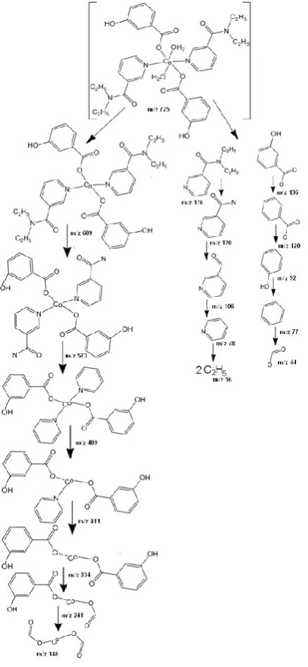
The thermal decomposition pathway of the [Co(m -hba)2(Dena)2(H2O)2] complex mass spectrum was recorded
(Fig. 2) by using direct insertion probe pyrolysis mass spectrometry method. The molecular ion peak is detected at 723 m/z in the mass spectrum recorded. The mass spectrum obtained is relatively complex. Beside the most abundant peaks, much fewer abundant peaks were observed on the spectrum depending probably on the nature of ligands. Scheme 2 summarizes the fragmentation pattern of the complex (1). The mass spectra data support to thermal decomposition.
Figure 2. Mass spectrum of the [Co(m-hba)2(dena)2(H2O)2]
complex
Data collection, structure solution, and refinement
Suitable crystals of C34H42CoN4O10 were selected for data
collection which was performed on a STOE IPDS II diffractometer equipped with a graphite-monochromatic Mo-Kα radiation (λ = 0.71073Ǻ) at 296 K. Other details of cell data, data collection, and refinement are summarized in Table 1. The structures were solved by direct methods using SHELXS-97 and refined by full-matrix least-squares methods on F2 using SHELXL-97 25 from within the
WINGX26 suite of software. All non-hydrogen atoms were
refined with anisotropic parameters.
The molecular structure of C34H42CoN4O10 (1) and the
atom-labelling scheme are shown in Fig. 3. The compound C34H42CoN4O10 crystallizes in the space group P-1 with
Z’=1.
Figure 3. A view of a [Co(m-hba)2(Dena)2H2O)2] molecule
showing the atom-numbering scheme. Displacement ellipsoids are drawn at 30% probability level. [Symmetry codes: (i) 1-x, 1-y, 1-z; (ii) 1-x, 2-y, 1-z.]
Table 1 Experimental details of the compound [Co(m -hba)2(Dena)2H2O)2].
Crystal data [Co(m-hba)2(Dena)2H2O)2]
Chemical formula C34H42CoN4O10
Mr 725.65
Cell setting, space group Triclinic, P -1 Temperature (K) 296
a, b, c (Ǻ) 8.7288 (3), 14.2213 (4), 15.4307 (5)
α, β, γ (º) 108.270 (2), 92.473 (3), 102.549 (3)
V (Ǻ3) 1762.64 (10)
Z 2
Dx(Mg m-3) 1.367
µ (mm-1) 0.55
Crystal form, colour Needle, pink Crstal size (mm) 0.50 x 0.40 x 0.13 Absorption correction Integration
Tmin 0.719
Tmax 0.938
No. of measured, independent and observed reflections
43297, 7292, 5526
Criterion for observed reflection I > 2σ(I)
Rint 0.051
θmax (º) 26.5
R[F2> 2σ(F2)], wR(F2), S 0.044, 0.122, 1.05
No. of reflections 7292 No. of parameters 457
(Δ/σ)max 0.001
Table 2.Hydrogen-bond parameters (Å, °) of C34H42CoN4O10.
D-H· · ·A D-H H…A D…A D-H…A Chain Motif Direction O4-H4···O5v 0.82 1.93 2.685 (3) 152 C (11) - [110]
O8-H8A···O3vi 0.82 2.02 2.803 (3) 161 - R
21(6) -
O9-H9A···O3vi 0.79 (4) 1.93(4) 2.707 (3) 166 (4) - R
11(6) -
O9-H9B···O7 0.82 (4) 1.93(4) 2.744 (3) 177 (4) - R22(14) -
O10-H10A···O7 0.76 (4) 1.89(4) 2.641 (3) 169 (4) - S(6) - O10-H10A···O6 0.76 (4) 2.57(4) 2.938 (3) 111 (4) - S(4) - O10-H10B···O1 0.82 (4) 1.99(4) 2.775 (4) 159 (4) C22(16) R22(14) [010]
C20-H20···O3vi 0.93 2.54 3.226 (3) 131 C
22(18) R21(6) [010]
C29-H29···O4 0.93 2.47 3.302 (3) 149 C22(18) R22(16) [010]
Cg2 = C1-C6. Symmetry codes: (i) x, y−1, z; (ii) −x+2, −y+1, −z+1; (iii) x, −y+3/2, z+1/2; (iv) 1-x, y-1/2, 3/2-z; (v) −x+2, −y+2, −z+1; (vi) −x+1, −y+1, −z+1.
Scheme 2. Mass spectral fragmentation pattern of the [Co(m -hba)2(Dena)2H2O)2].
Water H atoms were located in difference maps and refined subject to a DFIX restraint of O-H = 0.83(2)Ǻ, and with Uiso(H) =1.5Ueq(O). All other H atoms were located
from difference maps and then treated as riding atoms with C-H distances of 0.93-0.97Ǻ and O-H distances of 0.82Ǻ, and with Uiso(H) =1.2Ueq(C, O). Molecular diagrams were
created by using ORTEP-III.27 Geometric calculations were
performed with PLATON.28 The molecules are linked by
intramolecular hydrogen bonding, and graph-set notation29
was employed to describe the patterns of hydrogen bonding. Details of hydrogen-bond dimensions are given in Table 2.
The asymmetric unit of the complex C34H42CoN4O10
contains two crystallographically independent molecules. Each CoII ion is located at a centre of symmetry and is
coordinated by two O atoms from two equivalent carboxylate groups, two O atoms from aqua ligands, and two pyridyl N atoms (Table 3).
Table 3 Selected bond lengths (Å) and angles (°) for the C34H42CoN4O10
Symmetry codes: (i) −x+1, −y+1, −z+1; (ii) −x+1, −y+2, −z+1.
The C34H42CoN4O10 (1) molecules are linked into sheets
by a combination of seven O-H···O hydrogen bonds and two C-H···O hydrogen bonds (Table 2). Within the selected asymmetric unit, intramolecular H10A···O6 and O10-H10A···O7 hydrogen bonds produce S(4) and S(6) motifs. Atom O4 in the molecule at (x, y, z) acts as hydrogen-bond donor, via atom H4, to atom O5 in the molecule at (−x+2, −y+2, −z+1), so forming a C(11) chain running parallel to the [110] direction (Fig. 4). Atoms C20 and O8 in the molecule at (x, y, z) act as hydrogen-bond donors, via atoms H20 and H8A, to atom O3 in the molecule at (−x+1, −y+1, −z+1), so forming an R21(6) ring. The combination of
O-H···O and C-O-H···O hydrogen bonds produces R22(14)R22(16)
rings (Fig. 5).
Co2-O10 2.116(2) N1-Co1 2.154(2) O6-Co2 2.0571(19) O2-Co1 2.0882(16) N3-Co2 2.176(2) Co1-O9 2.1037(19) O2-Co1-O9i 90.08(7) O2i-Co1-N1 92.75(8)
O2i-Co1-O9i 89.92(7) O9i-Co1-N1 85.61(8)
O10-Co2-N3 87.32(9) O9-Co1-N1 94.39(8) O10-Co2-N3ii 92.68(9) O6ii-Co2-N3ii 88.59(8)
O2-Co1-N1 87.25(8) O6ii-Co2-O10 90.51(8)
Figure 4. Part of the crystal structure of C34H42CoN4O10, showing the formation of a chain along [110] generated by O-H· · ·O hydrogen
bonds. [Symmetry code: (v) −x+2, −y+2, −z+1.]
Figure 5. Part of the crystal structure of C34H42CoN4O10, showing the formation of R22(14)R22(16) rings. H atoms not involved in these
interactions have been omitted for clarity. [Symmetry code: (vi) −x+1, −y+1, −z+1].
Magnetic Studies
Magnetic properties have been recorded at 300 K on crushed crystalline samples of the complex. The magnetic behavior of the complex is shown in Figure 7. The variable-field magnetization data were collected in the magnetic variable-field (Oe) range -30000 to 30000 G. The MT value at 300 K is 3
cm3Kmol-1. The satisfaction magnetization, remanent
magnetization and coercivity values were calculated 0.2579 emu, 0.00191 emu and -95.149 Oe, respectively from the hysteresis loop of the complex. The magnetization curve of the complex recorded at room temperature is given in Figure 6.
Conclusion
Thermal decomposition takes place in three steps: dehydration of two moles of aqua ligands, elimination of the two moles of neutral N,N-diethylnicotinamide ligands, and decomposition of the two moles of anionic m -hydroxybenzoato ligands. In the reaction crucible, the CoO metal oxide remains as a decomposition product. According to thermal analysis data, releasing of the diethylnicotin-
Figure 6. The magnetization curve of C34H42CoN4O10. (1)
amide and m-hydroxybenzoato ligands is an endothermal process and resembles to thermal decomposition of diethylnicotinamide complexes reported by previous investigators.6,8,9,22,30 An earlier releasing of the
In the complex (1), all the ligands are coordinated to the metal ion in monodendate mode. The IR spectra of the intermediate products showed similar results. The (COO-)
sym.
peak is located at 1677 cm-1 for the complex (1). The (COO
-asym.peak is observed at 1444 cm-1. The shift () between
asym.and sym. bands of COO- group is 233 cm-1 for the
complex which is higher than for the sodium salt of m -hydroxybenzoic acid (163 cm-1) in which the monodentate
carboxylate group exists.22,24,Hiba! A könyvjelző nem létezik.
The mass spectra gave the expected fragmentation peaks of the complex.
The asymmetric unit of the complex C34H42CoN4O10
contains two crystallographically independent molecules. Each CoII ion is located at a centre of symmetry and is
coordinated by two O atoms from two equivalent carboxylate groups, two O atoms from aqua ligands, and two pyridyl N atoms (Table 3). The geometry around the CoII ion is that of a distorted octahedron, the equatorial
plane of which (O2, O2i, O6, O6ii, O9, O9i, O10 and O10ii)
is formed by two carboxylate O atoms and two aqua O atoms [symmetry codes: (i) 1-x, 1-y, 1-z; (ii) 1-x, 2-y, 1-z]. The axial positions are occupied by two pyridyl N atoms (N1, N1i, N3 and N3ii).The significant difference between
the Co-O bond distances in the equatorial plane and Co-N bond distances in the axial positions has also been observed in another cobalt complex11, 30 [11,31]. The benzene and
pyridine rings are almost planar, maximum deviations from the least-squares planes being 0.0052(20)Å for atom C4, 0.0174(17) Å for atom C20, 0.0124(19) Å for atom C8 and 0.0141(17) Å for atom N3.
Supplementary material
Crystallographic data for the structures reported in this paper have been deposited with the Cambridge Crystallographic Data Center: CCDC-837593 contains the supplementary crystallographic data for this paper. These data can be obtained free of charge from The Cambridge Crystallographic Data Centre, www.ccdc.cam.ac.uk/data request/cif.
Acknowledgements
The authors wish to acknowledge Hitit University Rectorate and the Faculty of Arts and Sciences, OndokuzMayis University, Turkey, for the use of the Stoe IPDS-II diffractometer (purchased from Grant No. F279 of the University Research Fund).
References
1
Casassas, E., Izquierdo-Ridorsa, A., Tauler, R., J. Chem. Soc., Dalton Trans., 1990, 2341.
2 Leban, I., Kozlevcar, B., Sieler, J., Segedin, P., Acta Cryst., 1997, C53, 1420.
3 Motz, C., Roth, H. P., Kirchgessner. M., Trace Elem. Electroly., 1995, 12, 1.
4 Petrovcic, N., Kozlevcar, B., Golic, L., Leban, I., Segedin, P., Acta Cryst., 1999, C55, 176.
5 Devereux, M., Curran, M., McCann, M., Casey, R. M. T., McKee,
V., Polyhedron, 1996, 15, 2029.
6Icbudak, H., Heren, Z., Kose, D. A., Necefoğlu, H., J. Therm. Anal. Cal., 2004, 76(3), 837.
7 Sahin, O., Buyukgungor, O., Kose, D. A., Necefoğlu, H., Acta Cryst., 2007, C63, m510.
8 Öztürkkan, E. F., Köse, D. A.,Necefoglu, H., Uzun, I., Asian J. Chem., 2007, 19(6), 4880.
9 Köse, D. A., Kaya, A.,Necefoğlu, H., Russian J. Coord. Chem., 2007, 33(6), 422.
10 Köse, D. A., Necefoğlu, H., J. Therm. Anal. Cal., 2008, 93(2),
509.
11 Sahin, O., Buyukgungor, O., Kose, D.A., Necefoğlu, H., Acta Cryst., 2008, C64, m317.
12 Yeşilel, O. Z., Olmez, H., Uçar, I., Bulut, A., Kazak, C., Z. Anorg. Allg. Chem., 2005, 631, 3100.
13 Chenoweth, M. B., Pharmocol. Rev., 1956, 8, 57.
14Sorenson, J. R. J., In: Siegel, H., (ed.) Metal Ions in Biologicals, vol.14, Marcel-Dekker,New York, 1982.
15 Reid, J., Watson, R. D., Cochran, J. B., Sproull, D. H., British. Med. J., 1951, 1, 321.
16 Olmez, H., Arslan, F., Icbudak, H., J. Therm. Anal. Calor., 2004,
76, 793.
17 Geraghty, M., Sheridan, V., McCann, M., Devereux, M., McKee,
V., Polyhedron., 1999, 18, 2931.
18 Kose, D.A., Zumreoglu-Karan, B., Unaleroglu, C., Şahin, O.,
Buyukgungor, O., J. Coord. Chem., 2006, 17, 2125.
19 Kose, D.A.,Içbudak, H.,Necefoglu, H., Hacettepe J. Biol. Chem., 2007, 35(2), 123.
20 Wolodkiewicz, W., Brzyska, W., Polish J. Chem.,1997, 71, 16. 21 Erdelyiová, A., Györyová, K., Gyepes, R., Halás, L., Kovárová,
J., Polyhedron, 2009, 28, 131.
22Kose, D. A., Necefoglu, H., Icbudak, H., J. Coord. Chem., 2008, 61(21), 3508.
23 Sutton, D., Electronic spectra of transition metal complexes. McGraw-Hill, London, 1968.
24 Zelenák, V., Vargová, Z., Györyová, K., Spectrochim.Acta A, 2007, 66, 262.
25 Sheldrick, G. M., Acta Cryst., 2008, A64, 112. 26 Farrugia, L. J., J. Appl. Cryst., 1999, 32, 837. 27Farrugia, L. J., J. Appl. Cryst., 1997, 30, 565.
28 Spek, A. L., PLATON–A Multipurpose Crystallographic Tool,
Utrecht University, Netherlands, 2005.
29 Bernstein, J., Davis, R. E., Shimoni, L., Chang, N. L., Angew. Chem. Int. Ed. Engl., 1995, 34, 1555.
30 Şahin, O., Büyükgüngör, O., Köse, D. A., Özturkkan E. F.,
Necefoglu, H., Acta Cryst., 2007, C63, m243.
![Figure 3. A view of a [Co(m-hba)2(Dena)2H2O)2] molecule showing the atom-numbering scheme](https://thumb-us.123doks.com/thumbv2/123dok_us/7838743.2090702/3.595.301.542.144.344/figure-view-dena-molecule-showing-atom-numbering-scheme.webp)
![Figure 4. Part of the crystal structure of C34H42CoN4O10, showing the formation of a chain along [110] generated by O-H· · ·O hydrogen bonds](https://thumb-us.123doks.com/thumbv2/123dok_us/7838743.2090702/5.595.314.559.513.679/figure-crystal-structure-showing-formation-chain-generated-hydrogen.webp)
