647
© 2018 by the Serbian Biological Society How to cite this article: Liu B, Ma Z, Gai X, Sun Y, Wang Y, He S, Gao Z. Analysis of putative sclerotia maturation-related gene expression in Rhizoctonia solani AG1-IA. Arch Biol Sci. 2018;70(4):647-53.
Analysis of putative sclerotia maturation-related gene expression in Rhizoctonia solani
AG1-IA
Bo Liu1,2, Zhoujie Ma1, Xiaotong Gai1, Yanqiu Sun1, Yanfeng Wang2, Shidao He1 and Zenggui Gao1,*
1Institute of Plant Immunology, Shenyang Agricultural University, Shenyang, Liaoning, China
2College of Life Sciences, Yan’an University, Yan’an, Shaanxi, China
*Corresponding author: [email protected]
Received: January 6, 2018; Revised: May 1, 2018; Accepted: May 4, 2018; Published online: Jun 14, 2018
Abstract: Rhizoctonia solani AG1-IA (R. solani AG1-IA) is a major soil-borne fungal pathogen of maize that causes signifi-cant yield losses in all maize-growing regions worldwide. The sclerotium produced by R. solani AG1-IA can overwinter in grass roots or diseased plants and infect crops the following year. The molecular mechanism underlying sclerotium forma-tion in R. solani is poorly understood. In this study, we constructed the cDNA library of the R. solani AG1-IA pathogenic strain WF-9, from which 329 high-quality expressed sequence tags (ESTs) were obtained. Of the 250 clustered unigenes, 12 genes were selected for further expression analysis during the three stages of sclerotial growth (mycelium, initiation of sclerotium, and maturation of sclerotium). The results of expression analysis support the previously suggested roles of chitin synthase D and exo-beta-1,3-glucanase in facilitating sclerotial growth through preservation of water content and energy. In addition, cytochrome P450, NADPH oxidase, catalase (CAT), acyl-CoA oxidase 1 (ACOX1), mitogen-activated protein kinase (MAPK), mitogen-activated protein kinase HOG1 (HOG 1), and the G-protein α subunit play important roles in balancing reactive oxygen species (ROS) levels during sclerotial development. The findings of this study can help understand the molecular mechanism of sclerotial development further.
Key words: Rhizoctonia solani AG1-IA; expressed sequence tags; qRT-PCR; sclerotial formation; sheath blight of maize; cDNA library
INTRODUCTION
Rhizoctonia solani (R. solani Kuhn) is a soil-borne
fun-gus belonging to the Basidiomycete group that can infect hundreds of crops, including maize, potato, rice, and soybean, leading to severe crop losses [1]. It has been classified into 14 anastomosis groups (AG-1 to AG-13 and AG-BI) [2], with three intraspecific groups (ISG) forming the R. solani AG1 subgroup. R. solani
AG1 subgroup is known to cause many diseases such as sheath blight and aerial blight [3]. Sheath blight of maize is one of the most serious and widespread crop diseases worldwide. In recent years, the increase in the planting density of maize and in the amount of nitrogen fertilizers used has made the microclimate conditions conducive to the frequent occurrence of sheath blight [4,5]. Sheath blight shows an annual ag-gravating trend and has become the major disease that restricts the continuous increase in maize yield [6].
Pathogenic differentiation is prevalent in plant pathogenic fungi. The study of pathogenic differen-tiation plays a significant role in the research of path-ogenic fungi, including composition, diversity and genetic differentiation, resistance identification, breed-ing of disease-resistant varieties and rational distribu-tion of varieties. Previous studies have reported dif-ferences in composition of fusion groups of R. solani
Sclerotium is a structure formed from aggregated mycelia. In 1954, Townsend and Willetts [9] divided the development of sclerotia into three stages: ini-tiation, development, and maturation. The initiation stage is marked by the appearance of sclerotium with white aerial mycelia entangled around the edges of the culture medium. In the development stage, the sclerotium is further entangled and an increase in size can be observed along with a clear or tan-colored liq-uid secretion on the surface. The maturation phase involves surface delineation and melanin deposition in peripheral rind cells, followed by internal consoli-dation [9].
Kwon [10] identified 55 proteins that were differ-entially expressed during sclerotial maturation using matrix-assisted laser desorption/ionization (MALDI)-time of flight (TOF) mass spectrometry (MS). These proteins can be categorized into 10 categories based on their functions, including carbon metabolism, cell defense, and amino acid metabolism. Among them, the expression of two vacuole function-related pro-teins was confirmed to be highly increased, as deter-mined by quantitative real-time reverse transcription-polymerase chain reaction (qRT-PCR). The proteins were found to be closely related to sclerotial growth [10]. The disruption of Rga1 (G-protein α subunit) led to decreased vegetative growth and pathogenicity. This demonstrated that Rga1 plays a key role in the growth of sclerotium [11]. NADPH oxidase 1 (NOX 1) is thought to be involved in superoxide anion genera-tion, fungal growth, and cell differentiation [12]. Nox 1 silencing in Sclerotinia sclerotiorum and loss of ROS in the mutant suppressed sclerotium formation [13].
A cDNA library provides the foundation for studying the gene expression of a specific organ, spe-cific tissue, and spespe-cific growing period [14-16]; a full-length cDNA library is an important resource to identify, screen, and analyze genes and to obtain full-length genes [17-19]. To date, few studies have inves-tigated the response of sclerotial formation of R. solani
AG1-IA in maize. We established the cDNA library
of R. solani AG1-IA to obtain additional molecular
information about it and to better understand gene expression during sclerotium growth. The findings of this study may be useful in developing new methods of disease control and prevention.
MATERIALS AND METHODS Pathogenicity determination
An appropriate amount of hypha was selected from the storage tube of R. solani AG1-IA, transplanted onto potato dextrose agar (PDA) and incubated at 25°C for three days for activation of culture. The ac-tivated strain was again transplanted onto the sur-face of PDA and incubated at 25°C for three days. A mycelial plug (5 mm) was excised from the periph-ery of the resulting colony after 4 days and prepared for inoculation [20].
The maize varieties were Liaoning single 565 (Liaoning Academy of Agricultural Sciences) and East Asia single 5 (Corn Breeding Office of Liaon-ing Province East Asian Seed Science Institute). The experiment was carried out at the seedling stage; the inoculation method in the greenhouse at 25°C was used, and the inoculation was performed on the leaf of maize. The prepared mycelial plug was inoculated into the first leaf sheath of maize seedlings, which in-cludes two corn varieties, and each one was inoculated into 10 plants. The soil surface was kept wet by spray moisturizing daily. Corn growth and disease develop-ment were observed daily, and disease investigation was pursued 15 days later using the formula:
disease index = Σ (disease grade × strains) / (max-imum disease series × total surveyed strains) × 100.
RNA extraction and cDNA synthesis
Construction of R. solani AGI-IA cDNA library
The sclerotium initiation stage was used to construct a cDNA library. The double-stranded cDNA was puri-fied by the QIAquick PCR purification kit (QIAGEN, Germany), and the cDNA was fractionated by CHRO-MASPIN + TE-1000 column and 500-3000-bp frag-ments were recovered. The cDNA was ligated with the pSMART2IFD linearized vector provided with the kit [18] and incubated in a PCR TProfessional Thermo-cycler (Biometra, Germany) at 50°C for 15 min. Then, the cDNA was transferred to ice and 2 μL of the ligated product was electroporated into DH 5α competent cells and cultured at 37°C for 1 h. Next, lysogeny broth (LB) culture medium (prewarmed to 37°C) was added. The transformation product (5 μL) was added to 95 μL of
LB liquid medium, and 40 μL of 100 mmol•L-1
isopro-pyl β-D-1-thiogalactopyranoside (IPTG) and 40 μL of
20 mg•mL-1 X-Gal were uniformly applied on LB agar
plates (with 100 μg•mL-1 ampicillin). The mixed
bacte-rial solution was coated evenly on the culture plate and then placed upside-down in a 37°C incubator for a 12-h culture. The growth of the R. solani AG1-IA isolated colony was checked and the recombination rate (pro-portion of leukoplakia) was calculated [21].
Processing of R. solani AG1-IA EST library
Four hundred clones were randomly picked from the cDNA library plates and sent to BGI China for 5΄ sequencing. The 3730xl DNA analyzer (Applied Biosystems, USA) was used for sequencing. The Phred package (http://www.phrap.org/consed/consed.html) was used to convert the sequencing peak figure files into sequence and quality files. It removes unloaded sequences, low-quality sequences (Q-value<13) (low-quality bases of head and tail), vector sequences, and sequences whose length was less than 100 bp. CAP3 [22] (http://seq.cs.iastate.edu/cap3.html) was used to splice the obtained EST sequences to generate contigs and singletons [23], and statistical data were produced. For annotations, all genes were searched against the Nr database using BLASTx, with 10-5 as the
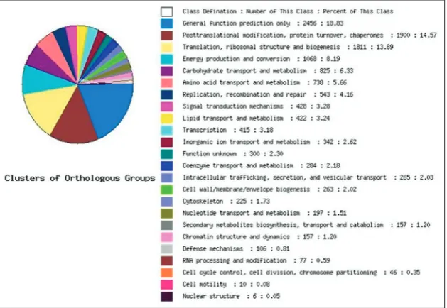
E-value cut-off point. The sequences with the highest similarities were retrieved from the NCBI web server (https://blast.ncbi.nlm.nih.gov), and the functions of the genes were retrieved from the Clusters of Ortholo-gous Group (COG) database [24,25].
Candidate gene expression analysis by qRT-PCR
The primer sequences and related information are shown in Table S1. According to the SYBR Premix Ex Taq Kit (TaKaRa, Shiga Pref, Japan) protocol, the reactions were run on an CFX-96 (BioRad, Hercules, CA, USA) using a 20-μL reaction system. The reac-tion condireac-tions were as follows: initial denaturareac-tion at 95°C for 3 min, followed by 39 cycles of denatura-tion at 95°C for 30 s and annealing for 30 s at 60°C. All experiments were performed in triplicate and two repeated tests. The relative expression levels were ana-lyzed and normalized to 18S rRNA transcript levels using the CFX_Manager analysis software. Mapping was performed by SigmaPlot 12.5.
RESULTS
Virulence of strains
We measured the isolates of R. solani AG1-IA in the anastomosis group, and the results showed that sapro-phytic colonization and pathogenicity between the isolates were significantly different. Two corn cultivars developed typical symptoms of sheath blight upon infection with R. solani AG1-IA. The tested isolates of
R. solani AG1-IA showed differences in pathogenicity
(Table 1), with isolate WF-9 displaying the strongest pathogenicity and isolate DR-28 displaying the weak-est pathogenicity (an average disease index of 79.69 and 23.44, respectively).
Gene annotation
chitin synthase D, cellulase, protein phosphatase, and cell wall-related hydrolase. In addition, we identified genes with a higher frequency of expression, namely, heat shock protein, ribosomal protein, aminopepti-dase, cytochrome P450, mannoprotein, elongation factor, ATP synthase, and glycosyl hydrolase family proteins.
Functional annotation and classification
All the functions of ESTs were an-notated (NCBI database search using BLASTX). The functions in-cluded transcription, signal trans-duction, metabolism, cellular com-ponents, as well as some unknown functions. The 250 ESTs were sub-sequently analyzed using the COG database, and 19 ESTs (17.27%) were found to be involved in trans-portation of carbohydrates and metabolism, and 18 ESTs (16.36%) were found to be involved in trans-lation, ribose structure, and bio-genesis (Fig. 1). Within the cellular component categories “cell” and “cell part” were the most abundant terms. In molecular function cat-egories “binding” and “catalytic activity” were the most abundant terms. In biological process cat-egories “metabolic process” was the most abundant term (Fig. 2). According to gene function analysis, the functions of R. solani AG1-IA genes included tran-scriptional regulation, signal transduction, transport, metabolism, defense, energy, cell division and chro-mosomal structure, protein and nucleic acid synthesis,
Table 1. Result of the pathogenicity test of different isolates on two corn cultivars.
Isolates Liaoning single 565 East Asia single 5Maize Cultivars (Disease Index)Average Isolates Liaoning single 565 East Asia single 5 AverageMaize Cultivars (Disease Index)
JR-80 78.13 71.88 75.01 JR-42 37.50 46.88 42.19
TR-37 68.75 62.50 65.63 DR-78 56.25 53.13 54.69
JR-89 46.88 50.00 48.44 DR-28 25.00 21.88 23.44
DR-84 34.38 43.75 39.07 JR-87 40.63 53.13 46.88
WF-10 59.38 53.13 56.26 SY-69 75.00 71.88 73.44
JR-90 31.25 37.50 34.38 DR-31 31.25 28.13 29.69
WF-9 81.25 78.13 79.69 DR-24 62.50 59.38 60.94
SY-39 65.63 53.13 59.38 TR-43 46.88 53.13 50.01
DR-35 78.13 62.50 70.32 SY-67 50.00 50.00 50.00
DR-30 75.00 65.63 70.32 SY-72 37.50 43.75 40.53
WF-8 56.25 62.50 59.38 TR-54 56.25 43.75 50.00
JR-80, TR-37, JR-89, etc. is the number of isolates; Liaoning single 565 and East Asia single 5 are maize varieties; disease index = Σ (disease grade × strains)/(maximum disease series × total surveyed strains) ×100; disease grading was as follows: grade 0 – no disease in whole plant; grade 1 – the 4th leaf sheath and above leaf sheath under ear; grade 2 – 3rd leaf sheath and above leaf sheath under ear; grade 3 – 2nd leaf sheath and above leaf sheath
under ear; grade 4 – the 1st leaf sheath under the ear and above the leaf sheath disease; grade 5 – ear and above leaf sheath disease.
Fig. 1. Functional classification and identification of characteristics of Rhizoctonia solani
cell components, and other aspects of fungal growth, and development that reflect the gene expression of
R. solani AG1-IA.
Validation of genes involved in sclerotia formation of R. solani AG1-IA by qRT-PCR
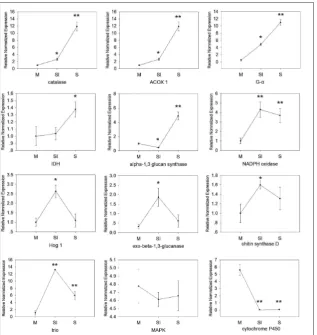
Twelve genes (Acyl-CoA oxidase 1, catalase, alpha-1,3 glucan synthase, mitogen-activated protein kinase HOG1, isocitrate dehydrogenase 1, triose-phosphate isomerase, cytochrome P450, G-protein α subunit, exo-beta-1,3-glucanase, mitogen-activated protein kinase, chitin synthase D, NADPH oxidase) were selected for qRT-PCR analysis. The results showed that during the three stages of sclerotial formation (mycelium, ini-tiation of sclerotium, and maturation of sclerotium), five genes (catalase, acyl-CoA oxidase 1, G-protein α subunit, isocitrate dehydrogenase 1, alpha-1,3 glucan synthase) showed increased expres-sion; five genes (NADPH oxidase, mitogen-activated protein kinase HOG1, exo-beta-1,3-glucanase, chi-tin synthase D, triose-phosphate iso-merase) initially showed increased expression, followed by decreased expression, and two genes (mitogen-activated protein kinase, cytochrome P450) showed decreased expression (Fig. 3).
DISCUSSION
Georgiou hypothesized that scle-rotium development in filamen-tous fungi is caused by oxidative stress. According to the hypothesis, when oxygen enters the cells of a sclerotium-forming fungi, the cells are incapable of handling the high concentrations of ROS and form an unstable hyperoxidant state [26]. Oxidative stress activates a signal transduction pathway, causing cells to differentiate to isolate oxy-gen molecules. Simultaneously, the cells undergo a series of synergistic mechanisms, including increase in
Fig. 3. Quantitative RT-PCR analysis of expression of 12 genes inthe three sclerotial de-velopmental stages (M, SI, S) of Rhizoctonia solani AG1-IA. M – mycelium; SI – initiation of sclerotium; S – maturation of sclerotium. The asterisk indicates that the mean value is significantly different from that of the mycelium stage (*P<0.05; **P<0.01).
Fig. 2. Different gene ontology (GO) terms that indicate gene
consumption of oxygen molecules, limiting the entry of oxygen molecules, or generating antioxidants to balance peroxide concentration. During ROS produc-tion, cytochrome P450, NADPH oxidase and catalytic extracellular O2 generate superoxide radicals and use these as a base to generate a series of byproduct ROS. Superoxide radicals are converted into hydrogen per-oxide and oxygen by superper-oxide dismutase (SOD) to balance the highly oxidized substances in the cells [26]. Acyl-coenzyme oxidase 1 (ACOX1) catalyzes the first rate-limiting step in peroxisomal beta-oxidation of medium and very long straight-chain fatty acids [27]. In this study, cytochrome P450 and NADPH oxidase 1 are highly expressed in the mycelium and initiation stages, suggesting that the early develop-mental stage of sclerotium generates ROS. In contrast, CAT and ACOX1 showed increased expression in the maturation stage, suggesting that the maturation stage increases the consumption of oxygen molecules and limits oxygen molecules from entering the cells to bal-ance the peroxide content.
The signal transduction pathway induced by oxi-dative stress is mainly regulated by MAPK, HOG 1 and the G-protein α subunit. A conserved family of fungal MAPKs (in Saccharomyces cerevisiae Hog1,
S. pombe Spc1, and possibly in filamentous fungi)
is activated by many stress signals, including oxida-tive stress [28]. R. solani with a knockout G-protein α subunit showed a change in morphology, and the ability to form sclerotium was completely lost [11]. In the present study, the changes in expression levels of MAPK, HOG 1, and G-protein α subunit were evi-dent, which is in accordance with their suggested role in ROS-induced signal transduction pathway.
Cell walls are composed of β-glucan and chitin, and the enzymes involved in their metabolism are chitin synthase D, exo-beta-1,3-glucanase, and pro-tein, which have been hypothesized to preserve water content and energy [29]. In this study, chitin synthase D and exo-beta-1,3-glucanase were increased in ex-pression in the initiation stage, suggesting that these two genes play a role in the thickening of cell walls.
In conclusion, we constructed a cDNA library of
R. Solani AG1-IA and analyzed 12 genes that play
dif-ferent roles in the difdif-ferent stages of sclerotial growth. Our study shows that the genes with significant
chang-es in exprchang-ession may be associated with the formation of sclerotia. The findings of this study can be used as information that may be useful for developing meth-ods for the prevention and control of diseases by R.
solani AG1-IA.
Funding: This work was supported by the National Key
Tech-nology Research and Development Program (2016YFD0300704, 2017YFD0300704), and the National Natural Science Foundation of China (Grant No. 31751005).
Author contributions: Bo Liu, Zhoujie Ma and Zenggui Gao
con-ceived and designed the experiments; Xiaotong Gai and Shidao He contributed reagents and materials; Bo Liu collected and interpreted the data; Bo Liu and Yanqiu Sun analyzed the data statistically; Bo Liu wrote the paper; Yanfeng Wang and Zenggui Gao provided a guideline for the work.
Conflict of interest disclosure: The authors declare no conflict of interest.
REFERENCES
1. Zheng A, Lin R, Zhang D, Qin P, Xu L, Ai P, Ding L, Wang Y, Chen Y, Liu Y. The evolution and pathogenic mechanisms of the rice sheath blight pathogen. Nat Commun. 2013;4:1424. 2. Ogoshi A. Ecology and pathogenicity of anastomosis and
intraspecific groups of Rhizoctonia solani Kuhn. Annu Rev Phytopathol. 1987;25(1):125-43.
3. Sattari A, Fakheri B, Noroozi M. Breeding for resistance to sheath blight in rice. Intl J Farm Alli Sci. 2014;3(9):970-79. 4. Gautam H, Bhardwaj M, Kumar R. Climate change and its
impact on plant diseases. Curr Sci. 2013;105(12):1685-91. 5. Renfro B, Ullstrup A. A comparison of maize diseases in
tem-perate and in tropical environments. PANS. 1976;22(4):491-8. 6. Prasanna B, Pixley K, Warburton ML, Xie CX. Molecular marker-assisted breeding options for maize improvement in Asia. Mol Breed. 2010;26(2):339-56.
7. Bruce A, McDonald, Celeste L. Pathogen population genet-ics, evolutionary potential, and durable resistance. Annu Rev Phytopathol. 2002;40(1):349-79.
8. Moni ZR, Ali MA, Alam MS, Rahman MA, Bhuiyan MR, Mian MS, Iftekharuddaula KM, Latif MA, Khan MA. Mor-phological and genetical variability among Rhizoctonia solani isolates causing sheath blight disease of rice. Rice Sci. 2016;23(1):42-50.
9. Townsend BB, Willetts HJ. The development of sclerotia in certain fungi. Trans Br Mycol Soc. 1954;37(3):213-21. 10. Kwon YS, Kim SG, Chung WS, Bae H, Jeong SW, Shin SC,
Jeong MJ, Park SC, Kwak YS, Bae DW. Proteomic analysis of
Rhizoctonia solani AG-1 sclerotia maturation. Fungal Biol. 2014;118(5-6):433-43.
pathoge-nicity in Rhizoctonia solani. World J Microbiol Biotechnol. 2008;24(3):345-51.
12. Peraza L, Hansberg W. Neurospora crassa catalases, singlet oxygen and cell differentiation. Biol Chem. 2002;383(3-4):569-75.
13. Kim HJ, Chen C, Kabbage M, Dickman MB. Identification and characterization of Sclerotinia sclerotiorum NADPH oxi-dases. Appl Environ Microbiol. 2011;77(21):7721-9. 14. Soares MB, Bonaldo MF, Jelene P, Su L, Lawton L, Efstratiadis
A. Construction and characterization of a normalized cDNA library. Proc Nati Acad Sci. 1994;91(20):9228-32.
15. Gasic K, Hernandez A, Korban SS. RNA extraction from different apple tissues rich in polyphenols and polysaccha-rides for cDNA library construction. Plant Mol Biol Rep. 2004;22(4):437-8.
16. Wiemann S, Mehrle A, Bechtel S, Wellenreuther R, Pep-perkok R, Poustka A, Consortium GC. cDNAs for functional genomics and proteomics: the German Consortium. C R Biol. 2003;326(10):1003-9.
17. Zhu Y, Machleder E, Chenchik A, Li R, Siebert P. Reverse transcriptase template switching: A SMART™ approach for full-length cDNA library construction. Biotechniques. 2001;30(4):892-7.
18. Gunning P, Ponte P, Okayama H, Engel J, Blau H, Kedes L. Isolation and characterization of full-length cDNA clones for human alpha-, beta-, and gamma-actin mRNAs: skeletal but not cytoplasmic actins have an amino-terminal cysteine that is subsequently removed. Mol Cell Biol. 1983;3(5):787-95. 19. Seki M, Narusaka M, Ishida J, Nanjo T, Fujita M, Oono Y,
Kamiya A, Nakajima M, Enju A, Sakurai T. Monitoring the expression profiles of 7000 Arabidopsis genes under drought, cold and high-salinity stresses using a full-length cDNA microarray. Plant J. 2002;31(3):279-92.
20. Rioux R, Manmathan H, Singh P, De los Reyes B, Jia Y, Tavantzis S. Comparative analysis of putative pathogenesis-related gene expression in two Rhizoctonia solani pathosys-tems. Curr Genet. 2011;57(6):391-408.
21. Zhulidov P, Bogdanova E, Shcheglov A, Shagina I, Wagner L, Khazpekov G, Kozhemyako V, Lukyanov S, Shagin D. A method for the preparation of normalized cDNA libraries enriched with full-length sequences. Russ J Bioorganic Chem. 2005;31(2):170-7.
22. Huang X,Madan A. CAP3: A DNA Sequence Assembly Pro-gram. Genome Res. 1999;9(9):868-77.
23. Martin DM, Bohlmann J. Identification of Vitis vinifera (−)-α-terpineol synthase by in silico screening of full-length cDNA ESTs and functional characterization of recombinant terpene synthase. Phytochem. 2004;65(9):1223-9.
24. Tatusov RL, Galperin MY, Natale DA, Koonin EV. The COG database: a tool for genome-scale analysis of protein functions and evolution. Nucleic Acids Res. 2000;28(1):33-6.
25. Tatusov RL, Fedorova ND, Jackson JD, Jacobs AR, Kiryutin B, Koonin EV, Krylov DM, Mazumder R, Mekhedov SL, Nikol-skaya AN. The COG database: an updated version includes eukaryotes. BMC Bioinformatics. 2003;4(1):41.
26. Georgiou CD, Patsoukis N, Papapostolou I, Zervoudakis G. Sclerotial metamorphosis in filamentous fungi is induced by oxidative stress. Integr Comp Biol. 2006;46(6):691-712. 27. Morais S, Knoll-Gellida A, André M, Barthe C, Babin PJ.
Conserved expression of alternative splicing variants of per-oxisomal acyl-CoA oxidase 1 in vertebrates and develop-mental and nutritional regulation in fish. Physiol Genomics. 2007;28(3):239.
28. Aguirre J, Rios-Momberg M, Hewitt D, Hansberg W. Reac-tive oxygen species and development in microbial eukaryotes. Trends Microbiol. 2005;13(3):111-8.
29. Willetts HJ, Bullock S. Developmental biology of sclerotia. Mycol Res. 1992;96(10):801-16.
Supplementary Data
Supplementary Tables S1. and S2.