Journal of Dental Biomaterials. 2015;2(3)
Original Article
The Effect of Time Intervals on Heat Transfer to the Implant-Bone
Interface during Preparation of a Titanium Abutment: An in Vitro
Study
Raoofi S.a, Behboud Z.a
a Department of Periodontology, School of Dentistry ,Shiraz University of Medical Sciences, Shiraz, Iran.
ARTICLE INFO Abstract
Article History
Received 5 April 2015 Accepted 20 July 2015
Statement of Problem: Thermal injury during dental implant placement and restoration is a clinical concern as it may cause bone damage and compromise osseointegration. The threshold level for heat-induced cortical bone necrosis is 47°C for 60 seconds.
Objectives: To measure the amount of heat transferred to the implant-bone interface when a two-piece or one-piece abutment was prepared in vertical and horizontal direction using various time intervals.
Materials and Methods: Three groups of samples (n = 24), one-piece and two-piece implant and natural teeth, were used in this study to compare the amount of heat transferred to the implant-bone interface. This study used cooling system in the 10, 20, 30, and 60 seconds time intervals. The Thermo-couples (K type) were attached to each sample at the crestal, middle and api-cal points. To have a similar condition with the oral cavity, each implant was embedded separately in transparent acrylic resin in a 37°C water bath. To have a constant cutting pressure, the turbine was fixed on the stable stand and a 100 g counterweight hanged to it. Then, the bath was fixed in front of it and cutting started at vertical and horizontal directions for 10, 20, 30, 60 seconds.
Results: The maximum decrease from 37°C was observed in two-piece im-plant at the apical point (3.95°C) after 60 seconds and the minimum decrease was seen in one-piece implant at the crestal point (0.6°C) after 60 seconds. Also the minimum increase was observed in the natural teeth at the apical point (0.15°C) at 10 seconds and the maximum temperature increase was seen in one-piece implant at the apical point (1.95°C) at 20 seconds.
Conclusions: Within the limitation of this study, it was concluded that to reduce the thermal damage on the bone tissue, an intermittent cut up to 20 seconds is acceptable. Cutting one-piece implant caused more heat transfer than that of two-piece implant.
Key words: Abutment Implant
Osseointegration Heat transfer Bone
Corresponding Author:
Zeinab Behboud, Department of
Periodontology, School of Dentistry, Shiraz University of Medical Sciences, Shiraz, Iran.
Tel: +98-9173065835
Email:
Cite this article as:Raoofi S, Behboud Z. The Effect of Time Intervals on Heat Transfer to the Implant-Bone Interface during Preparation of a Titanium Abutment: An in Vitro Study. J Dent Biomater, 2015;2(3): 103-109.
Introduction
The origins of Osseointegration go back to the early 1950’s when the Swedish professor, Per-Ingvar Branemark first began conducting experimenting with titanium implant chambers to study the blood flow in rabbit bone. He discovered that the bone had integrated so completely with the implant that the chamber could not be removed. Brånemark called the discovery "Os-seointegration”. The cornerstone of successful implant therapy is osseointegration. Damage to the adjacent osseous tissues can occur at any point during the resto-ration of the implant, including abutment contouring, implant indexing, and prosthesis repair [1,2]. The re-sultant heat transfer to the implant-bone interface may cause irreversible tissue damage. Thermal injury to the implant-bone interface may lead to bone necrosis and loss of osseointegration [3]. Research has shown that the threshold level for heat-induced cortical bone necro-sis is 47°C for 1 minute, and that excessive frictional heat generated during osteotomy preparation can impair the turnover activity of the bone tissue by causing hy-peremia, necrosis, fibrosis, osteocytic degeneration, and increased osteoclastic activity [4]. After cutting, a zone of devitalized bone forms around the outer walls of the osteotomy, and the extent of the necrotic zone will vary exponentially based on the magnitude of the cutting temperature. Denaturation of the bone proteins causes necrosis, which results in soft tissue encapsulation of the implant, thereby preventing integration and causing implant failure. Surgical techniques were developed to control heat generation to prevent thermal injury to the bone [5,6]. Mason et al.’s study examined the amount of heat transferred to the implant-bone interface when a zirconia crown was drilled to access the screw channel or section a crown with a high-speed dental turbine. Temperature change was recorded for each specimen at the cervical and apical aspect of the implant with ther-mocouples and a logging thermometer. Within the limi-tations of their investigation, the use of cooling system with a high-speed dental turbine to remove a ceramic-veneered zirconia crown resulted in a temperature in-crease at the implant-bone interface insufficient to cause irreversible damage. Also, they discovered a lack of cooling system which might yield a temperature in-crease capable of producing irreversible damage at the
coronal aspect of the implant [7]. Huh, et al. worked on a zirconia/alumina abutment in vitro. The abutments were connected to implants and embedded in an acrylic resin block in a 37 degrees C water bath. The abutments were reduced by 1 mm in height over a period of 1 mi-nute with a high-speed turbine and then polished for 30 seconds with a low-speed turbine, both with and with-out an air/water coolant. Temperature were recorded via thermocouples at the cervical, middle and apical part of the implant surfaces. Preparation of a zirconia/ alumina abutment caused an increase in temperature within the implant, but this temperature increase did not reach the critical levels discribed in the implant literature [8].
Won Joo studied one-piece implant and discovered that Zimmer One-Piece Implants significantly reduced abutment preparation time and heat concentrations in the crestal bone region. Use of water-cooling system was crucial for preventing thermal damage to the bone for both implant designs. [9]. Gross, et al. examined abutment reduction with medium- and extra-fine-grain diamond and tungsten burs. Titanium-alloy abutments connected to a titanium-alloy cylindrical implant em-bedded in an acrylic-resin mandible in a 37 degrees C water bath were reduced horizontally and vertically. They figured out that abutment reduction with medi-um-grit diamonds using intermittent pressure and normal turbine coolant was unlikely to increase suffi-cient interface-temperature leading irreversible bone damage and compromise osseointegration [10]. In this study, the effect of heat generation was examined at the implant surface caused by one-piece and two-pieces with diamond burs in a high-speed dental tur-bine. As mentioned, the temperature threshold limit was 47°C for 60 seconds, so the time was divided into four sections (0-10, 10-20, 20-30, 30-60) to study the procedure of heat transfer in samples. What distin-guishes this study from others is comparing one and two-piece implant and the procedure of heat transfer in them.
Materials and Methods
In this study, three types of samples –one-piece im-plant (Intralock, ILA solid abutment,4*13 mm) , two-piece implant (Intralock, Straight body , short collar DT4013STI, 4*13 mm), and natural teeth (single root
premolar by the same length with other samples) were used to compare the amount of heat transferred to the implant-bone interface. Thermocouples (K type) were attached to each implant at the crestal, middle and apical points [11,12]. To have a similar condition in the oral cavity, each implant was embedded separately in transparent acrylic resin in a 37 degrees C water bath. Because of distinct tissue, the natural teeth were considered as the comparator. Thermometer (TES 1303) and an electric dental turbine (KavoIntramatic; Kavo Dental) with a modified round-end taper dia-mondrotary instrument (856LK CFC, 180- micron grit diamond bur; Brasseler USA) were used. A new rotary instrument was used for each test. The turbine was operated at 20000 rpm. The temperature of the water bath was controlled by a thermostat (DENA) and checked by two thermometers. In order to avoid the destructive heat effects of the samples on each other, each sample was fixed in acrylic cube separately.
Then these cubes were fastened at the side wall of the bath. It prevents the turbine coolant from affecting the temperature of water in the bath [13-15]. To have a constant cutting pressure, the turbine was fixed on the stable stand and a 100 gr counterweight was attached
to it. Then the bath was fixed in front of it. At first we did the pilot study to check and define the threshold time for heat induced bone necrosis without cooling system. As it was figured out, the limitation was bro-ken shortly after 60 seconds. So, the cutting time was divided into separate intervals up to 60 seconds. Then as a common method, the study was done using the cooling system. Cutting started at vertical and hori-zontal directions for 10, 20, 30, and 60 seconds. Burs were replaced after each cutting. Temperature read-ings were recorded from 4 thermocouples: apical, crestal, middle, and ambient temperatures (at a loca-tion remote from the testing apparatus to record the ambient temperature).
For each variable type, 24 units were tested, 12 for cutting the vertical direction and 12 for cutting the horizontal one. The initial temperatures for each ther-mocouple were used as the control temperatures to determine the temperature change [16-20].
Results
In this study, multivariate repeated measures ANOVA was used to determine any significant difference in
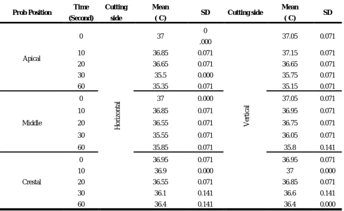
Table 1: Summary of average temperature changes and standard deviation in natural teeth by the time, probing position and cutting side.
Prob Position Time (Second)
Cutting side
Mean
(ºC) SD Cutting side
Mean
(ºC) SD
Apical
0
H
o
ri
zon
tal
37 0
.000
V
erti
cal
37.05 0.071
10 36.85 0.071 37.15 0.071
20 36.65 0.071 36.65 0.071
30 35.5 0.000 35.75 0.071
60 35.35 0.071 35.15 0.071
Middle
0 37 0.000 37.05 0.071
10 36.85 0.071 36.95 0.071
20 36.55 0.071 36.75 0.071
30 35.55 0.071 36.05 0.071
60 35.85 0.071 35.8 0.141
Crestal
0 36.95 0.071 36.95 0.071
10 36.9 0.000 37 0.000
20 36.55 0.071 36.85 0.071
30 36.1 0.141 36.6 0.141
60 36.4 0.141 36.4 0.000
Table 2: Mean and standard deviation of temperature changes in one-piece implant by the time, probing position and cutting side.
Prob Position Time (Second)
Cutting side
Mean
(ºC) SD
Cutting side
Mean
(ºC) SD
Apical
0
H
o
ri
zo
n
tal
36.95 0.071
V
erti
cal
37.05 0.071
10 37.95 0.071 38.1 0.141
20 38.75 0.071 38.95 0.071
30 38.15 0.071 38.45 0.071
60 36.95 0.071 37.4 0.000
Middle
0 37 0.000 37 0.000
10 37.85 0.071 37.9 0.141
20 38.25 0.071 38.65 0.071
30 38 0.000 38.5 0.000
60 36.75 0.071 37.85 0.071
Crestal
0 37 0.000 37 0.000
10 36.85 0.071 37.05 0.071
20 36.6 0.141 36.85 0.071
30 36.45 0.071 36.7 0.000
60 36.4 0.000 36.65 0.071
the data. The results revealed that:
1) The effect of time on temperature changes at all points - apical, middle and crestal - was significant (p = 0.001), so the difference between the
tempera-ture of each time (0,10,20,30,60) was significant and this occurred for all points.
2) The effect of time on temperature changes at all points, separately in each sample group, was
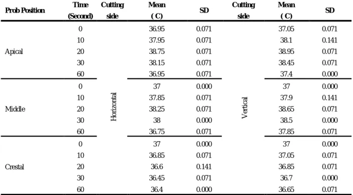
sig-Table 3: Mean and standard deviation of temperature changes in two-piece implant by the time, probing position and cutting side.
Prob Position Time
(Second) Cutting side
Mean
(ºC) SD Cutting side
Mean
(ºC) SD
Apical
0
H
o
ri
zon
tal
37.05 0.071
V
erti
cal
37 0.000
10 37.2 0.000 37.2 0.000
20 36.75 0.071 36.55 0.071
30 36.2 0.141 35.75 0.071
60 33.8 0.141 33.05 0.071
Middle
0 36.95 0.071 37 0.000
10 37.05 0.071 36.85 0.071
20 36.05 0.071 35.9 0.141
30 35.95 0.071 35.35 0.071
60 34.6 0.141 33.8 0.141
Crestal
0 37.05 0.071 36.95 0.071
10 36.85 0.071 37.05 0.071
20 36.2 0.000 36.5 0.000
30 35.7 0.141 35.6 0.141
Chart 1: Estimated marginal means at apical, middle and crestal points in the horizontal and vertical sides. Colors show: Green: one-piece implant – Yellow: two-piece implant – Blue: teeth.
nificant (p = 0.001), so the result of the statistical test on each sample group (one-piece, two- piece implant, and natural teeth) separately showed that temperature change by time was significant, as well.
The results of one-piece implant at the apical point in the horizontal cut showed increased temperature that decreased by time, but in other points, the temper-ature decreased. Maximum tempertemper-ature was 38.95 and
minimum was 36.4. Temperature changes in the verti-cal cut were more (Table 2).
Results from two-piece implant showed that, ex-cept the crestal and middle points in the vertical and crestal points in the horizontal cut, the temperature of the other modes first increased and then decreased. At the mentioned points, the temperature decreased. Maximum temperature was 37.2 and minimum was
33.05 (Table 3).
The results of the horizontal and vertical cuts of three samples in three points and at five intervals were compared (Chart 1).
Discussion
As revealed in the pilot study, when a high-speed den-tal turbine was used without the cooling system to cut, the heat transfer to the implant-bone interface exceed-ed the thermal injury threshold for the bone (47°C, or an increase of 10°C) shortly after 60 seconds. This procedure aimed to determine the minimum time in-tervals that passed from 47°C. In contrast, Huh et al. [8] reported a maximum temperature of 41.22°C at the cervical aspect of an implant when the abutment was reduced by 1.0 mm without the cooling system.
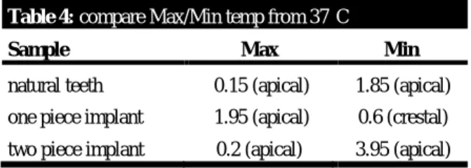
As can be seen in Table 4, by cutting with a cooling system, the temperature limitation increased up to about 2°C. So the findings of this in vitro investigation con-firmed that the use of cooling system was a significant factor in heat transfer to the implant-bone interface.
Table 4: compare Max/Min temp from 37°C
Sample Max Min
natural teeth 0.15 (apical) 1.85 (apical) one piece implant 1.95 (apical) 0.6 (crestal) two piece implant 0.2 (apical) 3.95 (apical)
*V shows vertical, h shows horizontal, s means second.
So, using a suitable cooling system can be an ac-ceptable offer to prevent the thermal injury caused in abutment preparation. This theory has been confirmed by other studies, such as the study of Mason and Gross [7,10].
However, if cooling system was not used, the threshold of thermal injury to the bone was increased, with the potential for irreversible damage. When the abutment type, cutting method, and the side of the implant were evaluated, the temperature change did not exceed the threshold of thermal injury for the bone when cooling system was used.
Gross et al. [10] and Huh et al. [8] prepared titani- um and zirconia abutments for 30 seconds and found that the amount of heat transferred to the interface was
not sufficient to cause bony alterations. Similarly, the present investigation found that the amount of heat transferred to the implant-bone interface was not suf-ficient to cause irreversible damage to the bone if sep-arated cutting time intervals were used during the abutment preparation.
In a similar study by Nissan et al. [18], impression plaster was used to index implant copings, and the heat transfer was sufficient to cause permanent bone changes at the cervical aspect of the implant (maxi-mum temperature of 50.4°C) but not at the apical por-tion. Similar to the present investigation, when the temperature change at the cervical portion of the im-plant was not sufficient to cause irreversible bone changes, the apical temperature change was maximal.
In this way, as shown in Table 4 and Chart 1, it was found that although the cutting area was closer to the crestal point, due to the cooling system, the maximum temperature changes often affected the apical point.
The thermal properties of titanium would suggest higher temperature changes compared to natural teeth as shown in Table 4. Comparing temperature changes of samples from 37°C, found the most increase in one-piece implant and the most decrease in two-one-piece im-plant. The heat transfer law confirmed that it depended on two factors: temperature gradient and the area of surface transfer) [20,21].
Checking the result of two-piece implant showed that – in some modes - the temperature at first had a partial increase, and then decreased. The increases hap-pened; this increase in one- piece implant was more.
As seen in this study, the maximum temperature in-creased from 37°C occurred in 20 seconds (1.95°C), So it suggested that an intermittent cut up to 20 seconds did not cause temperature changes more than 1.95°C and these changes are acceptable (less than 10°C). Gross et al. [10] reported that intermittent cutting with cooling system caused an increase of not more than 0.5°C. Thus intermittent cutting with cooling system appeared to be the optimum clinical technique required to induce minimal temperature changes if abutment or occlusal reduction was attempted intra - orally.
Conclusions
• Cutting in separate time intervals (up to 20 se-conds) has a great effect on the reduction of heat generated from preparing abutment. So we didn’t have a considerable increase in temperature by us-ing this method.
• The trend of heat transfer in each sample was dif-ferent from another. We found the most increase in one-piece implant and the most decrease in two-piece implant in the apical point, from the base point 37°C.
• One-piece implant transferred the heat generated from cutting preparation more than two- piece im-plant.
• Results from horizontal and vertical cut were al-most similar.
References
1. Palma-Carrio C, Maestre-Ferrin L, Penarrocha-Oltra D, et al. Risk factors associated with early failure of dental implants: a literature review. Med Oral Patol Oral Cir Bucal. 2011;16:514-517.
2. Kazemi M, Jalali H, Egtedari M, et al. Acrylic resin polymerization in direct contact to the abutment and the temperature at the bone-implant interface: a pilot in vitro study. J Oral Implantol. 2012;38:595-601.
3. Sharon E, Shapira L, Wilensky A, et al. Efficiency and Thermal Changes during Implantoplasty in Relation to Bur Type. Clin Implant Dent Relat Res. 2013;15:292-296. 4. Okayasu K, Wang HL. Decision tree for the
manage-ment of periimplant diseases. Implant Dent. 2011;20: 256-261.
5. Augustin G, Zigman T, Davila S, et al. Cortical bone drilling and thermal osteonecrosis. Clin Biomech. 2012; 27: 313-325.
6. Trisi P, Berardini M, Falco A, et al. Insufficient cooling system induces peri-implant bone resorption: an in vivo histologic analysis in sheep. Clin Oral Implants Res. 2014; 25:696-701.
7. Mason AG, Sutton A, Turkyilmaz I. An investigation of heat transfer to the implant-bone interface when drilling through a zirconia crown attached to a titanium or zirco-nia abutment. J Prosthet Dent. 2014 ;17: 1119–1125. 8. Huh JB, Eckert SE, Ko SM, et al. Heat transfer to the
implant-bone interface during preparation of a
zirco-nia/alumina abutment. Int J Oral Maxillofac Implants. 2009; 24:679-683.
9. Joo W, Phd – senior research engineer, Heat generation in one-piece implants during abutment preparation with high- speed cutting instruments. Zimmer dental Inc., Carlsbad, CA. 2008.
10.Gross M, Laufer BZ, Ormianar Z. An investigation on heat transfer to the implant-bone interface due to abutment preparation with high-speed cutting instru-ments. Int J Oral Maxillofac Implants. 1995;10:207– 212.
11.Lundskog J. Heat and bone tissue: an experimental in-vestigation of the thermal properties of bone and thresh-old levels for thermal injury. Scand J Plast Reconstr Surg. 1972;9:1-80.
12.Eriksson A, Albrektsson T, Grane B, et al. Thermal injury to bone: a vital-microscopic description of heat effects. Int J Oral Surg. 1982;11:115-121.
13.Eriksson AR, Albrektsson T. Temperature threshold levels for heat-induced bone tissue injury: tissue injury: a vital-microscopic study in the rabbit. J Prosthet Dent. 1983;50:101-107.
14.Eriksson AR, Albrektsson T. The effect of heat on bone regeneration: an experimental study in the rabbit using the bone growth chamber. J Oral Maxillofac Surg. 1984; 42: 705-711.
15.Eriksson RA, Adell R. Temperature during drilling for the placement of implants using The osseointegration technique. J Oral Maxillofac Surg. 1986;44:4-7. 16.Li S, Chien S, Branemark PI. Heat shock-induced
necro-sis and apoptonecro-sis in osteoblasts. J Orthop Res. 1999; 17: 891–899.
17.Zarb GA, Albrektsson T, Baker G, et al. Osseointegra-tion: on continuing synergies in surgery,prosthodontics, and biomaterials. Chicago:Quintessence; 2008:23-25. 18.Nissan J, Gross M, Ormianer Z, et al. Heat transfer of
impression plasters to an implant-bone interface. Im-plant Dent. 2006; 15: 83-88.
19.Eriksson AR, Albrektsson T, Albrektsson B. Heat caused by drilling cortical bone temperature measured in vivo in patients and animals. Acta Orthop Scand. 1984; 55: 629-631.
20.Chacon GE, Bower DL, Larsen PE, et al. Heat produc-tion by 3 implant drill systems after repeated drilling and sterilization. J Oral Maxillofac Surg. 2006; 64: 265-269.