Bacterial Conjugation and its Inhibition:
The Hows and Whys of Conjugation and What Can be Done to Control It
Scott A. Lujan
A dissertation submitted to the faculty of the University of North Carolina at Chapel Hill in partial fulfillment of the requirements for the degree of Doctor of
Philosophy in the department of Biochemistry and Biophysics.
Chapel Hill, NC 2008
Approved by
Abstract
SCOTT A. LUJAN: Bacterial Conjugation and its Inhibition: The Hows and
Whys of Conjugation and What Can be Done to Control It
(Under the direction of Matthew R. Redinbo, Ph.D.)
Conjugation is the primary vehicle for the horizontal transfer virulence factor
genes, such as antibiotic resistance, within and between bacterial strains. In
certain epicenters, such as hospitals in less developed parts of the world,
immune-compromised patients and misuse of antibiotics combine to select for
the development and dissemination of these pathogenicity factors via
conjugation. Inhibition of conjugation would prove a boon for curbing the creation
and spread of new virulent or multi-drug resistant strains. DNA relaxases are the
keystone proteins of each conjugative system. TraI is the relaxase of the F
plasmid, the archetypal model system for conjugation. Toward revelation and
inhibition of relaxase mechanisms, I used bioinformatics and limited proteolysis
to find and identify new domains on the F TraI enzyme. I then solved TraI/DNA
co-crystal structures that showed a novel DNA binding mode. Based on structure
comparisons, sequence conservation, and mutant activity studies, I proposed a
mechanism for TraI activity that required the existence of a dual phosphotyrosine
TraI inhibitors. I determined the relaxase structure in complex with one such
inhibitor, bound as predicted. We showed that several of these compounds are
potent in cell inhibitors of conjugation that often selectively kill
conjugation-capable cells, a novel antibiotic paradigm. We showed that oral treatment of
gnotobiotic mice with two inhibitors, clodronate and etidronate, decreased the
gastrointestinal F+ bacterial load twentyfold without apparent side effects. Beyond
the model system, etidronate showed selective in cell potency versus cells
harboring a known clinical resistance-bearing R100 plasmid. Etidronate and
clodronate are already clinically approved for the treatment of bone loss in
humans. Toward more general relaxase inhibition phylogenetic analyses and
studies with other medically relevant relaxases, including viral replicative
Dedication
For my parents, who got me to this point,
Acknowledgements
I would like to thank Rebekah Potts, Heather Ragonese, and Dr. Laura
Guogas for all of their help in the execution of my crazy schemes. I would also
like to thank Sarah Kennedy, Debi Haisch, Dhruthi Patel, Heather Bethea, and
Lori Hannula for their assistance on the bench in recent years and Drs. Sompop
Bencharit and Diem-Thu Lesher for their assistance and training early on. Joseph
Lomino, Dr. Eric Ortlund, and Dr. Mike Miley, you listened, aided, and
occasionally abetted, for which you have my appreciation. Thanks to Maureen
Bower and the staff of the UNC Gnotobiotic Mouse facility, under the auspices of
the Center for Gastrointestinal Biology and Disease, for the care and handling of
our rodent subjects. Likewise, thanks to Dr. Balfour Sartor for mice, expertise,
and facility time. My distinguished committee, Drs. Richard Wolfenden, Jan
Hermans, Brenda Temple, Gary Pielak, and Steve Matson, thank you for your
support and advice. Steve and Gary, you listened and advised as if I was a
member of your own labs, for which I am grateful. Finally, to Dr. Matthew
Redinbo, every time I asked for a little more rope, you gave it to me. You risked
time, funding, and stress on me and on this project. I hope someday to be to be
Table of Contents
List of Tables ... x
List of Figures ...xi
1 Background and Significance ... 1
1.1 Humanity at War ... 2
1.2 Conjugation ... 3
1.3 TraI: Keystone of F Plasmid Conjugation ... 5
1.4 All in the Family: The Relaxase Domain ... 6
1.5 Something New has been Added: The TFG Domain ... 7
1.6 Summary ... 9
1.7 Figure Legends ... 11
1.8 References ... 16
2 Inhibition of F Plasmid TraI: Structure, Kinetics, & in cell assays ... 18
2.1 Introduction ... 19
2.2 Results ... 21
2.3 Discussion ... 29
2.4 Materials and Methods ... 32
2.4.1 Protein Expression and Purification ... 32
2.4.2 Oligonucleotides ... 32
2.4.4 Functional Assays ... 34
2.4.5 Kinetic Assays ... 36
2.4.5.1 Kinetic Assay Formulations ... 36
2.4.5.2 Fluorescent Kinetic Assays ... 37
2.4.5.3 Kinetic Data Processing ... 38
2.4.5.4 Calculation of Kinetic Constants ... 39
2.4.6 Mating and Cell Toxicity Assays ... 41
2.4.6.1 Mating, selection on solid substrate ... 41
2.4.6.2 Cell Toxicity, selection on solid substrate ... 41
2.4.6.3 Fluorescent Mating & Toxicity, selection in liquid media . 42 2.5 Figure Legends ... 43
2.6 References ... 61
3 Further Relaxase Inhibition: Expanded Library, R100, & in vivo Assays ... 66
3.1 Introduction ... 67
3.2 Results ... 69
3.2.1 In cell inhibitor library ... 69
3.2.2 Lethality with a clinical resistance plasmid ... 71
3.2.3 Decrease in total bacterial load and F+ fraction... 71
3.3 Discussion ... 73
3.3.1 Qualitative SAR analysis ... 73
3.3.2 Bisphosphonates in other systems ... 76
3.4 Materials and Methods ... 78
3.4.2 Fluorescent Mating & Toxicity, selection in liquid media ... 78
3.4.3 Care and handling of gnotobiotic mice ... 79
3.4.4 Plasmid fraction and bacterial load ... 79
3.5 Figure Legends ... 82
3.6 References ... 88
4 Advent of TraI: Phylogenetics of Relaxase Evolution ... 91
4.1 Introduction ... 91
4.2 Results ... 93
4.2.1 Alignment of diverse bacterial relaxases ... 93
4.2.2 Relaxase phylogenetics ... 93
4.2.3 Motif I duplication ... 94
4.3 Discussion ... 95
4.3.1 Ancestral relaxase host ... 95
4.3.2 Evolution of multi-tyrosine relaxases ... 96
4.3.3 Multiple tyrosines and fused helicases ... 96
4.4 Materials and Methods ... 98
4.4.1 Initial homology searches ... 98
4.4.2 Semi-automated sequence annotation ... 100
4.4.3 Initial multiple sequence alignment ... 101
4.4.4 Tree building ... 101
4.5 Special Acknowledgement ... 101
4.6 Figure Legends ... 102
5 Appendix 1: Attempted Real-Time Relaxase Kinetics ... 108
5.1 Results and Discussion ... 109
5.1.1 Attempted continuous kinetic assays ... 109
5.2 Materials and Methods ... 110
5.2.1 Oligonucleotide design ... 110
5.2.2 Oligonucleotide substrate validation ... 110
5.2.3 Attempted Kinetic Assays ... 111
5.2.4 Data Analysis ... 112
5.3 Figure Legends ... 113
5.4 References ... 116
6 Appendix 2: Progress Toward Further F Plasmid Protein Structures ... 117
6.1 TraI constructs ... 117
6.2 The F relaxosome ... 118
6.3 References ... 120
7 Appendix 3: Progress Toward a Comprehensive Real-Time Mating Assay .... 121
7.1 Previous attempts ... 121
7.2 Three fluors and complete characterization ... 122
List of Tables
Table 2-1. Structure Statistics ... 58
Table 2-2. Comparison of Magnesium-Binding Sites ... 59
Table 2-3. Projected Bisphosphonate Doses ... 60
Table 3-1. EC50s from relative cell counts ... 84
Table 4-1. ClustalX alignment input variables ... 104
List of Figures
Figure 1-1 Bacterial Conjugation... 13
Figure 1-2 Relaxase/Nickase Helicases ... 14
Figure 1-3 Models of Proposed TraI/TraD Interaction ... 15
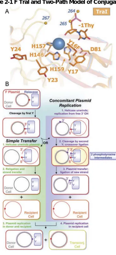
Figure 2-1 F TraI and Two-Path Model of Conjugation ... 48
Figure 2-2 Relaxase Inhibition by PNP ... 49
Figure 2-3 Bisphosphonates Examined for Relaxase Inhibition ... 50
Figure 2-4 Effects of F TraI inhibitors ... 51
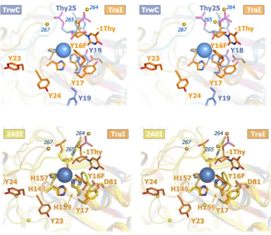
Figure 2-5 Superimposed TraI and TrwC active sites ... 52
Figure 2-6 TraI 300 wild type vs. TraI 300 H159E Cleavage Activity ... 53
Figure 2-7 Stereo ribbon cartoon of the N300 active ... 54
Figure 2-8 The N300 Y16F active site of the 3.0 Å structure with PNP ... 55
Figure 2-9 N300 inhibition ... 56
Figure 2-10 EC50 ranges ... 57
Figure 3-1 Compounds tested for this and previous studies ... 85
Figure 3-2 F+ fraction during 0.1x bisphosphonate treatment ... 86
Figure 3-3 F+ fraction and bacterial loads during 1x bisphosphonate treatment ... 87
Figure 4-1 Maximum parsimony phylogram of bacterial relaxases ... 103
Figure 5-1 Probable equilibration artifact ... 114
1 Background and Significance
In many underdeveloped areas around the globe, antibiotic resistance
among virulent bacterial strains is reaching crisis proportions. Bacterial
conjugation is a major player in the spread of antibiotic resistance within and
between bacterial strains. The F-plasmid conjugation system is used as a model
system for general bacterial conjugation. TraI, also known as DNA Helicase I, is
a keystone protein for F-plasmid conjugation: it possesses both helicase and
relaxase activities necessary for the plasmid mobilization phase of conjugation
(1). Transfer assays show that certain TraI truncations maintain nicking and
unwinding activity but abrogate strand transfer (1). This suggests that TraI plays
a crucial role beyond the mobilization phase. The mechanisms of TraI activity are
not well understood, nor are its extended roles. F TraI relaxase mechanisms
have been postulated based on the functions of TraI homologues, including TrwC
of the R388 IncW plasmid and TraI of the RP4 IncP plasmid. However,
architectural and biochemical data suggest that F TraI relaxase mechanisms vary
somewhat from those of TrwC and a great deal from those of RP4 TraI (2, 3).
work will test these suggestions and reveal the structural basis for TraI function
as a model for R-plasmid relaxase/helicases.
1.1 Humanity at War
Horizontal transfer of genetic information among bacteria is accomplished
mainly through conjugation (4, 5). It is usually mediated by a conjugative plasmid
residing in the cytoplasm or integrated into the genomic DNA of the donor cell.
There is an energy cost associated with hosting a plasmid: plasmid repair,
translation of plasmid proteins, and copy number maintenance all use valuable
cellular resources. As with laboratory strains transformed with artificial vectors,
daughter cells that lack foreign plasmids are at a selective advantage versus
those that are hosts. This helps curb the spread of plasmid borne antibiotic
resistance and virulence factors. Problems arise when a bacterial population,
some individuals of which are plasmid hosts, is subjected to selective pressure,
such as antibiotic treatments. The selective balance then shifts to daughter cells
that inherit plasmid copies. This in itself is not a major concern for healthcare
providers, as the modern suite of antibiotics allows some flexibility in
circumventing resistance. However, continued selective pressure eventually
encourages the development of new forms of resistance, which can then be
disseminated via conjugative plasmids (4, 6). It is then probable that some
bacteria will gain multiple forms of resistance, sometimes to entire classes of
drugs (4). These multi-drug resistant (MDR) strains can become flexible enough
to thwart antibiotic treatment altogether, becoming so-called “super bugs”. In
is the case with MDR strains of mycobacterium tuberculosis in Russia, Peru,
certain African nations, and parts of China (7, 8). Resistance-bearing conjugative
plasmids have been found in many of mankind’s historical nemeses, including
Haemophilus influenzae (bacterial meningitis), Salmonella typhi (typhoid fever),
Vibrio strains (cholera and its relatives), and Yersenia pestis (plague) (6).
Similarly, previously avirulent strains may be pathogenized by the introduction of
virulence factors via conjugation. Such a process has been implicated by the
localization of Salmonella typhimurium derived virulence genes on an F-like
plasmid in pathogenic E. coli O157:H7 and by F-plasmid transfer genes on the
pFra virulence plasmid of Yersenia pestis strain G8786 (9, 10). Medically, the
potential benefits of understanding and fighting antibiotic resistance and
virulence factors spread via conjugative plasmids cannot be overestimated.
1.2 Conjugation
Parallels have been drawn between conjugative mobilization and the rolling
circle form of replication seen in many viruses. Indeed, all of the relaxases
discussed herein are considered members of the Mob class of nickases, which
falls (along with the Rep class) into the Rolling Circle Replication (RCR)
superfamily of magnesium dependent nucleases (11). Though any true
evolutionary linkage is unclear, conjugative plasmids, like viruses, are subject to
selective pressure. Thus, many conjugative systems include genes that provide
selective advantages to host cells. In E. coli, the R series of plasmids is
into incompatibility groups, such as IncF for F-plasmid and R100, IncP for RP4
and RK2, IncW for R388, and Ti-type IncN for R46 (6). Most R plasmids also
code for a repressor that limits their conjugation rate. The F-plasmid is similar to
the rest of the R series - nearly identical to plasmid R100 - with the notable
exception that the repressor gene of F lacks a start codon (14). F may thus be
considered a constitutively active R100 mutant. As such, I will often refer to F
and R systems interchangeably. Wild type F also lacks resistance or virulence, a
rare example of a system that persists on efficiency alone, a true parasite
conferring no selective advantage to its hosts. F-plasmid must thus rely solely
upon its conjugation proficiency to propagate successfully. Among the most well
characterized of all bacterial conjugation systems, The F-plasmid system is an
ideal model system. My long term goal was to characterize the mechanism of
plasmid to identify methods to inhibit the propagation of antibiotic
resistance and bacterial virulence factors.
There are four major phases in bacterial conjugation (Figure 1):
1. Contact: a physical connection is made between the donor and recipient
cells, often through sex pili. The connection must span the cellular
membranes and cell walls of both cells in order to create a conduit between
opposing cytoplasmic spaces (12).
2. Mobilization: the conjugative plasmid is prepared for transfer. The plasmid is
nicked and separated into component strands, which requires unwinding of
the duplex DNA. Mobilization results in a single-stranded circular non-transfer
3. Transfer: one linear strand is moved to the cellular membrane and passed
into the intercellular conduit. Some systems include a “pilot” protein that
guides the strand through the conduit to the recipient cytoplasm (15, 16).
4. Replication: the transferred strand is replicated and either re-circularized or
integrated into the recipient genome. The non-transferred strand is also
replicated, possibly in conjunction with Step 2 unwinding (12).
Specific protein systems are involved in each phase of conjugation, usually
in the form of large, dynamic complexes. Pilus proteins initiate contact with the
recipient cell and may help conduct the transferred strand across intercellular
space. Adapter proteins are important for strand transfer, forming the link
between extracellular pilus assemblies and the cytoplasm of the donor cell (12).
Mobilization is conducted by a complex dubbed the relaxosome. The relaxosome
is responsible for the initiation, execution, and termination of strand transfer. In
the F-plasmid, the relaxosome is composed of three essential proteins (TraI,
TraY, IHF) and a fourth (TraM) required for maximal activity (17-19).
1.3 TraI: Keystone of F Plasmid Conjugation
TraI, also known as DNA Helicase I, possesses both relaxase
(transesterase) and helicase activities. TraI performs the initial nick that begins
plasmid transfer, unwinds and separates the nucleotide strands, and has other
proposed activities during both the mobilization and transfer phases of
conjugation (20). This makes TraI a key protein in F-plasmid conjugation and
1.4 All in the Family: The Relaxase Domain
The details of the diverse Mob family relaxase mechanisms are poorly
understood. Multiple sequence alignments show that some
relaxase/transesterase/nickases possess one catalytic tyrosine residue (TraA
and VirD2 of Rhizobia and Agrobacteria pTi plasmids; TraI of plasmid RP4 IncQ)
while other have a constellation of conserved tyrosines (TraA of Corynebacteria
plasmids; TrwC of plasmid R388 IncW; TraI of plasmid R100 IncF plasmids). The
IncF/W constellation of two tyrosine pairs follows a pattern of Yy-X3-26-YY, where
‘y’ denotes either a tyrosine or a phenylalanine in some IncW relaxases (21).
(Fig. 2) The tyrosine pairs are thought to be responsible for alternating
transesterifications during sequential initiations/terminations of the mobilization
phase in a rolling-circle manner as described for bacteriophage phi X174 (11, 12,
22). During the nicking (transesterification) reaction, a catalytic tyrosine forms a
covalent bond with a phosphate of the nicked strand (1, 13). It has been
postulated that R388 TrwC exploits this covalent bond and acts a pilot protein
that guides the transferred strand from donor to recipient (12, 15). This was
suggested through analogy with known pilot proteins of Agrobacterium
(Ti-system) and RP4 TraI (RP4). However, experimental evidence is against an
F-plasmid pilot, and at 1756 residues in length and 191 kDa in mass, TraI is far
larger than known pilot proteins, an unlikely candidate for transmembrane
secretion (15, 16). Mutant data with R388 TrwC show that all four tyrosines show
varying degrees of capability for creating the initial nick (3). The exact reason for
Mechanisms for TraI relaxase activity have been posited in analogy with R388
TrwC, for which an abundance of mutant activity data exist (3). More recent
mutational data on TraI activity appear to contradict many of the TrwC results,
suggesting that either the mechanisms differ or that a much more complicated
story is in the offing (2). Crystallographic data exist for apo TraI (23) and DNA
bound TrwC (24), but both structures leave much to be desired. The TraI
structure lacks the thirty residues of the DNA binding loop (disordered), and the
TrwC structures lack residues and DNA proximal to the active site (disordered
and not present, respectively).
1.5 Something New has been Added: The TFG Domain
The central 600 residues of F TraI (situated between the relaxase and
helicase domains) and the carboxyl terminal 300 residues have long been a
mystery. Transfer is lost upon their truncation or deletion, and activity is not
recovered upon reconstitution of the helicase and relaxase domains (1). These
regions lack any previously defined domain architecture and there are no
homologous regions in IncP, Ti, or even the closely related IncW incompatibility
groups (R388 TrwC consists of a relaxase domain and a helicase domain
connected by a short linker with no C-terminal extension) (1). I have now shown,
via computational analyses, that there resides in TraI residues 300 to 600 a
Rossman fold domain, similar in structure to the GTPase domain of Thermus
aquaticus Ffh, dubbed the TraI Ffh GTPase-like domain (TFG) (25). This
does the transferred strand move to the inner membrane and couple with the
conjugative secretion system? The answer hinges on an understanding of the
function of Ffh.
Ffh is a member of the bacterial Signal Recognition Particle (SRP). The
SRP is charged with the task of translocating ribosomes to the inner membrane
upon translation of membrane targeting sequences (26). Ffh has two domains:
the M domain, in SRP parlance, which recognizes and binds targeting sequences
and a small SRP RNA; and an N/G domain, consisting of an N-terminal four-helix
bundle (N) and a C-terminal GTPase subdomain (G). After signal recognition and
binding, the complex (Ffh, the ribosome, tRNA, nascent polypeptide, Ffh bound
GTP, and a small SRP RNA) translocates to the inner membrane, where the Ffh
N/G domain complexes with an adapter protein, FtsY. FtsY also consists of two
domains: a C-terminal membrane associating domain; and an N-terminal N/G
domain. The N/G domains interact along a large interface that includes both
GTPase active sites. After the nascent polypeptide (signal sequence) passes to
the secretion system (membrane pore), simultaneous GTP hydrolysis occurs,
mediating Ffh/FtsY dissociation (27, 28).
The SRP system suggests a novel activity for TraI only if an FtsY-type
adapter protein exists in the IncF. TraD is an F encoded hexameric membrane
protein that interacts with TraI, non-specific DNA, and the conjugal secretion
system. TraD has a short transmembrane portion and a large Rossman type
domain (with a subset of canonical NTPase sequences), capped by a four-helix
site) faces out into the cytoplasm (based on homology modeling versus R388
TrwB (29, 30)). This makes TraD an excellent candidate to play FtsY to TraI’s
Ffh. (Fig. 3).
1.6 Summary
My scientific priorities have shifted since my original thesis proposal. A
crystal structure of the TFG domain would show unequivocally whether or not
this domain is structurally homologous to the FtsY family of small GTPases (Aim
2). Likewise, a co-crystal structure of TraI/TraD or TraI/TraD/DNA, would explore
a novel TraI function in the transfer phase of conjugation (Aim 3). However, these
aims are left to future work, as work on Aim 1 (below) and the original long-term
goal of conjugation inhibition proved to be wildly more successful than originally
anticipated.
An understanding of bacterial conjugation is essential for humanity’s long
war with our microbial adversaries. The F-plasmid conjugation system is an ideal
model for a large family of medically relevant conjugative plasmids. As the
keystone of the F-plasmid system, structural knowledge of the various domains
of TraI has been invaluable in the development of weapons in our continued
fight.
I originally proposed (Aim 1) to acquire structural data showing the relative
partial occupancies of the four F TraI tyrosines coordinated to either small
oligonucleotides or intermediate/transition state analogues in order to shed light
and structures of the R388 homologue, TrwC (24), allowed me to propose a
multi-tyrosine relaxase mechanism (31). This in turn allowed me to find small
molecule inhibitors of relaxase activity, which I showed to be potent both in vitro
and in cell. I then solved the structure of the relaxase domain in complex with
DNA and one of these inhibitors, bound as predicted. I then showed that some of
these inhibitors, including two that are already approved for human use to treat
unrelated conditions, selectively kill donor cells both in vitro and in a gnotobiotic
mouse gut. Studies with plasmid systems closely related to the F plasmid have
been promising. Should inhibition prove viable for broad host range plasmids and
viruses with replicative relaxases, and should human safety and drug potency
1.7 Figure Legends
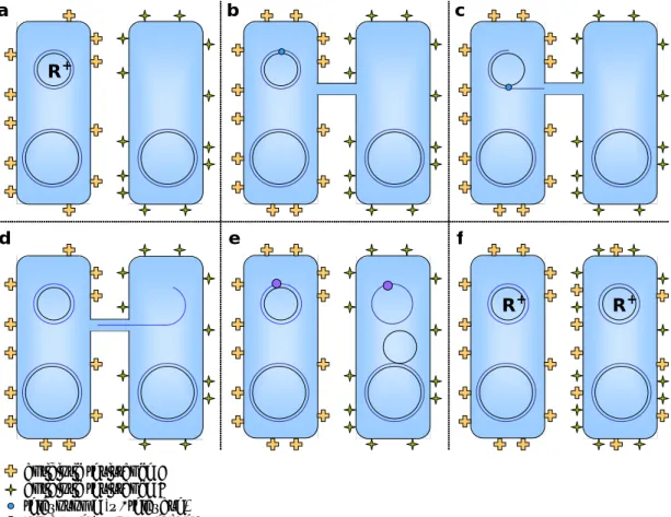
Figure 1. Bacterial Conjugation
In a simplified schematic of bacterial conjugation, the left-hand cell is R+(the
donor), the right-hand cell is initially R- (the recipient).
A. Pre- conjugation: the donor has plasmid encoded drug resistance, the
recipient has genomic drug resistance.
B. Contact: a conduit forms for DNA transfer; a mating signal prompts
relaxosome formation on the plasmid.
C. Mobilization: the plasmid is nicked, unwound, transported to the
membrane, and replicated.
D. Transfer: the nicked strand is transported across the conduit and into the
recipient.
E. Replication: the conduit is terminated and the transferred strand is either
circularized (religated) and replicated (as shown) or integrated into the
recipient genome (not shown).
F. Expression: Plasmid genes, including those for new antibiotic resistance,
are expressed in the recipient, now R+.
Figure 2. Relaxase/Nickase Helicases.
A representative set of proteins containing relaxase and helicase domains.
Relaxases in which these are not coupled (such as IncP RP4 TraI) are not
bacterial conjugative plasmids from related incompatibility (Inc) groups. Color
variations within a domain indicate approximate sub-domains.
A. These F-type proteins have a relaxase domain with four catalytic tyrosines
(Yx4), a proposed Ffh GTPase-like domain (TFG), and a helicase domain
with a characteristic DESS Walker B motif.
B. IncW relaxases have a relaxase domain with three or four catalytic
tyrosines and a helicase domain with a characteristic DEAX Walker B
motif.
C. Ti-type relaxases (involved in bacteria-to-plant conjugation) have a smaller
helicase domain with an R388-like DEAX Walker Bmotif. Agrobacterium
and Rhizobium relaxases have one catalytic tyrosine (IncP-like);
Corynebacteria bear an IncF-like four-tyrosine motif.
Figure 3. Models of Proposed TraI/TraD Interaction
A. TraI and TraD during plasmid mobilization. Hexameric TraD is
represented in grey. TraI domains colored as in Fig. 2: relaxase red, TGF
blue, and helicase green. TraI helicase action pumps the nicked DNA
strand (cyan) through the Type IV Secretion System (T4SS) pore complex
in the direction of the blue arrow.
B. A homology model of the TGF/TraD interaction. The TGF domain (cyan)
is modeled against Ffh. TraD (blue) is modeled against TrwB. Putative
NTP binding loops are red. On the right, a second monomer in the TraD
. antibiotic resistance A
antibiotic resistance B relaxosome (w/ relaxase) replisome (w/ polymerase)
a b c
d e f
R +
R+ R +
Figure 1-2 Relaxase/Nickase Helicases
pKM101 (R46) TraI
Relaxase Helicase
1 306 309 590 960 1476
R388 TrwC Xanthomonas TrwC Pseudomonas putida TraC Nostoc Tra F TraI R100 TraI pSLT TraI
Vibrio vulnificus TraI Photobacterium profundum TraI Corynebacteria Rhodococci Mycobacteria Agrobacteria Rhizobia TFG Sinorhizobia Mezorhizobia 1756 IncF-type Yx4 IncW-type Yx3 or 4
Ti-type Yx1 or 4 a
b
Figure 1-3 Models of Proposed TraI/TraD Interaction
A
1.8 References
1. Byrd, D. R., Sampson, J. K., Ragonese, H. M., & Matson, S. W. (2002) J Biol Chem277, 42645-42653.
2. Sampson, J. K. & Matson, S. W. (2004) (University of North Carolina, Unpublished).
3. Grandoso, G., Avila, P., Cayon, A., Hernando, M. A., Llosa, M., & de la Cruz, F. (2000) J Mol Biol295, 1163-1172.
4. Collignon, P. J. (2002) Med J Aust177, 325-329.
5. Wilkins, B. M. (2002) Environ Microbiol4, 495-500.
6. Waters, V. L. (1999) Front Biosci4, D433-456.
7. Espinal, M. A. (2003) Tuberculosis (Edinb)83, 44-51.
8. Escalante, P., Ramaswamy, S., Sanabria, H., Soini, H., Pan, X., Valiente-Castillo, O., & Musser, J. M. (1998) Tuber Lung Dis79, 111-118.
9. Makino, K., Ishii, K., Yasunaga, T., Hattori, M., Yokoyama, K., Yutsudo, C. H., Kubota, Y., Yamaichi, Y., Iida, T., Yamamoto, K., et al. (1998) DNA Res5, 1-9.
10. Golubov, A., Neubauer, H., Nolting, C., Heesemann, J., & Rakin, A. (2004)
Infect Immun72, 5613-5621.
11. Dyda, F. & Hickman, A. B. (2003) Structure (Camb)11, 1310-1311.
12. Llosa, M., Gomis-Ruth, F. X., Coll, M., & de la Cruz Fd, F. (2002) Mol Microbiol45, 1-8.
13. Howard, M. T., Nelson, W. C., & Matson, S. W. (1995) J Biol Chem270,
28381-28386.
14. Yoshioka, Y., Ohtsubo, H., & Ohtsubo, E. (1987) J Bacteriol169, 619-623.
15. Rees, C. E. & Wilkins, B. M. (1990) Mol Microbiol4, 1199-1205.
16. Rees, C. E. & Wilkins, B. M. (1989) J Bacteriol171, 3152-3157.
17. Inamoto, S., Fukuda, H., Abo, T., & Ohtsubo, E. (1994) J Biochem (Tokyo)
116, 838-844.
19. Ragonese, H. M. & Matson, S. W. (2004) (University of North Carolina, Unpublished).
20. Matson, S. W., Sampson, J. K., & Byrd, D. R. (2001) J Biol Chem276,
2372-2379.
21. Lujan, S. & Redinbo, M. (2004) (University of North Carolina, Unpublished).
22. van Mansfeld, A. D., van Teeffelen, H. A., Baas, P. D., & Jansz, H. S. (1986) Nucleic Acids Res14, 4229-4238.
23. Datta, S., Larkin, C., & Schildbach, J. F. (2003) Structure (Camb)11,
1369-1379.
24. Guasch, A., Lucas, M., Moncalian, G., Cabezas, M., Perez-Luque, R., Gomis-Ruth, F. X., de la Cruz, F., & Coll, M. (2003) Nat Struct Biol10,
1002-1010.
25. Ramirez, U. D., Minasov, G., Focia, P. J., Stroud, R. M., Walter, P., Kuhn, P., & Freymann, D. M. (2002) J Mol Biol320, 783-799.
26. Doudna, J. A. & Batey, R. T. (2004) Annu Rev Biochem73, 539-557.
27. Egea, P. F., Shan, S. O., Napetschnig, J., Savage, D. F., Walter, P., & Stroud, R. M. (2004) Nature427, 215-221.
28. Focia, P. J., Shepotinovskaya, I. V., Seidler, J. A., & Freymann, D. M. (2004) Science303, 373-377.
29. Gomis-Ruth, F. X., Moncalian, G., de la Cruz, F., & Coll, M. (2002) J Biol Chem277, 7556-7566.
30. Gomis-Ruth, F. X. & Coll, M. (2001) Int J Biochem Cell Biol33, 839-843.
2 Inhibition of F Plasmid TraI: Structure, Kinetics, & in
cell assays
Conjugative transfer of plasmid DNA via close cell-cell junctions is the
main route by which antibiotic resistance genes spread between bacterial strains.
Relaxases are essential for conjugative transfer and act by cleaving DNA strands
and forming covalent phosphotyrosine linkages. Based on data indicating that
multi-tyrosine relaxase enzymes can accommodate two phosphotyrosine
intermediates within their divalent metal-containing active sites, we hypothesized
that bisphosphonates would inhibit relaxase activity and conjugative DNA
transfer. We identified bisphosphonates that are nanomolar in vitro relaxase
inhibitors. Furthermore, we utilized cell-based assays to demonstrate that these
compounds are highly effective at preventing DNA transfer and at selectively
killing cells harboring conjugative plasmids. Two potent inhibitors, clodronate
and etidronate, are already clinically approved to treat bone loss. Thus, the
inhibition of conjugative relaxases is a potentially novel antimicrobial approach,
one that selectively targets bacteria capable of transferring antibiotic resistance
2.1 Introduction
Conjugative elements are responsible for the majority of horizontal gene
transfers within and between bacterial strains (reviewed (1)), as first described
for the Escherichia coli F plasmid by Lederberg and Tatum in 1946 (2).
Conjugative DNA transfer is also the central mechanism by which antibiotic
resistance and virulence factors are propagated in bacterial populations
(reviewed (3)). Indeed, it is well established that antibiotic resistance can be
rapidly acquired in clinical settings, and that such acquisition is critically
dependent on conjugative DNA transfer (reviewed (4)). Small molecule inhibition
of conjugation could prove to be a powerful method for curbing the generation
and spread of multi-drug resistant strains. Past studies suggested that various
antibiotics, polycyclic chemicals, and crude extracts inhibit conjugation at
concentrations less than the antibacterial minimum inhibitory concentration
(MIC)(5-11); however, most of these effects have been attributed to
non-conjugation-specific inhibition of bacterial growth or DNA synthesis(12-15). This
study describes a bottom-up approach used to identify the first small molecule
inhibitors of conjugative DNA transfer that target an enzyme of the conjugative
system.
The DNA relaxase is a central enzyme in each conjugative system
(16-18), and thus is a prime target for inhibition. The conjugative relaxase initiates
DNA transfer with a site and strand specific single-stranded DNA (ssDNA) nick in
5’-phosphotyrosine intermediate (16, 19-23). The nicked T-strand moves from
the donor cell (plasmid+) to the recipient cell (plasmid-) via an intercellular
junction mediated by a Type IV secretion system (reviewed (19, 24, 25)). The
relaxase completes DNA transfer by reversing the covalent phosphotyrosine
linkage and releasing the T-strand. In the F plasmid, this relaxase is located in
the N-terminal domain of a large multifunctional protein, TraI (DNA Helicase I)
(22, 23, 26-28). Some conjugative relaxases utilize one active site tyrosine (e.g.,
IncQ RSF1010 MobA (29), IncP RP4 TraI (30, 31), IncI R64 NikA (32),
Agrobacterium Ti VirD2 (33), Tn5252 MocA/BmgA (34); where “R” indicates
plasmids that propagate antibiotic resistance). F-like relaxases (e.g., IncF R1 and
R100 plasmid TraIs (28), IncN R46 and pCU1 TraIs (35), IncW R388 TrwC (36),
Pseudomonas IncP9 pWW0 TraC (37)) maintain a conserved, bifurcated
constellation of two to five tyrosines near their N-termini. The most common
arrangement is four tyrosines (Y1-Y4; tyrosines 16, 17, 23, and 24 in F TraI), with
pairs Y1/2 and Y3/4 separated by a variable linker region. Crystal structures
show all four tyrosines are proximal to a bound metal ion (this study and others
(38-41)). Optimal relaxase cleavage, ligation and transfer of ssDNA require the
metal ion and two catalytic tyrosines, one from each pair (42). F TraI relaxase
shares significant sequence identity with relaxases of many R plasmids (e.g.,
98% with R100 TraI); thus, the F plasmid serves as a model system for
In this study, we first sought to understand the role that the relaxase
enzyme plays in the initiation and termination of DNA conjugation, and then
sought to use that information to identify potent relaxase-specific inhibitors. Our
results establish that the conjugative DNA transfer process can be selectively
disrupted by relaxase-targeted compounds, including some clinically-approved
drugs. This is a potentially novel antimicrobial approach, one that could be used
to purge from microbial populations the bacteria capable of propagating antibiotic
resistance genes.
2.2 Results
We determined the 2.4 Å crystal structure of the 300-residue N-terminal
relaxase domain of F plasmid TraI (N300) with a tyrosine 16 to phenylalanine
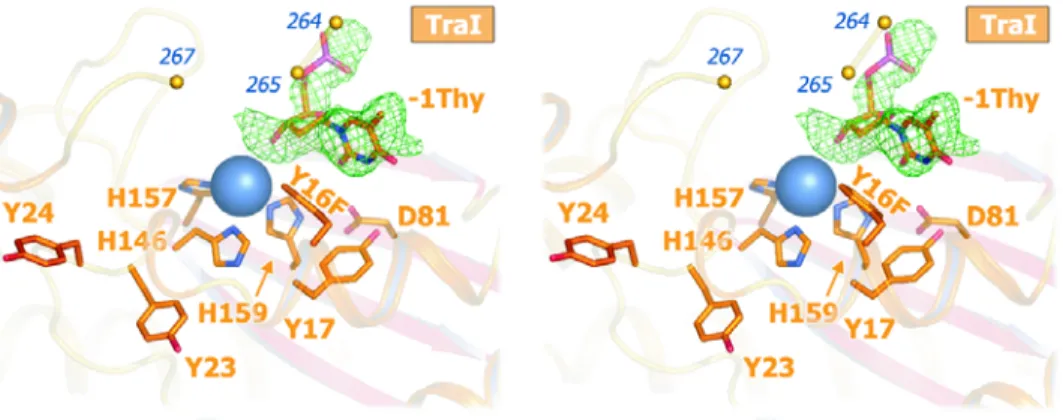
mutation (Y1 of F TraI; Y16F) (Fig. 1a; Table 2). The structure of N300 is similar
to those of a 330-residue F TraI fragment (39, 41) (N330) and the relaxase
domain of R388 TrwC (38, 40) (Fig. 5). Crystallization required a 9-base
single-stranded DNA (ssDNA) oligonucleotide consisting of the F oriT nick site
sequence. In spite of the Y16F mutation, which reduces N300 DNA cleavage
600-fold, we observed electron density for just one DNA base in the active site
(Fig. 1a). We interpreted this as the oriT thymidine immediately upstream of the
scissile phosphate (-1 Thy). We also observed density for a bound metal ion,
chelated by three conserved histidine sidechains (softligands, i.e. uncharged
and high polarizability). Despite unusual soft chelation, we interpret the bound
preference for charged ligands) based on bond lengths, electron density, and
octahedral coordination. A survey of magnesium-binding proteins in the Protein
Data Bank revealed that the chelation of Mg2+ by neutral residues is diagnostic of
a site that simultaneously binds to multiple phosphate groups (Table 2). Mutation
of the metal-chelating residue histidine 159 to glutamic acid (thus reducing the
effective charge of the metal site) eliminated relaxase activity (Fig. 6). These
data indicate that the 2+ charge on the bound metal ion is critical to relaxase
function.
We then considered models for conjugative DNA transfer that would
require the observed 2+ metal ion and two catalytically-competent tyrosine
residues (Fig. 1b). In the case of Simple Transfer (green), a single catalytic
tyrosine is sufficient for successful intercellular DNA transfer. However, if the
free 3’-hydroxyl product of relaxase-mediated DNA cleavage becomes a
substrate for Concomitant Plasmid Replication (CPR; purple), analogous to
rolling circle replication (43), then two tyrosines would be required to resolve the
replicative oriT intermediate and release the T-strand to complete transfer. CPR
events explain the reported low frequency generation of greater than unit-length
conjugative plasmids (44). Although CPR may not be the primary conjugative
pathway, plasmids with relaxases capable of resolving CPR intermediates would
be expected to have a selective advantage. Single-tyrosine relaxases could
achieve resolution of CPR intermediates through relaxase multimerization (45) or
undesirable replication products would also confer oriT specific recombinase
activity between plasmids (47, 48) or between tandem oriT repeats on the same
plasmid (46, 49). However, for our purposes, a key prediction that arose from this
model is that multi-tyrosine relaxases are capable of accommodating two
phosphotyrosine intermediates simultaneously within their active sites. The need
to handle dual phosphotyrosine intermediates would explain the key features of
the TraI relaxase outlined above: the presence of two catalytically-competent
tyrosines and the requirement for a metal ion with an obligate 2+ charge.
We examined the catalytic role of the relaxase active site divalent metal
ion, which is coordinated by three conserved histidine residues and has been
interpreted as either magnesium or zinc (40, 41). While zinc coordination by
histidines is common, the exclusive chelation of a hard magnesium ion by soft
imidazole side chains is unusual. However, magnesium may be essential to
relaxase function, as the activity of several relaxases are enhanced by Mg2+ (29,
50). As stated in the main body of this study, a survey of magnesium-binding
proteins in the Protein Data Bank (51) revealed that the chelation of Mg2+ by
neutral residues is diagnostic of polyphosphate binding (Table 2). We tested
whether the 2+ charge on the metal ion bound to the F TraI relaxase was
required for enzyme function by mutating one of the three coordinating histidine
residues (H159) either to a neutral glutamine residue (H159Q) or to an acidic
glutamic acid residue (H159E). The nearly isostructural H159Q mutation had no
approximately 95% of wild type TraI. The H159E mutation, however, which would
reduce the effective charge on the bound metal ion to 1+, essentially eliminated
enzyme function, generating a 12-fold decrease in cleavage activity, no
measurable crossover activity, and a three order-of-magnitude decrease in
conjugative transfer activity. Thus, a 2+ charge on the bound metal ion is
essential for the catalytic activity of the TraI relaxase, particularly the generation
of DNA crossovers and successful conjugative DNA transfer.
Based on the prediction that multi-tyrosine relaxases employ two
simultaneous phosphotyrosine intermediates coordinated to one magnesium ion,
we hypothesized that simple bisphosphonates would bind the magnesium center
and thus inhibit F TraI relaxase activity and conjugative transfer. To test this
hypothesis, a kinetic assay using fluorophore-labeled oriT ssDNA for cleavage by
the F TraI relaxase domain (TraI N300) was developed to complement existing
radiolabel-based techniques (52, 53). Imidodiphosphate (PNP), a simple and
relatively stable bisphosphonate, was the first compound examined in this assay
(Fig. 2a). We found that PNP is a nanomolar inhibitor of TraI relaxase activity in
vitro. Analysis of cleavage velocity curves revealed that PNP is a mixed-type
(specifically, noncompetitive) inhibitor of TraI, with apparent competitive (Kic,app)
and uncompetitive (Kiu,app) inhibition constants of 2.0-2.4 nM and 2.7-3.5 nM,
respectively. Thus, a simple bisphosphonate serves as a potent inhibitor of a
To determine whether PNP derives its inhibitory power by binding to the
TraI relaxase active site, TraI N300 Y16F crystals were soaked with PNP and the
x-ray structure determined to 3.0 Å resolution (N300+PNP; Table 1). As in the
N300 structure, three of the six magnesium octahedral coordination positions are
filled by histidine side chains, the fourth by the 3’-hydroxyl of the scissile
thymidine, and the fifth is occluded by the Y16F side chain (Fig. 2b). Unlike the
N300 structure, a 5σ simulated-annealing omit electron density peak appears in
the sixth coordination position (Fig. 8), indicating the binding of a single PNP
phosphate group within 3.7 Å of the magnesium ion. The second PNP
phosphate was not observed, due either to disorder or, more likely, to hydrolysis
by a water molecule activated by the adjacent 2+ metal (54-57). The N300+PNP
structure supports the conclusion that PNP inhibits TraI by binding to the
relaxase catalytic site.
The N300+PNP structure revealed a novel phosphate binding site, which
allowed us to model the second phosphotyrosine intermediate in the relaxase
active site. By rotating the first alpha helix (αA; lower right corner Fig. 1a) to
match the orientation observed in the R388 TraI homologue, TrwC (40), and
extending helicity through tyrosine 24 (Y4 of F TraI), tyrosine 23 (Y3 of F TraI)
reorients such that its side chain hydroxyl overlaps with the N300+PNP
phosphate position (Fig. 2c). In this orientation, tyrosine 24 also makes an
aromatic stacking interaction with the side chain of tryptophan 278 (W278), a
how two phosphotyrosines, one at Y16 and one at Y23, can be accommodated
within the active site, and in the process fulfill the octahedral coordination
geometry of a bound magnesium ion.
A variety of compounds were then examined for their ability to inhibit the F
TraI relaxase in vitro. A coarse screen (200 nM concentrations and pH 7.4) of
eleven bisphosphonates and three negative controls (sodium chloride, dibasic
potassium phosphate, and ampicillin, none of which exhibited relaxase inhibition)
yielded five additional inhibitors: methylenediphosphonic acid (PCP);
iminobis(methylphosphonic acid) (PCNCP); etidronic acid (ETIDRO); clodronic
acid (CLODRO); and 1,2-bis(dimethoxyphosphoryl)benzene (PBENP) (Fig. 3).
Results from this screen reveal that effective inhibitors have two phosphonate
moieties separated by three or fewer atoms and have no additional negative
charge at pH 7.4. Four clinically-approved bisphosphonates were tested; these
drugs are used to treat bone loss by inhibiting farnesyl diphosphate synthase
(reviewed (58)). The simplest, ETIDRO and CLODRO, inhibited the TraI
relaxase, while pamidronic acid and neridronic acid (PAMDRO, NERDRO,
respectively), which have an alkyl-amine side chain, did not. Two other inhibitors
identified, PCP and PNP, have been used as radioisotope carriers in humans
(59, 60). Pyrophosphate was not examined due to its rapid hydrolysis in aqueous
solution. The simplest inhibitors, PCP, ETIDRO and CLODRO, were then
characterized further using a kinetic assay and exhibited purely competitive
the PNP results, these data validate the prediction that F-like conjugative
relaxases can accommodate two phosphotyrosine intermediates simultaneously
within their active sites. Significantly, these data also establish that
bisphosphonates (including clinically-approved compounds) potently inhibit the in
vitro relaxase activity of F TraI with Ki values in the nanomolar range.
We next asked whether PNP could impact conjugative DNA transfer
between living bacterial cells. F+E. coli were mated with F-E. coli in dilute
media and in the presence of increasing concentrations of PNP. The resulting
mixture was applied to agar plates with antibiotic selection for transconjugants
(newly formed F+ cells). Colony counts revealed that PNP inhibited DNA transfer
with a half-effective concentration (EC50) of approximately 10 µM, the lowest
concentration tested in this assay (Fig. 4a). We also found that PNP selectively
kills F+ donor cells with an EC50 of <10 µM (compared to low millimolar EC50
against F- recipient cells). This suggests that the TraI relaxase sensitizes F+ cells
to a toxic effect of PNP. Minimal cell growth was observed in control mating
mixtures, so decreases in donor cell count relative to controls are attributed
primarily to cell death rather than to a lack of cell growth. Thus,
inhibitor-dependant decreases represent bacteriacidal rather than bacteriastatic effects.
Strains containing an F plasmid lacking the TraI gene (F+/TraI-) or containing an
F plasmid with the four active site tyrosines mutated to phenylalanine (F+/TraI
4Y-F) behaved like F- cells in this assay, in that they were resistant to the lethal
of a catalytically active TraI relaxase. Thus, the simple bisphosphonate PNP
enters living bacteria, inhibits conjugative DNA transfer, and selectively kills cells
in an active relaxase-dependent manner.
A fluorescence-based 96-well assay was then employed to examine
further these effects and to screen additional compounds for the ability to impact
cell survival and DNA transfer in living bacterial cells. This higher-throughput cell
enumeration assay utilized an oxygen-quenched fluorophore imbedded within a
hydrophobic gel; the concentration of live (oxygen consuming) cells is
proportional to fluorescence (61). The six compounds effective at inhibiting TraI
relaxase activity in vitro (PNP, PCP, PCNCP, PBENP, CLODRO, ETIDRO; Fig.
3), along with two controls (PAMDRO and K2HPO4 [PO4]) were examined for
their impacts on F+ and F- cell survival and on DNA transfer (Fig. 4b, 4c).
PAMDRO and PO4, which had no effect on TraI relaxase activity in vitro, showed
little effect on cell survival and DNA transfer in these cell-based assays,
exhibiting EC50 values greater than the highest concentrations tested (10 mM
and 100 mM, respectively) and no selectivity for F+ over F- cells. In contrast, all
six potent in vitro TraI relaxase inhibitors were also effective in living E. coli cells.
EC50 values for inhibiting F+ donor cell survival ranged from 10 µM (ETIDRO) to
16 nM (PCNCP), and for inhibiting conjugative DNA transfer from 31 µM (PNP) to
110 nM (CLODRO). These compounds have little effect on F- recipient cells,
with EC50 values ranging from 0.34 mM (ETIDRO) to >100 mM (PBENP), which
and PBENP were more effective at inhibiting F+ cell survival, ETIDRO and
CLODRO were more effective at inhibiting DNA transfer, and PCP and PCNCP
were effective against both F+ cell survival and DNA transfer (Fig. 4b, 4c; Fig. 3).
The ranges for EC50 values (Fig. 4b) were derived considering the error bars
present in the survival curves (Fig. 4c). Thus, for the most extreme case of
ETIDRO, the median EC50 for transfer inhibition is 330 nM with a range of 1.1 µM
to <10 pM (Fig. 10). Taken together, however, these data establish that
relaxases can be inhibited with nanomolar affinity within living bacterial cells and
that this inhibition both limits DNA transfer and selectively kills microbes
harboring conjugative plasmids.
2.3 Discussion
This study outlines a potentially novel antimicrobial paradigm that
specifically targets the DNA relaxase enzyme required to initiate and terminate
the process of bacterial conjugation. The compounds identified could be used
along or in combination with existing antibiotics to treat recalcitrant bacterial
infections. Other antibiotics and natural extracts have been reported to disrupt
conjugative DNA transfer and the presence of plasmids within actively dividing
bacterial cells(5-15). In each case, however, the macromolecular target of those
compounds was not understood and their mechanism of action has not been
determined. We took a “bottom up” approach targeted DNA conjugation by
considering first the mechanism and role of a single enzyme (the DNA relaxase)
relaxase active site). While our data was collected on the relaxase from the
well-established F plasmid that was first identified in 1946(2), the F conjugative
machinery shares up to 99% sequence identify with R plasmids that transfer
antibiotic resistance in the wild. Thus, our approach is likely to be effective
against a range of plasmids involved in propagating a range of resistance genes
and virulence factors, many of which play an important role in clinical infections
(62-64).
In addition to inhibiting DNA transfer, we show that simple
bisphosphonates selectively purge populations of bacteria containing a
conjugative plasmid with an active relaxase enzyme. However, because TraI is
F plasmid encoded, not an essential E. coli enzyme, and was not expected to
play a significant role in isolated F+ cells, this relaxase-dependent cell lethality
was a surprise. Several mechanisms could be envisioned to explain this
observation, and future studies will be required to distinguish between them. One
possibility is that relaxases engage in cycles of plasmid DNA cleavage and
religation that are uncoupled from mating and conjugative DNA transfer, and that
the disruption of that process results in a competitive disadvantage relative to
cells without conjugative plasmids. Indeed, inspection of Fig. 4C reveals that a
direct competition appears to exist between plasmid propagation and donor cell
survival (note particularly PNP, PCNCP, PBENP, CLODRO). F+ donor cell
survival is enhanced at higher bisphosphonate concentrations to the detriment of
classic “zero-sum game” in which toxic relaxase-specific bisphosphonate
inhibitors pit the interests of endosymbiont plasmids against those of their
bacterial hosts.
We show that the clinically-approved bisphosphonates etidronate
(Didronel®) and clodronate (Bonefos®), but not other bisphosphonate
therapeutics, are potently effective at killing F+ cells and preventing conjugative
DNA transfer. These particular compounds could also be combined with existing
antibiotics to create potent antimicrobial cocktails. Etidronate and clodronate
exhibit low absorption (65, 66) and can be administered at high oral doses (Table
3). Extrapolating from our results, approved doses of etidronate and clodronate
would be expected kill >90% of plasmid+ cells and to stop >80% of conjugative
transfer within the gastrointestinal track. Such results are relatively mild, given
the large bacterial populations present in the gastrointestinal tract or at wound
sites, but may be enough shift the balance toward success in a variety of
recalcitrant clinical infections, especially given the prevalence of conjugative
plasmids within multi-drug resistant bacterial strains. The treatment of skin
infections, primary sites of nosocomial antibiotic resistance transfer, using the
topical applications of bisphosphonates may also be effective. In summary, this
study establishes conjugative relaxases as a unique antimicrobial target. Our
results suggest that approved therapeutics could have an immediate impact,
propagation during clinical treatment of bacterial infections, and in extending the
lifetime of our antibiotic arsenal.
2.4 Materials and Methods
2.4.1 Protein Expression and Purification
An amino-terminal 300 residue F plasmid TraI construct, bearing a
tyrosine to phenylalanine mutation at position 16 (N300 Y16F), was cloned into
IMPACT® vector pTYB2 (New England Biolabs) for expression as a C-terminal
intein-chitin-binding-domain (CBD) fusion. Protein was expressed in either E. coli
BL21 (DE3)/pLysS or HMS174 (DE3)/pLysS and was purified as per the
standard IMPACT® protocol. Briefly, cellular extracts were prepared and
incubated with 1 mL of Chitin Resin (New England Biolabs) per liter of cell
culture. The resin was washed and incubated with 50 mM dithiothreitol (DTT)
overnight to cleave the relaxase from its CBD tag. The DTT laden eluent was
extensively dialyzed in 20 mM NaCl and 20 mM Tris-HCl (pH 7.5). The resulting
N300 Y16F was >95% pure by SDS-PAGE, and was concentrated to 3 mg/mL
for crystallization in 50 mM NaCl, 10% glycerol, and 10 mM Tris-HCl (pH 7.5)
prior to flash-freezing in liquid nitrogen for storage at -80 °C. Protein for functional
and kinetic assays was concentrated to 42.3 µM in 150 mM NaCl, 50% glycerol,
and 10 mM Tris-HCl (pH 7.5) for long-term storage at -80 °C.
2.4.2 Oligonucleotides
A 9-base single-strandedDNA oligonucleotide (9mer) derived from the F
plasmid oriT (5’-GGT GT^G GTG-3’, where ^ is the scissile phosphate) was
Center Nucleic Acids Core Facility. Labeled oligonucleotides for fluorescence
fluorescent kinetic assays were synthesized by Integrated DNA Technologies
(IDT). For the initial kinetic assay the substrate oligonucleotide was a 5’-biotin
(bio) labeled 29mer (“b29”; 5’-bio-TTT GCG TGG GGT GT^G GTG CTT TTG
GGT GG-3’). The complementary fluorescent probe oligonucleotide was a
5’-6-carboxyfluorescein (56FAM™) labeled 15mer (“downF”; 5’-56FAM-CC ACC CAA
AAG CAC C-3’). The b29 substrate molecule was derived from the F plasmid
oriT. The downF probe is complementary to the downstream portion of b29. The
melting temperature was 50.8° C for downF versus b29 (15 base-pairs) , as
calculated with IDT OligoAnalyzer™ 3.0 with default parameters. For later kinetic
assays, the substrate oligonucleotide was a 5’-biotin (bio) labeled and 3’-56FAM
labeled 27mer (“Bio19'8FAM”; 5’-bio-CT TGT TTT TCG TGG GGT GT^G GTG
CTT T -3’). The Bio19'8FAM substrate molecule has a larger portion of the F
plasmid oriT than does the b29 oligonucleotide. Oligonucleotides for site directed
mutagenesis were synthesized by Integrated DNA Technologies.
2.4.3 Crystallization and Structure Determination
N300 Y16F crystals grew in a DNA-dependent manner in 75 mM sodium
nitrate, 14% w/v PEG 3350, 10 mM spermine, and 110 µM 9mer. These rods
were cryoprotected via a two-second dip in 150 mM sodium nitrate, 35% w/v
PEG 3350, and 10 mM spermine and flash cooled in liquid nitrogen for storage
and transport. Crystals employed for the PNP-bound structure were soaked for
N,N-imidobisphosphonate (PNP) and flash cooled. Rods 200 × 30 × 20 µm in size
were generated by hanging drop vapor diffusion (>35 days of growth) and
diffracted to between 2.9 Å and 3.4 Å in-house. Data sets were collected at the
Advanced Photon Source (APS) at Argonne National Laboratoy (ANL), at
Southeast Regional Collaborative Access Team (SER-CAT) Sector 22 Insertion
Device Beamline (22-ID) and the General Medicine and Cancer Institutes
Collaborative Access Team (GM/CA-CAT) Sector 23 Insertion Device Beamline
(23-IDin; for the PNP complex). Crystals were of space group of P212121 and
contained two protein monomers in the asymmetric unit (Table 1). X-ray
diffraction data were indexed and scaled with the HKL2000 or MOSFLM (CCP4)
(67). Initial phases were determined by molecular replacement in Molrep (CCP4)
(67) with the apo-TraI structure (Protein Data Bank accession 1P4D (41)) as a
search model. Model adjustment was completed with O (68) and σa-weighted
electron density maps (69), and structures were refined using torsion angle
dynamics and the maximum likelihood target as implemented in CNS (70).
Structure figures were constructed in PyMol v0.98 (71).
2.4.4 Functional Assays
Both wild-type and mutant TraI proteins (either full length protein or
TraI-N300) were examined in oligonucleotide cleavage (DNA nicking), strand transfer
(DNA religation) and liquid mating (DNA transfer) assays. The oligonucleotide
cleavage reaction mixture (10 µl) contained 50 mM Tris-HCl (pH 7.5), 150 mM
(unless otherwise stated). Reactions were assembled at room temperature and
incubated at 37°C for 20 minutes. Reactions were stopped by the addition of
SDS to 0.2%, and incubation was continued at 37°C for 10 minutes. Ten µl 85%
formamide, 50 mM EDTA, 0.1% dyes were added to the reaction, the products
were denatured at 100°C for 3 minutes and analyzed on a 16% polyacrylamide, 8
M urea denaturing gel. The gels were electrophoresed at 25 watts in 1xTBE (90
mM Tris-borate and 2 mM EDTA) and visualized using a PhosphorImager
(Molecular Dynamics). Markers were prepared as described previously (72).
Strand transfer reactions were performed in a manner similar to the
oligonucleotide cleavage assay except after the 20-minute incubation, 1 pmol of
a second unlabeled oligonucleotide of differing length containing the F plasmid
nic site was added to the reaction and incubation was continued at 37°C for 1
hour. The reaction was stopped and analyzed using the procedure described
above. Liquid mating assays were performed as previously described (26)
except HMS174 cells were utilized instead of HMS174 (DE3) to reduce the
constitutive expression of the complementing protein. Briefly, cells containing
pOX38T∆TraI and the appropriate complementing plasmid were grown overnight
in the presence of appropriate antibiotics. Overnight cultures were used to
inoculate cultures that were grown at 37oC to mid-log phase (2-3 hours) in the
absence of antibiotics. Donor cells were mixed (1:10) with recipient cells,
incubated at 37oC and then plated to select for transconjugants and
counterselect for donors and recipients. Site-directed mutations in the traI gene
protocol supplied by Stratagene. pTYB2-traIN300 served as the template for
PCR. The resulting clones were sequenced to confirm the presence of the
engineered mutations and the absence of unintended mutations. A unique 700
bp NdeI-StuI fragment of traI containing the engineered mutations was removed
from pTYB2-traIN300 and ligated into the full length traI gene in pET11c-traI that
had been digested at unique NdeI and StuI sites to create the mutant
pET11c-traI derivatives that were at utilized in genetic complementation assays.
2.4.5 Kinetic Assays
2.4.5.1 Kinetic Assay Formulations
Reaction Buffer: 6.42 mM MgCl2, 20.5% glycerol, 153.9 mM NaCl, and
51.3 mM Tris-HCl pH 7.5. Streptavidin Wash Buffer (Tris buffered saline,
TBS/Tween): and 150 mM NaCl, 25 mM Tris-HCl pH 7.5, and 0.05% Tween-20.
Stopping Buffer: 1.2% sodium-dodecyl sulfate (SDS), and 300 mM EDTA pH 10.
TE Buffer: 1 mM EDTA, and 10 mM Tris-HCl pH 7.4. Fluorescence Buffer: 80%
glycerol, 200 mM Tris-HCl pH 8.0. Short-term N300 stock (for storage at -20 °C):
50% Reaction Buffer, 49.8% glycerol, 0.2% long-term protein solution (84.6 nM
final N300 concentration). All 5x stocks except Probe stocks were diluted in
Reaction Buffer. 5x Enzyme Stock: 8.4 mL short-term N300 stock diluted to 2.02
nM N300. 5x Inhibitor Stocks: imidodiphosphate (PNP) at 0-50 nM in Reaction
Buffer. 5x Substrate Stock: b29 oligonucleotide was diluted to 19.6-158.5 mM
each, by 3-fold serial dilutions. 5x Probe Stocks: 23.5-190.2 mM (1.2-fold molar
involving downF were assembled and performed in low-light conditions. Solutions
and microtiter plates containing downF were kept in foil-lined containers at all
times to prevent photobleaching. In Later kinetic assays, different substrate
dilution schemes were used, Fluorescence Buffer was abandoned in favor of
Streptavidin Wash Buffer, and the Stopping Buffer SDS concentration was
reduced to 0.2%.
2.4.5.2 Fluorescent Kinetic Assays
Two oriT derived oligonucleotides, b29 and Bio19'8FAM, were designed
for binding and cleavage by TraI based on past studies (73) in two phases. The
Initial phase includes the initial uninhibited characterization of N300
oligonucleotide cleavage, all inhibition studies with PNP, and a blind rough
screen of all listed inhibitor candidates (Fig. 3). The Later phase used improved
oligonucleotide design and included the inhibitor characterizations of PCP,
ETIDRO, and CLODRO. The overall method is similar to those described
previously for the study of TraI and R388 TrwC (27). Reactions were assembled
from 16 µL of 5x Substrate Stock, 16 µL of 5x Inhibitor Stock, and 32 µL of
Reaction Buffer. 80 µL reactions were initiated with 16 µL of 5x Enzyme Stock
and raised to 37 °C. 10 µL samples were removed at eight time points and
stopped. In Initial assays, reactions were stopped with 10 µL of Stopping Buffer
at room temperature and placed on a 100 °C heat block for one minute, spiked
with 2 µL of 5x Probe Stock while still hot, and incubated at 37 °C for 10 minutes
were stopped with 2 µL of Stopping Buffer at 80 °C. Samples were diluted to 65
µL with Streptavidin Wash Buffer and transferred to streptavidin-coated microtiter
plates (Thermo Electron Biobind™ Assembly white 96-well plates for Initial
assays; Reacti-Bind™ High Binding Capacity black 384-well plates for Later
assays). Plates were incubated at 37 °C for 45 minutes (Initial assays) or one
hour (Later assays). Plates were washed with at least 5-fold excess Streptavidin
Wash Buffer (in an inverted position to prevent excess biotinylated species from
transferring between wells; in Later assays this step occurred after reading the
plate to determine total oligonucleotide content). Washed wells were then filled
with either 65 µL of Fluorescence Buffer (for optimum 6-FAM™ fluorescence;
Initial assays) or with one well volume of Streptavidin Wash Buffer (Later
assays). Plates were read in a BMG Labtech FLUOstar Optima (Initial assays) or
Pherastar (Later assays ) with a 490 nm excitation and 520 nm emission filters
(10 nm bandpass) with the gain optimized for maximum signal.
2.4.5.3 Kinetic Data Processing
All timecourses were fitted by nonlinear regression with un-weighted
least-squares methods using SigmaPlot 8.0 (also used for graph construction).
Timecourses were manually culled for very high signal (incomplete washing) or
very low signal (physical damage to streptavidin coating). Timecourses were
scaled to reflect fractional cleavage and replicate timepoints were averaged (n =
three-parameter exponential decay equation (Equation 1), and converted into units of
concentration.
Equation 1:
bt ae y signal = 0 + −
where t is time in seconds, y0+a is the initial signal, b is a constant that
determines the shape of the curve. Integration of the overall rate equation for
fitting of timecourses was prohibitive. The derivative of the decay equation at
zero time was taken as the negative of the initial reaction velocity (v0; equals ab
from Equation 1). Michaelis/Menton curves (v0 versus substrate concentration)
were constructed for each inhibitor concentration.
2.4.5.4 Calculation of Kinetic Constants
Competitive (Kic) and uncompetitive (Kiu) inhibition constants were
estimated from Vmaxapp and Kmapp values obtained with one of two methods: first,
velocity curves were fitted to the simple modern Michaelis/Menton equation
(Equation 2) by nonlinear regression with un-weighted least-squares method in
SigmaPlot 8.0; second, the Cornish-Bowden/Eisenthal scale-free direct linear
plot method was applied as implemented in the Exploratory Enzyme Kinetics
Macro for SigmaPlot 8.0 (74-78). These methods produced virtually identical
results, so in Later assays, only the former method was used.
where Kmapp is the apparent Michaelis constant and Vmaxapp is the maximum
reaction velocity. For PNP, competitive (Kic) and uncompetitive (Kiu) inhibition
constants were estimated from extrapolation of the negative of ordinate
intercepts of Kmapp/Vmaxapp-versus inhibitor concentration and 1/Vmaxapp
-versus-inhibitor concentration, respectively. Definitions for Vmaxapp and Kmapp (Equations
4-5) were obtained by comparing the equation for mixed inhibition (Equation 3)
with the Michaelis/Menton equation (Equation 1) and setting Kic and Kiu
individually to infinity.
Equation 3: ) / 1 ( ) / 1 ( max iu ic
m I K S I K
K S V v + + + =
where v is reaction velocity, S is substrate concentration, and I is inhibitor
concentration.
Equation 4, 5:
iu app K I V V / 1 max max + = , ic m app m K I K V K / 1 / max + =
where Vmax is the uninhibited maximum velocity and Km is the uninhibited
Michaelis constant. For inhibitors other than PNP, no uncompetitive inhibition
was observed. Kic for these inhibitors was estimated by fitting inhibited velocity
curves to Equation 3 with Vmax and Km set as determined in uninhibited assays