The Journal of Experimental Medicine
J. Exp. Med. © The Rockefeller University Press • 0022-1007/2004/11/1315/10 $8.00 Volume 200, Number 10, November 15, 2004 1315–1324
1315
G
13 Mediates a Signal That Is Essential for Proliferation
and Survival of Thymocyte Progenitors
V. McNeil Coffield,
1,3Whitney S. Helms,
2,3Qi Jiang,
2,3and Lishan Su
1,2,31Curriculum in Genetics and Molecular Biology, 2Department of Microbiology and Immunology, 3Lineberger
Comprehensive Cancer Center, University of North Carolina at Chapel Hill, Chapel Hill, NC 27599
Abstract
G protein signaling via the G12 family (G12 and G13) has not been well studied in T cells. To investigate whether G12 and G13 are involved in thymopoiesis, we expressed the regu-lator of G protein signaling domain of p115RhoGEF to inhibit G12 and G13 during thy-mopoiesis. Fetal thymus organ cultures seeded with p115DH-expressing progenitor cells showed impaired thymopoiesis with a block at the CD4CD8CD44CD25 (DN3) stage. Using G13 or G12 minigenes, we demonstrated that G13, but not G12, is required for thymopoiesis. T progenitor cells expressing p115DH showed reduced proliferation and in-creased cell death. T cell receptor stimulation of the fetal thymus organ cultures did not rescue the block. Overexpression of the antiapoptotic gene Bcl2 rescued the defect in DN3 cells and partially rescued T cell development. Therefore, G13-mediated signaling is necessary in early thymocyte proliferation and survival.
Key words: thymopoiesis • G protein • p115RhoGEF • RhoA • RGS
Introduction
Developing T cells undergo a highly regulated process of selection in the thymus before exiting the organ and mi-grating to secondary lymphoid tissues. Hematopoietic stem cells or common lymphoid progenitor cells move to the fetal liver during early gestation or the bone marrow later in development and then home to the thymus (1, 2) to begin the process of committing to the T cell lineage (3). The most abundant cells in a normal thymus are double positive (DP) thymocytes, which express both CD4 and CD8 recep-tors. DP cells go through a process of positive selection whereby the TCR chain rearranges. Only T cells that produce a TCR capable of recognizing either MHC I or MHC II and transducing a signal are able to survive. The surviving thymocytes then proceed through negative selection to eliminate cells that respond too strongly to the presentation of self antigen, preventing the release of autoreactive single positive mature T cells into the periphery (4, 5). However, before thymocytes express CD4 and CD8 and begin the crucial process of selection they must progress through four double negative (DN) stages of development defined by the expression of CD44 and CD25: CD44CD25
(DN1), CD44CD25 (DN2), CD44CD25 (DN3), and CD44CD25 (DN4) (6–8). DN2 stage cells initiate rearrangement of the TCR chain, which is completed during the DN3 stage. TCR pairs with the pre–T chain to form a functional pre-TCR (9). Successful chain rear-rangement as measured by efficient signaling through the pre-TCR results in significant expansion of the DN3 cells and down-regulation of CD25 as the cells move to DN4 (7, 10). IL-7 and c-kit signaling are also important for survival of DN thymoyctes, whereas Notch signaling is required for differentiation of both DN and DP thymocytes at several stages of development (11). In addition, other signaling path-ways, including G protein–coupled receptors (GPCRs), are implicated in controlling thymocyte survival, differentia-tion, and proliferation (12–14).
G protein–coupled receptors play a key role in enabling T cells to react to a variety of external signals ranging from developmental cues to chemotactic molecules. GPCRs transduce signals through their direct interaction with mem-bers of the heterotrimeric G protein families: Gi, Gs, Gq, and G12. Research into GPCR function in T cells
V. McNeil Coffield and W.S. Helms contributed equally to this work. Address correspondence to Lishan Su, Lineberger Comprehensive Cancer Center, Dept. of Microbiology and Immunology, School of Medicine, University of North Carolina, Chapel Hill, NC 27599-7295. Phone: (919) 966-6654; Fax: (919) 966-8212; email: [email protected]
G13 Is Essential for Thymopoiesis 1316
has focused primarily on a subfamily of GPCRs known as chemokine receptors. Chemokine receptor signaling pri-marily utilizes the Gi pathway, which plays a particularly important role in both development and inflammation (15). Thymic treatment with pertussis toxin, an inhibitor of Gi, results in the specific depletion of the DP population (12). Other G protein signaling cascades, including the G12 family, are also important for peripheral T cell func-tion but remain largely unexplored during thymopoiesis. The G12 family consists of two members, G12 and G13, which have been reported to mediate actin poly-merization during T cell activation (16). Differences in function between these two family members are not well defined, although G13 knockout embryos die at day 9 due to defects in angiogenesis, whereas G12 knockouts are viable (16, 17). Furthermore, recent work showed that G2A knockout mice, a GPCR that is known to work spe-cifically through the G13 pathway, develop a late onset autoimmune disorder (18, 19).
The p115RhoGEF protein couples G13 signaling to RhoA activation. This protein contains both a regulator of G protein signaling (RGS) domain enabling specific bind-ing to either G12 or G13, resulting in the activation of their GTPase activity, and a Dbl homology (DH) domain that acts as a guanine exchange factor to specifically activate the small GTPase RhoA (20–22). Both p115RhoGEF and RhoA have been shown to affect T cell function. Periph-eral T cells in p115RhoGEF knockout mice hyperprolifer-ate even in the absence of antigenic stimulation (23). Ex-tensive research has demonstrated that RhoA is important in both lymphocyte trafficking and T cell development. Experiments using the transgenic lck C3 transferase mouse, which express the RhoA inhibitor C3 transferase under the control of the lck promoter, revealed a specific require-ment for RhoA at the DN2 and DN3 stages for survival (24). C3 transferase expressed under the control of the CD2 promoter, which is active at a later stage of thy-mocyte development than that of lck, results in a defect in differentiation at DN3 (25).
The clear importance of RhoA in thymocyte develop-ment and G13’s specific link to RhoA activation led us to question whether signaling through G13 or G12 specifi-cally plays a role in thymopoiesis. To address this question, we constructed a retroviral vector expressing a mutant of p115RhoGEF (p115DH), which lacks the DH do-main (amino acids 466–547) and, therefore, cannot activate RhoA signaling. This mutant acts as a dominant negative of p115RhoGEF, able to bind both G12 and G13 through the RGS domain to prevent them from transducing a sig-nal, thus terminating GPCR signaling (26–29). We trans-duced fetal liver (FL) progenitor cells with vector control or p115DH retroviruses and seeded the cells into a thy-mocyte-depleted fetal thymic lobe. Analysis of cells gener-ated from the p115DH seeded mouse fetal thymus organ cultures (FTOCs) showed a developmental block by DN3. Using G12 and G13 minigenes, we demonstrated that this block was mediated specifically by G13. FTOCs seeded with p115DH FL progenitor cells also exhibited
reduced prethymocyte proliferation and increased levels of cell death. Experiments using Bcl2-tg FL progenitor cells rescued the defect in DN3, demonstrating a role for G13 in mediating a survival signal during early thymopoiesis.
Materials and Methods
Animals. All animals were housed at University of North
Carolina-Chapel Hill in a sterile animal facility. FL progenitor cells were harvested from embryonic day (E)14 or E15 fetuses. Fe-tal thymic lobes were harvested from E15 fetuses. The Institu-tional Animal Care and Use Committee approved all experiments.
FTOC. FTOC media contains RPMI 1640 (GIBCO BRL)
supplemented with l-glutamine, 10% FBS, 50 M 2-mercapto-ethanol, 2 mM l-glutamine, 1% nonessential amino acid (GIBCO BRL), 10 mM Hepes, 1 mM sodium pyruvate solution (GIBCO BRL), 100 U/ml penicillin, and 100 mg/ml Streptomycin. After dissection, E15 thymic lobes were cultured at 37C on membranes (Millipore), so that the lobe was in contact with 5% CO2. The
lobes were cultured for 5 d in FTOC media supplemented with 2-deoxyguanosine (2-dG; 1.35 mM; Sigma-Aldrich) (30). The lobes were then washed four times (for 1 h per wash) with PBS/2% FBS and seeded with GFP E14/15 FL progenitor cells. To seed lobes, 105 GFP E14/15 FL cells were cocultured in a Terasaki well with
a single thymic lobe for 48 h (31). The lobes were washed in PBS/ 2% FBS and placed onto culture membranes for the indicated times. The media was changed every other day.
Retrovirus Production and Infection. 293T cells were
trans-fected with plasmids encoding HSPG retroviral DNA, VSVg, and gag/pol as described (32). E14/15 FL progenitor cells were cultured for 24 h in FTOC media supplemented with 50 ng/ml SCF, IL-6, and 10 ng/ml IL-11 (Peprotech) before transduction. Retroviral infections by spin inoculation were performed in 2 mL Eppendorf tubes by mixing 2.5 106 cells (500 L FTOC
media with cytokines), viral supernatant (500 L), and 8 g/ml polybrene. Cells were incubated at 22C for 20 min and centri-fuged (2,000 g) for 3 h. Cells were sorted for green fluorescent protein (GFP) expression 48 h after transduction.
FACS. GFP-positive FL progenitor cells were sorted on
ei-ther a Cytomation MoFlo FACS or Becton Dickinson FACS Ad-vantage. Each lobe was crushed (pestle and eppendorf tube), fil-tered, thymocytes were counted, washed with PBS/2% FBS, and stained with the indicated antibodies. Total cells harvested per lobe were analyzed using a Becton Dickinson FACS Caliber. Hoechst 33342 staining was performed as described previously (33) and analyzed on a Cytomation MoFlo FACS. Cell death was measured by harvesting FTOC lobes after 2 wk in culture to pre-vent progression of thymopoiesis beyond DN3 and stained for CD25 and annexin V (34). CD4, CD8, and CD3 antibodies were purchased from Caltag. CD25, CD44, and annexin V antibodies were purchased from BD Biosciences.
Western Blot. Transduced Jurkat cells (106 cells) or mouse
fe-tal liver cells (105 cells) were lysed and resolved on a 10%
Results
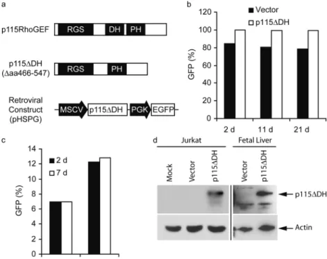
The p115DH Protein Is Not Cytotoxic when Stably Ex-pressed in Cell Lines and Hematopoietic Progenitor Cells. To investigate the G12/13 pathway, we used a dominant negative p115RhoGEF (p115DH) in a retroviral vector. The NH2 terminus of wild-type p115RhoGEF encodes an
RGS domain that functions as a GTPase-activating protein, which specifically inactivates G12 and G13 signaling (20, 22, 28). The DH domain encodes a guanine nucle-otide exchange factor (GEF) specific to RhoA activation and a plextrin homology (PH; reference 36) domain (35) critical to plasma membrane localization. The deletion of the DH domain (amino acids 466–547) in p115RhoGEF (p115DH) inhibits the protein’s ability to signal down-stream to RhoA (26, 27, 29, 35). The remaining RGS and PH domains enable the mutant to inactivate G12/13 sig-naling and localize properly to the cytoplasmic membrane, respectively. The p115DH construct is expressed from a murine stem cell virus LTR, and transduced cells were monitored through a phosphoglycerate kinase–driven en-hanced GFP marker (Fig. 1 a) (37).
Jurkat T cells transduced with p115DH exhibited sta-ble levels of GFP expression over 3 wk in culture (Fig. 1 b). Mouse E15 FL progenitor cells transduced with p115DH retained stable levels of GFP expression at least 1 wk in vitro (Fig. 1 c). Jurkat and mouse fetal liver cells demonstrated stable expression of the p115DH protein at days 21 and 7 postretroviral transduction, respectively (Fig. 1 d). This data demonstrates that expression of p115DH and subsequent inhibition of G12/13 signaling does not negatively affect survival and growth of Jurkat T cells or primary mouse FL progenitor cells.
G12/13 Signaling Is Critical for Thymocyte Development. Although RhoA, a critical component of many different receptor signaling cascades, including multiple families of G protein–coupled receptors, has been shown to mediate proliferation and survival signals during thymopoiesis, it is not clear which upstream signaling pathways are involved in the process. The heterotrimeric G protein G13 has been clearly shown to be a specific activator of RhoA (28, 38). To address the question of whether G12/13 signal-ing played a role in thymocyte development, we expressed the mutant p115DH protein that specifically inactivates G12/13 signaling (26–28) in the pHSPG retroviral vec-tor (Fig. 1 a). We used a reconstituted mouse FTOC sys-tem. E15 FL progenitor cells were transduced, sorted for GFP expression, and seeded into 2-dG–treated E15 thy-mic lobes. The lobes were cultured for 3 wk on mem-branes and analyzed for cell number and expression of GFP, CD4, and CD8. We observed a significant reduction (P 0.005) in the percentage and number of GFP cells in all lobes seeded with p115DH FL progenitor cells but no reduction in the GFP population (Fig. 2, a and b). Al-though the percentages of GFP CD4CD8, CD4CD8, CD4CD8, and CD4CD8 cells were similar between vector and p115DH (Fig. 2 c), the p115DH-seeded lobes showed an overall decrease in the numbers of GFP thymocytes in each population, significant in CD4CD8, CD4CD8, and CD4CD8 populations (Fig. 2 d). There was no reduction in the number of any of the GFP cell populations (Fig. 2 e). This data indicates a crit-ical role for G12/13 signaling during early thymocyte development.
G13 Is Essential for Thymopoiesis 1318
G12/13 Signaling Is Important for Early Thymopoiesis. When the thymocytes were analyzed for DN subsets, the FTOC system revealed that the expression of p115DH in FL progenitor cells generated a lower percentage of cells preferentially in the DN3 population (Fig. 3 a). However, there was a significant reduction in the number of DN2 and DN3 cells derived from p115DH-seeded FTOC lobes and a general reduction of DN1 and DN4 cells (Fig. 3 b). These results directly demonstrate an essential role for G12/13 signaling in early thymopoiesis, most dramatically at the DN3 stage.
G13 But Not G12 Signaling Is Required for Early Thymopoiesis. The expression of the eleven amino acid COOH terminus, termed minigene, of a G protein func-tions as a specific dominant negative inhibitor of its respec-tive G protein by competing for the GPCR binding site (39–45). To determine if G12 or G13 signaling was spe-cifically required for thymopoiesis, we expressed either a
Figure 2. The expression of p115DH inhibits T cell development. 2-dG–depleted E15 thymic lobes were seeded with transduced E14/15 FL progenitor cells and cultured for 3 wk. 24 lobes from four different experiments were analyzed for the percentage (a and b) and the number (b) of GFP and GFP cells. The percentage (c) and number of GFP (d)
and GFP (e) CD4CD8, CD4CD8, CD4CD8, and CD4CD8 cells generated per lobe were also analyzed. Bars indicate SE; **P 0.005 compared with vector.
G12 or G13 minigene fused to GFP in the FTOC sys-tem. The G13 minigene, but not the G12 minigene, paralleled the p115DH phenotype by significantly reduc-ing the total number of GFP thymocytes (Fig. 4 a). Early thymocyte populations were also inhibited in the G13 minigene reconstituted FTOCs similarly to the phenotype seen using p115DH (Fig. 4, b and c). The combination of the p115DH and G13 minigene results suggests that the
block in thymopoiesis is a result of the specific inhibition of G13 signaling.
The Inhibition of G13 Signaling Results in Decreased DN Proliferation and Increased Cell Death. The reduction in the number of prethymocytes derived from the p115 DH-seeded FTOCs led us to question whether G13 signaling could be involved in proliferation or survival of these cells. Hoechst staining of p115DH-derived thymocyte progen-itor cells showed a decrease in the percentage of cells in S/G2 for all DN populations (Fig. 5 a). This decrease dem-Figure 4. The expression of the G13 minigene, but not the G12
mini-gene, inhibits early thymopoiesis. Six FTOC lobes for vector or p115DH were individually analyzed to determine the total number of GFP (a), total CD3CD4CD8 (b), and DN1, DN2, DN3, and DN4 (c) cells generated per lobe. The data is representative of two independent experiments. Bars indicate SE; *P 0.05 compared with vector.
Figure 5. The inhibition of G13 signaling results in decreased DN proliferation and increased cell death. 2-dG–depleted E15 thymic lobes were seeded with transduced E14/15 FL progenitor cells. (a) Duplicate pooled samples consisting of six lobes for both vector and p115DH were stained with Hoechst 33342 and CD3/CD4/CD8/CD25/CD44. At least 5 105 GFP cells were analyzed to determine the percentage of
G13 Is Essential for Thymopoiesis 1320
onstrated that inhibition of G13 signaling resulted in low-ered proliferation of prethymocytes, most obviously in DN1 and DN4 cells. To look at cell death, we harvested fetal liver seeded lobes at 2 wk and stained the cells with annexin V as a marker of apoptosis. At the 2 wk time point, few progenitor cells have progressed past DN3 (un-published data), allowing us to look at cell death exclu-sively in prethymocytes. We stained cells with anti-CD25 to separate DN1 from DN2/3. Cells harvested from p115DH-seeded lobes showed an increase in annexin V staining in both CD25 and CD25 populations compared with vector (Fig. 5, b and c). This data indicated that the thymic phenotype generated through the inhibition of G13 signaling was a combination of reduced DN prolifer-ation and increased cell death.
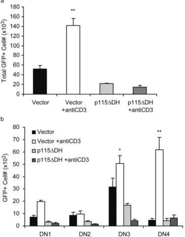
TCR Stimulation Fails to Compensate for the Loss of G13 Signaling in Thymopoiesis. RhoA signaling has been impli-cated to be downstream of the pre-TCR, though expsion of constitutively active RhoA is not sufficient to res-cue a defect in pre-TCR signaling (46, 47). In addition,
during the DN2/DN3 stages of development the pre-TCR signal serves as a checkpoint for further cell expan-sion. We asked whether enhancing TCR signaling could compensate for the loss of G13 signaling in thymopoiesis. FTOC assays were performed in the presence of anti-CD3 mAb during the last 7 d in culture. Vector control lobes treated with anti-CD3 demonstrated a significant increase in the total number of recovered thymocytes compared with untreated controls. However, p115DH lobes showed no difference between cultures treated with or without anti-CD3 (Fig. 6 a). Individual DN3 and DN4 populations showed significant increases in vector lobes treated with
Figure 6. TCR stimulation does not rescue the block in thymopoiesis. 2-dG–depleted E15 thymic lobes were seeded with transduced E14/15 FL progenitor cells and cultured for 2 wk. Lobes were stimulated with 10 g/ml of anti-CD3 mAb from day 14 to 21 in culture. Fresh media containing anti-CD3 was added every 2 d. Six lobes per sample were in-dividually analyzed to determine the total number of GFP (a) and DN1, DN2, DN3, and DN4 (b) cells generated per lobe. Bars indicate SE; *P 0.05; **P 0.005 as vector or p115DDH samples compared with unstim-ulated controls.
anti-CD3 compared with untreated, but again no increase was observed in p115DH lobes (Fig. 6 b). The block in G13 signaling cannot be overcome through exogenous TCR signaling, placing G13 signaling in a pathway dis-tinct from the pre-TCR signaling.
Overexpression of Bcl2 Rescues the Block in G13 Signaling during Thymopoiesis. The inhibition of RhoA in the lck C3 transferase transgenic mouse model revealed a critical role for RhoA signaling during DN3 cell development, which can be rescued by overexpression of Bcl2 (24). To address whether G13 signaling is also acting through a survival pathway, we used Bcl2-tg FL progenitor cells to reconstitute FTOCs. Wild-type littermate controls re-peated the initial observation that p115DH inhibited thy-mopoiesis and specifically reduced the number of both total and DN cells (Fig. 7, a and b). Bcl2-tg FL progenitor cells expressing p115DH showed a partial but significant res-cue of total thymopoiesis (Fig. 7 a) and most obviously of DN3 thymocytes (Fig. 7 c). This rescue defines a critical role for G13 signaling in the survival of early thymocyte progenitors specifically by the DN3 stage of development. Although expression of Bcl2 rescued DN3 development in p115DH-seeded FTOCs, we still observed a difference in cell number between vector and p115DH in the DN1 and DN4 populations (Fig. 7 c). The inability of DN1and DN4 populations to be rescued by Bcl2 may be explained by a reduction in prethymocyte proliferation (Fig. 5 a). These data demonstrate the critical role of G13 signaling in the survival of DN3 cells and in the proliferation of cells during DN1 and DN4.
Discussion
Although previous studies have clearly identified multiple roles for RhoA signaling in thymocyte development (24, 25, 48), it is not clear which upstream signaling pathways are involved in activating RhoA in the thymus. We have shown that blocking G13 signaling inhibits thymopoiesis by using a dominant negative mutant of p115RhoGEF (p115DH) to block G12 and G13 signaling in T cell progenitors. Cells expressing p115DH fail to expand to wild-type levels in FTOC assays. By using G12 and G13 minigenes (39–45), we were able to demonstrate that G13 but not G12 was involved in thymopoiesis. In addition, G13 signaling was shown to be important for the prolifer-ation and survival of thymocyte progenitors.
The phenotype observed using the p115DH mutant in our FTOC system is similar, although not identical, to the thymic phenotype seen in the lck-C3 transferase transgenic mouse. It has been reported recently that RhoA mediates a p53-dependent checkpoint at the DN3 stage of thymocyte development, which prevents cells with nonproductive TCR rearrangements from progressing to the DP stage (47). Though pre-TCR signaling is also important for pro-gressing past the p53-mediated checkpoint, evidence sug-gests that RhoA signaling acts independently of the TCR signaling pathway to affect survival (47). This is consistent with our finding that stimulation of the pre-TCR with
anti-CD3 does not rescue the defect in the G13-mediated proliferation and survival signals. Bcl2, a member of a large family of pro- and antiapoptotic factors, has been shown to prevent the death of immature thymocytes in response to many signals including dexamethasone treatment, CD3 stimulation, and radiation (49). By using FL progenitor cells constitutively expressing Bcl2 (50), we were able to partially rescue the effect of blocking G13 signaling on thymocyte development, indicating that G13 is transduc-ing a survival signal durtransduc-ing thymopoiesis.
However, this rescue does not rule out a role for G13 in proliferation of cells at different points in development. Hoechst staining of thymocytes derived from p115 DH-seeded FTOCs showed a decrease in the proliferation of cells at each DN stage, most obviously at DN1 and DN4. Interestingly, Bcl2 expression rescued the defect in DN3 but not DN1 and DN4 by p115DH. Further evidence for an important role for G13 in cell death is the increased annexin V staining in cells expressing p115DH. The lck-C3 transferase mouse has a similar phenotype—large amounts of apoptosis during DN2 and DN3 followed by a defect in the ability of surviving DN4 cells to proliferate (25, 47). Although the lck-C3 transferase mouse also shows an accu-mulation of cells at DN1 which we do not see, this may be the result of continuous reseeding of the thymus in the transgenic mouse that we are unable to account for in the FTOC system. Additionally, blocking G13 signaling does not preclude RhoA activation by other pathways, making differences between the phenotypes unsurprising. Never-theless, similarities between the phenotypes suggest that a critical G13 signal in early thymopoiesis uses RhoA acti-vation to mediate its effect.
to-G13 Is Essential for Thymopoiesis 1322
gether makes G2A an attractive candidate for activating the G13 signaling pathway in thymopoiesis. Preliminary data in our lab indicated that the addition of LPC to FTOCs stimulated thymocyte production during early thymocyte development (unpublished data). Alternatively, several re-ceptors closely related to G2A that also bind LPC, includ-ing the sphinclud-ingosylphosphorylcholine receptors (55), may be additional candidates relevant to G13-mediated survival signaling in early thymocytes.
Two other proteins that are known to be important in early thymocyte survival are IL-7 and c-kit (56). Although both IL-7/ and IL-7R/ mice exhibit impaired
mic cellularity, neither mutation completely blocks thy-mopoiesis (57, 58). Interestingly, Bcl2 is able to partially rescue the thymocyte phenotype in mice deficient in IL-7 signaling, though the rescue is incomplete (59). Lack of c-kit causes a decrease in thymocyte numbers as early as the DN1 stage (60). Evidence suggests that c-kit is involved in thymocyte proliferation since mice lacking c-kit signaling show a 50% decrease in BrdU incorporation compared with wild-type controls, although a definitive role for c-kit in thymocyte survival has not been established (56). The IL-7 and c-kit signaling pathways can compensate for each other in vivo since mice lacking both c-kit and IL-7 recep-tor common chain (c) fail to develop T lymphocytes (56). As described above, we see a partial rescue of thymopoiesis in p115DH-expressing cells in the presence of Bcl2. It is possible that G13 signaling may interact with one or both of these receptor kinase signaling pathways to promote sur-vival in developing thymocytes.
Our data shows the first evidence that G13-mediated signaling plays an important role in the proliferation and survival of thymocytes during development. This pathway may also have an important role in the T cell response in the periphery. The discovery that G13 signaling is able to influence thymocyte maturation not only increases our un-derstanding of T cell development but also opens novel av-enues for modulating thymopoiesis and investigating other points in the T cell life cycle where G13 might have an important regulatory role.
We thank Drs. C. Der, M. Kondo, and D. Siderovski, and members of the Su laboratory for discussion. We thank Drs. M. Hart, C. Der, and J. Domen for kindly providing the p115RhoGEF DNA, G12 and G13 minigenes DNAs, and Bcl2-tg mice, respectively; and Dr. L. Arnold (University of North Carolina-Chapel Hill) and Dr. M. Kondo (Duke University) for assistance in GFP cell sorting.
The project was partially supported by grants from the National Institutes of Health (5T32AI07273 to W.S. Helms, and AI5380402 and AI04840704 to L. Su).
The authors have no conflicting financial interests.
Submitted: 12 May 2004 Accepted: 20 September 2004
References
1. Itoi, M., H. Kawamoto, Y. Katsura, and T. Amagai. 2001. Two distinct steps of immigration of hematopoietic
progeni-tors into the early thymus anlage. Int. Immunol. 13:1203– 1211.
2. Cumano, A., and I. Godin. 2001. Pluripotent hematopoietic stem cell development during embryogenesis. Curr. Opin.
Immunol. 13:166–171.
3. Kondo, M., I.L. Weissman, and K. Akashi. 1997. Identifica-tion of clonogenic common lymphoid progenitors in mouse bone marrow. Cell. 91:661–672.
4. Robey, E., and B.J. Fowlkes. 1994. Selective events in T cell development. Annu. Rev. Immunol. 12:675–705.
5. Starr, T.K., S.C. Jameson, and K.A. Hogquist. 2003. Positive and negative selection of T cells. Annu. Rev. Immunol. 21: 139–176.
6. Godfrey, D.I., and A. Zlotnik. 1993. Control points in early T cell development. Immunol. Today. 14:547–553.
7. Fehling, H.J., and H. von Boehmer. 1997. Early alpha beta T cell development in the thymus of normal and genetically al-tered mice. Curr. Opin. Immunol. 9:263–275.
8. Shortman, K., and L. Wu. 1996. Early T lymphocyte pro-genitors. Annu. Rev. Immunol. 14:29–47.
9. von Boehmer, H., and H.J. Fehling. 1997. Structure and function of the pre-T cell receptor. Annu. Rev. Immunol. 15: 433–452.
10. Dudley, E.C., H.T. Petrie, L.M. Shah, M.J. Owen, and A.C. Hayday. 1994. T cell receptor beta chain gene rearrangement and selection during thymocyte development in adult mice.
Immunity. 1:83–93.
11. Di Santo, J.P., F. Radtke, and H.R. Rodewald. 2000. To be or not to be a pro-T? Curr. Opin. Immunol. 12:159–165. 12. Person, P.L., R. Korngold, and C. Teuscher. 1992. Pertussis
toxin-induced lymphocytosis is associated with alterations in thymocyte subpopulations. J. Immunol. 148:1506–1511. 13. Plotkin, J., S.E. Prockop, A. Lepique, and H.T. Petrie. 2003.
Critical role for CXCR4 signaling in progenitor localization and T cell differentiation in the postnatal thymus. J. Immunol.
171:4521–4527.
14. Rossi, D., and A. Zlotnik. 2000. The biology of chemokines and their receptors. Annu. Rev. Immunol. 18:217–242. 15. Johnson, E.N., and K.M. Druey. 2002. Heterotrimeric G
protein signaling: role in asthma and allergic inflammation. J.
Allergy Clin. Immunol. 109:592–602.
16. Offermanns, S., V. Mancino, J.P. Revel, and M.I. Simon. 1997. Vascular system defects and impaired cell chemokinesis as a result of Galpha13 deficiency. Science. 275:533–536. 17. Gu, J.L., S. Muller, V. Mancino, S. Offermanns, and M.I.
Si-mon. 2002. Interaction of G alpha(12) with G alpha(13) and G alpha(q) signaling pathways. Proc. Natl. Acad. Sci. USA. 99: 9352–9357.
18. Kabarowski, J.H., K. Zhu, L.Q. Le, O.N. Witte, and Y. Xu. 2001. Lysophosphatidylcholine as a ligand for the immuno-regulatory receptor G2A. Science. 293:702–705.
19. Le, L.Q., J.H. Kabarowski, Z. Weng, A.B. Satterthwaite, E.T. Harvill, E.R. Jensen, J.F. Miller, and O.N. Witte. 2001. Mice lacking the orphan G protein-coupled receptor G2A develop a late-onset autoimmune syndrome. Immunity. 14: 561–571.
20. Kozasa, T., X. Jiang, M.J. Hart, P.M. Sternweis, W.D. Singer, A.G. Gilman, G. Bollag, and P.C. Sternweis. 1998. p115 RhoGEF, a GTPase activating protein for Galpha12 and Galpha13. Science. 280:2109–2111.
Sternweis, J.D. Rothstein, T. Kozasa, and P.C. Sternweis. 2002. Mechanisms for reversible regulation between G13 and Rho exchange factors. J. Biol. Chem. 277:1174–1181. 23. Girkontaite, I., K. Missy, V. Sakk, A. Harenberg, K.
Ted-ford, T. Potzel, K. Pfeffer, and K.D. Fischer. 2001. Lsc is re-quired for marginal zone B cells, regulation of lymphocyte motility and immune responses. Nat. Immunol. 2:855–862. 24. Galandrini, R., S.W. Henning, and D.A. Cantrell. 1997.
Dif-ferent functions of the GTPase Rho in prothymocytes and late pre-T cells. Immunity. 7:163–174.
25. Cleverley, S., S. Henning, and D. Cantrell. 1999. Inhibition of Rho at different stages of thymocyte development gives different perspectives on Rho function. Curr. Biol. 9:657– 660.
26. Majumdar, M., T.M. Seasholtz, C. Buckmaster, D. Toksoz, and J.H. Brown. 1999. A rho exchange factor mediates thrombin and Galpha(12)-induced cytoskeletal responses. J.
Biol. Chem. 274:26815–26821.
27. Mao, J., H. Yuan, W. Xie, and D. Wu. 1998. Guanine nu-cleotide exchange factor GEF115 specifically mediates activa-tion of Rho and serum response factor by the G protein al-pha subunit Galal-pha13. Proc. Natl. Acad. Sci. USA. 95:12973– 12976.
28. Hart, M.J., X. Jiang, T. Kozasa, W. Roscoe, W.D. Singer, A.G. Gilman, P.C. Sternweis, and G. Bollag. 1998. Direct stimulation of the guanine nucleotide exchange activity of p115 RhoGEF by Galpha13. Science. 280:2112–2114. 29. Wang, L., H. Zhang, P.A. Solski, M.J. Hart, C.J. Der, and L.
Su. 2000. Modulation of HIV-1 replication by a novel RhoA effector activity. J. Immunol. 164:5369–5374.
30. Jenkinson, E.J., L.L. Franchi, R. Kingston, and J.J. Owen. 1982. Effect of deoxyguanosine on lymphopoiesis in the de-veloping thymus rudiment in vitro: application in the pro-duction of chimeric thymus rudiments. Eur. J. Immunol. 12: 583–587.
31. Kingston, R., E.J. Jenkinson, and J.J. Owen. 1985. A single stem cell can recolonize an embryonic thymus, producing phenotypically distinct T-cell populations. Nature. 317:811– 813.
32. Pear, W.S., G.P. Nolan, M.L. Scott, and D. Baltimore. 1993. Production of high-titer helper-free retroviruses by transient transfection. Proc. Natl. Acad. Sci. USA. 90:8392–8396. 33. Blaheta, R.A., M. Franz, M.K. Auth, H.J. Wenisch, and
B.H. Markus. 1991. A rapid non-radioactive fluorescence as-say for the measurement of both cell number and prolifera-tion. J. Immunol. Methods. 142:199–206.
34. Vermes, I., C. Haanen, H. Steffens-Nakken, and C. Reutel-ingsperger. 1995. A novel assay for apoptosis. Flow cytomet-ric detection of phosphatidylserine expression on early apop-totic cells using fluorescein labelled Annexin V. J. Immunol.
Methods. 184:39–51.
35. Hart, M.J., S. Sharma, N. elMasry, R.G. Qiu, P. McCabe, P. Polakis, and G. Bollag. 1996. Identification of a novel gua-nine nucleotide exchange factor for the Rho GTPase. J. Biol.
Chem. 271:25452–25458.
36. Lapham, C.K., M.B. Zaitseva, S. Lee, T. Romanstseva, and H. Golding. 1999. Fusion of monocyctes and macrophages with HIV-1 correlates with biochemical properties of CXCR4 and CCR5. Nature. 5:303–308.
37. Coffield, V.M., Q. Jiang, and L. Su. 2003. A genetic ap-proach to inactivating chemokine receptors using a modified viral protein. Nat. Biotechnol. 21:1321–1327.
38. Hart, M.J., W. Roscoe, and G. Bollag. 2000. Activation of
Rho GEF activity by G alpha 13. Methods Enzymol. 325:61– 71.
39. Gilchrist, A., M. Bunemann, A. Li, M.M. Hosey, and H.E. Hamm. 1999. A dominant negative strategy for studying roles of G proteins in vivo. J. Biol. Chem. 274:6610–6616. 40. Gilchrist, A., J.F. Vanhauwe, A. Li, T.O. Thomas, T.
Voyno-Yasenetskaya, and H.E. Hamm. 2001. G alpha mini-genes expressing C-terminal peptides serve as specific inhibi-tors of thrombin-mediated endothelial activation. J. Biol. Chem.
276:25672–25679.
41. Gilchrist, A., A. Li, and H.E. Hamm. 2002. G alpha COOH-terminal minigene vectors dissect heterotrimeric G protein signaling. Sci STKE 2002:PL1.
42. Gilchrist, A., A. Li, and H.E. Hamm. 2002. Design and use of C-terminal minigene vectors for studying role of hetero-trimeric G proteins. Methods Enzymol. 344:58–69.
43. Zhou, H., and K.S. Murthy. 2004. Distinctive G protein-dependent signaling in smooth muscle by sphingosine 1-phosphate receptors S1P1 and S1P2. Am. J. Physiol. Cell
Physiol. 286:C1130–C1138.
44. Le Page, S.L., Y. Bi, and J.A. Williams. 2003. CCK-A recep-tor activates RhoA through G alpha 12/13 in NIH3T3 cells.
Am. J. Physiol. Cell Physiol. 285:C1197–C1206.
45. Arai, K., Y. Maruyama, M. Nishida, S. Tanabe, S. Takaga-hara, T. Kozasa, Y. Mori, T. Nagao, and H. Kurose. 2003. Differential requirement of G alpha12, G alpha13, G alphaq, and G beta gamma for endothelin-1-induced c-Jun NH2-terminal kinase and extracellular signal-regulated kinase acti-vation. Mol. Pharmacol. 63:478–488.
46. Henning, S.W., and D.A. Cantrell. 1998. p56lck signals for regulating thymocyte development can be distinguished by their dependency on Rho function. J. Exp. Med. 188:931– 939.
47. Costello, P.S., S.C. Cleverley, R. Galandrini, S.W. Henning, and D.A. Cantrell. 2000. The GTPase rho controls a p53-dependent survival checkpoint during thymopoiesis. J. Exp. Med. 192:77–85.
48. Cantrell, D.A. 2002. Transgenic analysis of thymocyte signal transduction. Nat. Rev. Immunol. 2:20–27.
49. Rathmell, J.C., and C.B. Thompson. 2002. Pathways of apoptosis in lymphocyte development, homeostasis, and dis-ease. Cell. 109(Suppl.):S97–S107.
50. Domen, J., K.L. Gandy, and I.L. Weissman. 1998. Systemic overexpression of BCL-2 in the hematopoietic system pro-tects transgenic mice from the consequences of lethal irradia-tion. Blood. 91:2272–2282.
51. Kabarowski, J.H., J.D. Feramisco, L.Q. Le, J.L. Gu, S.W. Luoh, M.I. Simon, and O.N. Witte. 2000. Direct genetic demonstration of G alpha 13 coupling to the orphan G pro-tein-coupled receptor G2A leading to RhoA-dependent ac-tin rearrangement. Proc. Natl. Acad. Sci. USA. 97:12109– 12114.
52. Seasholtz, T.M., M. Majumdar, and J.H. Brown. 1999. Rho as a mediator of G protein-coupled receptor signaling. Mol.
Pharmacol. 55:949–956.
53. Sah, V.P., T.M. Seasholtz, S.A. Sagi, and J.H. Brown. 2000. The role of Rho in G protein-coupled receptor signal trans-duction. Annu. Rev. Pharmacol. Toxicol. 40:459–489. 54. Graler, M.H., and E.J. Goetzl. 2002. Lysophospholipids and
their G protein-coupled receptors in inflammation and im-munity. Biochim. Biophys. Acta. 1582:168–174.
G13 Is Essential for Thymopoiesis 1324
Pharmacol. 64:161–167.
56. Di Santo, J.P., and H.R. Rodewald. 1998. In vivo roles of receptor tyrosine kinases and cytokine receptors in early thy-mocyte development. Curr. Opin. Immunol. 10:196–207. 57. von Freeden-Jeffry, U., P. Vieira, L.A. Lucian, T. McNeil,
S.E. Burdach, and R. Murray. 1995. Lymphopenia in inter-leukin (IL)-7 gene-deleted mice identifies IL-7 as a nonre-dundant cytokine. J. Exp. Med. 181:1519–1526.
58. Peschon, J.J., P.J. Morrissey, K.H. Grabstein, F.J. Ramsdell, E. Maraskovsky, B.C. Gliniak, L.S. Park, S.F. Ziegler, D.E.
Williams, C.B. Ware, et al. 1994. Early lymphocyte expan-sion is severely impaired in interleukin 7 receptor-deficient mice. J. Exp. Med. 180:1955–1960.
59. Akashi, K., M. Kondo, U. von Freeden-Jeffry, R. Murray, and I.L. Weissman. 1997. Bcl-2 rescues T lymphopoiesis in interleukin-7 receptor-deficient mice. Cell. 89:1033–1041. 60. Rodewald, H.R., M. Ogawa, C. Haller, C. Waskow, and