Evaluations Of Severe Acute Respiratory Syndrome Coronavirus Therapeutics And A Viral Capacity For Plasticity And Escape.
Meagan Elise Bolles
A dissertation submitted to the faculty of the University of North Carolina at Chapel Hill in partial fulfillment of the requirements for the degree of Doctor of Philosophy
in the Department of Microbiology and Immunology.
Chapel Hill 2013
Approved by: Ralph S. Baric, Ph.D.
Mark T. Heise, Ph.D. Aravinda deSilva, Ph.D.
Eric Donaldson, Ph.D. William Funkhouser, M.D.
ABSTRACT
MEAGAN ELISE BOLLES: Evaluations Of Severe Acute Respiratory Syndrome Coronavirus Therapeutics And A Viral Capacity For Plasticity And Escape.
(Under the direction of Ralph S. Baric, Ph.D.)
The Severe Acute Respiratory Syndrome Coronavirus (SARS-CoV) emerged in 2002/2003,
causing the deaths of almost a tenth of the 8000 individuals infected worldwide before it was controlled by public health measures. While the 2003 epidemic strain is likely extinct, the importance of coronaviruses as
emergent zoonotic viruses was again realized with the emergence of a novel human coronavirus in Saudi Arabia in 2012. Despite a decade of research on SARS-CoV no approved vaccine or therapeutic yet exists,
and development of broadly neutralizing and effective therapeutics for coronaviruses remains a priority. Neutralizing antibodies targeting the Spike glycoprotein (S) are both necessary and sufficient for protection
against SARS-CoV, but the high genetic diversity and mutability of SARS-CoV in natural infections presents a challenge to both vaccine- and antibody-based therapeutics. Thus, an effective SARS-CoV
therapeutic should provide S-specific immunity that is nonetheless broad enough to counter heterologous and derivative S variants. This work was designed to assess immunization strategies towards SARS-CoV,
to explore the plasticity and neutralization networks of the Spike glycoprotein, and to assess the utility of molecular models to predict host range and antibody neutralization.
In the first study we explored the limitations of a doubly inactivated SARS-CoV vaccine, identifying a vaccine-induced immunopathology and emphasizing the importance of rigorous challenge
viruses and animal models that accurately recapitulate age-associated lung pathology. Second, in two collaborative studies we assessed multi-generational monoclonal antibodies designed to be broadly
neutralizing or escape resistant, and extended our characterization of the Spike receptor binding domain (RBD) as a highly plastic antiviral target. Finally, we characterized ten recombinant Combinatorial Escape
networks across the RBD. The tools developed this study will assist in the development of predictive models and standardized platforms for combination monoclonal antibody immunotherapies for emergent
viruses. These studies of SARS-CoV have extended our understanding of a key neutralizing target and have provided a valuable foundation for the rapid characterization of novel coronaviruses and potential
TABLE OF CONTENTS
LIST OF TABLES ... vii
LIST OF FIGURES ... viii
LIST OF ABBREVIATIONS ... x
CHAPTER 1: SARS-CoV and Emergent Coronaviruses: Viral Determinants of Interspecies Transmission ...11
1.1. Overview ...11
1.2. Introduction ...11
1.3. Coronavirus Phylogeny and Mechanisms of Genome Diversity ...12
1.4. Multiple incidents of cross-species transmission. ...15
1.5. SARS-related CoVs in Bats ...15
1.6. Genesis of an Epidemic ...18
1.7. Coronavirus Cross-Species Transmission: Role of Spike-Receptor Interactions in Viral Entry. ...19
1.8. Plasticity of the Spike glycoprotein ...22
1.9. Conclusions ...24
1.10. Contributions. ...26
CHAPTER 2: A Double-Inactivated SARS-CoV Vaccine Provides Incomplete Protection In Mice And Induces Increased Eosinophilic Pro-Inflammatory Pulmonary Response Upon Challenge ...27
2.1. Overview ...27
2.2. Introduction: ...28
2.3. Methods and Materials ...30
2.4. Results ...35
2.5. Discussion ...52
2.6. Contributions ...59
CHAPTER 3: Iterative Development of Potent and Broadly Neutralizing Antibodies Targeting the Spike Receptor Binding Domain. ...60
3.1. Overview ...60
3.2. Introduction ...61
3.3. Increased Antibody Affinity Confers Broad In Vitro Protection against Escape Mutants of Severe Acute Respiratory Syndrome Coronavirus ...65
3.3.2. Introduction ...65
3.3.3. Materials and Methods: ...66
3.3.4. Results ...68
3.3.5. Discussion ...72
3.4. Effects of Targeting Spike Protein Receptor Binding Domain on Neutralization Escape and Fitness of SARS-Coronavirus ...74
3.4.1. Overview ...74
3.4.2. Introduction ...75
3.4.3. Materials and Methods ...77
3.4.4. Results ...80
3.4.5. Discussion ...87
3.4.6. Contributions ...90
CHAPTER 4: Structural Plasticity and Antibody Escape in the SARS-CoV Spike Receptor Binding Domain. ...91
4.1. Overview ...91
4.2. Introduction ...92
4.3. Methods ...94
4.4. Results ...97
4.5. Discussion ...110
CHAPTER 5: Discussion and Future Directions. ...115
5.1. Summary ...115
5.2. SARS-Coronavirus whole inactivated vaccine studies ...116
5.2.1. Ongoing studies ...117
5.3. Live attenuated vaccine studies ...119
5.4. Iterative development and characterization of anti-spike antibodies ...120
5.5. Combinatorial Escape Viruses ...121
5.5.1. Future directions and ongoing studies ...122
5.6. SARS-Coronavirus ...124
LIST OF TABLES
Table
3.3.2: Amino acid changes in the RBD of spike protein found in the
LIST OF FIGURES
Figure
1.1: Spike Phylogeny of Representative CoVs and Models of SARS-CoV
Emergence. ... 14
1.2: Sequence changes over the SARS-CoV epidemic. ... 18
1.3: Crystal structures of coronavirus receptor binding domains (RBD) complexed with their receptors. ... 22
1.4: Experimental Evolution at the SARS S Glycoprotein RBD-Ligand Interface. ... 26
2.1: DIV vaccination and nonlethal heterologous challenge in aged animals. ... 37
2.2: Neutralizing antibody titers of vaccinated mice. ... 39
2.3: Morbidity and Mortality of lethal MA and zoonotic challenges following DIV immunization. ... 41
2.4: Pathology in young mice challenged with icMA15 or icHC/SZ/61/03-S. ... 43
2.5: Pathology following immunization and subsequent lethal challenge in aged Harlan mice. ... 44
2.6: Cytokine and chemokine mRNA expression profiles in aged mice. ... 46
2.7: Visual identification of eosinophils following lethal challenge. ... 47
2.8: Flow cytometry gating strategy for cell populations. ... 49
2.9: Flow cytometric analysis of additional lung immune cell populations in young and aged mice following immunization and subsequent lethal challenge. ... 50
2.10: Eosinophilia influx is conserved across Group 2b N-proteins. ... 52
3.3.3: Neutralization activity of the SK4 and RSK scAbs in comparison to 80R. ... 69
3.3.4: Cross neutralization studies of high-affinity scAbs with escape mutants with the D480A and D480Y mutations. ... 70
3.3.5: SK4 neutralization of escape variants. ... 72
3.4.2. Prophylactic treatment of SARS-CoV infections in 12-month-old aged BALB/c mice by 80R and Fm6 nAbs. ... 81
3.4.4. Broadly neutralization activity of nAbs, fm6 and Y112A. ... 82
3.4.5. Locations of neutralization escape variant mutations on the structure of the SARS-CoV RBD and effects on the binding to human ACE and viral growth. ... 84
3.4.6. Effect of neutralization escape on in vivo replication. ... 86
4.1: Combinatorial Escape Viruses. ... 98
4.2: Panel of antibody-escape profiles. ... 99
4.3: Predicted localized destabilization in CEV-9. ... 100
4.4: Predicting CEV viability ... 102
4.5: Growth curves to assess replication and virus production. ... 104
4.7: Expanded binding tract for mAb s230.15. ... 106
4.8: Mechanisms of escape from m396. ... 109
4.9: Co-neutralization with s227.14 and s230.15. ... 110
4.10: Summary of prediction methodologies. ... 114
5.1 IL-4 signaling phenotypes following lethal MA15 SARS-CoV infection. ... 118
5.2: MA-ExoN vaccination protects from lethal challenge. ... 119
LIST OF ABBREVIATIONS
Ab antibody
CEV Combinatorial Escape Virus CoV coronavirus
ExoN exonuclease
nAb neutralizing antibody
MA mouse-adapted mAb monoclonal antibody
RBD receptor binding domain S spike glycoprotein
SARS Severe Acute Respiratory Syndrome scFv single chain variable fragment
CHAPTER 1: SARS-CoV and Emergent Coronaviruses: Viral Determinants of Interspecies Transmission1
1.1.Overview
Most new emerging viruses are derived from strains circulating in zoonotic reservoirs.
Coronaviruses, which had an established potential for cross-species transmission within domesticated animals, suddenly became relevant with the unexpected emergence of the highly pathogenic human
SARS-CoV strain from zoonotic reservoirs in 2002. SARS-SARS-CoV infected approximately 8000 people worldwide before public health measures halted the epidemic. Supported by robust time-ordered sequence variation,
structural biology, well-characterized patient pools, and biological data, the emergence of SARS-CoV represents one of the best studied natural models of viral disease emergence from zoonotic sources. This
review article summarizes previous and more recent advances into the molecular and structural characteristics, with particular emphasis on host-receptor interactions, that drove this remarkable virus
disease outbreak in human populations.
1.2.Introduction
Coronaviruses have an established potential for cross-species transmission that became broadly
recognized with the emergence of a novel human coronavirus in 2002. Severe Acute Respiratory Syndrome
(SARS) was first identified as an atypical pneumonia in isolated patients in Guangdong Province, China. The disease, caused by SARS-coronavirus (SARS-CoV), spread into epidemic disease proportions
following key super spreader events that were associated with a novel respiratory virus introduction into a globalized community. SARS-CoV rapidly spread around the world, causing about 8,000 infections and 800 deaths worldwide, before aggressive public health intervention strategies contained the epidemic by
July 2003. The epidemic went through three distinct phases: early, middle, and late, as determined by
1 Meagan Bollesa, Eric Donaldsonb, Ralph Barica,b. Department of Microbiology and Immunologya and
Department of Epidemiologyb, University of North Carolina at Chapel Hill, Chapel Hill, North Carolina. First published in Current Opinion in Virology, December 2011, 1(6): 624-34.
molecular analysis. The decimating lethality of SARS-CoV emergence was borne largely by the elderly, in whom mortality rates approached 50% or more. Aggressive public health measures limited and eventually
ended the epidemic in July 2003, absent any effective therapeutics [1]. A subsequent explosion of coronavirus research identified SARS-CoV in several small carnivores (palm civets and raccoon dogs) of the Chinese wet markets and SARS-like CoV in the predicted reservoir host, horseshoe bats (genus
Rhinolophus). The vastly expanded CoV phylogeny includes two novel human coronaviruses (NL63 and
HKU1) and ultimately tripled the number of full length genome sequences available in GenBank. SARS-CoV was shown to use a novel host receptor, Angiotensin Converting Enzyme 2 (ACE2), for docking and
entry and the viral attachment protein, Spike, was extensively characterized both as a determinant of host specificity and as a therapeutic target. The more recent studies of coronaviruses have progressed to increased surveillance and characterization of numerous new coronaviruses circulating in bats, bids, and
other species, integrated bioinformatics and microbiological studies, and extensive evaluations of potential
therapeutics [2].
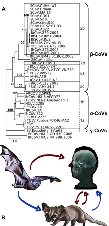
1.3.Coronavirus Phylogeny and Mechanisms of Genome Diversity
Following the SARS-CoV outbreak a surge in global coronavirus genome sequencing efforts
vastly expanded our insight into the CoV phylogeny and resulted in the definition of several
sub-classifications (Fig 1). The greatest contribution of new strains was derived from the newly discovered bat
coronavirus (BtCoV), which may be the source of most, if not all, mammalian CoVs [3-9]. The high diversity of coronaviruses is attributable to three viral traits [10]. The first characteristic is the potentially
high mutation rates associated with RNA replication, generally estimated as 10-3 to 10-5. Surprisingly, the estimated mutation rate for SARS-CoV and other coronaviruses approached 2x10⁻⁶ [11-13]. In contrast to
other RNA viruses, recent data suggests that coronaviruses encode an RNA proof-reading activity associated with the 3’ to 5’ exonuclease activity encoded within nsp14 [14]. It is not clear whether RNA
proof-reading fidelity is altered in changing environmental settings or during virus replication under stress related conditions, but such possibilities may allow for rapid virus evolution in changing ecologic
conditions [14]. Second, recombination frequencies within the coronavirus family have been calculated to be as high as 25% during mixed infection, likely the result of discontinuous RNA transcription and the
recombination between viral genomes and subgenomic replication complexes [15,16]. The role of
discontinuous transcription in recombination is supported by the higher rate of recombination at the 3’ ends
of viral genomes and by targeted RNA recombination techniques designed to genetically manipulate the 3’ end of the genome [17]. Although poorly studied, conservation of transcription regulatory sequence (TRS) sites across viral species may implicate these sequences as foci or hot spots of recombination [17]. Thirdly,
as the largest of the RNA viruses at ~27-31kb, coronaviruses have both increased opportunity for change
and room for modification, clearly evidenced by the presence of numerous unique open reading frames and protein functions encoded at the 3’ end of the genome [10]. These genomic characteristics allow for rapid
phylogenetic and receptor analysis studies suggest a direct emergence from bats to humans, with subsequent cross transmission between humans and civets (red arrow).
1.4.Multiple incidents of cross-species transmission.
Coronaviruses have a strong history of host shifting as evidenced by phylogenetic incongruences in the family tree [18]. In addition to SARS-CoV, two human coronaviruses, OC43 and
HCoV-229E, are now also recognized as having likely emerged from animal reservoirs. HCoV-OC43 and bovine coronavirus (BCoV), betacoronaviruses, have very high sequence similarity, suggesting a recent and
common origin (Fig 1). Molecular clock analysis of the Spike glycoprotein of both species estimates that HCoV-OC43 originated from a BCoV ancestor around 1890 [19]. Similarly, HCoV-229E likely emerged
from a bat alphacoronavirus approximately 200 years ago [20]. In an example of reverse zoonsis, porcine epidemic diarrhea virus emerged suddenly in the early 1980’s, most likely originating from HCoV-229E
[20]. Additionally, a coronavirus isolated in 1988 from a child with acute diarrhea, HECV-4408, was shown to be more closely related to bovine coronavirus (BCoV), indicating the continued introduction of
zoonotic coronaviruses into human populations [21]. The origins of HCoV-NL63 and HCoV-HKU1, the most recently discovered human coronaviruses, remain under study. The most recent example of zoonotic
emergence of a human coronavirus is the example of SARS-CoV, which had at least two independent emergence events from zoonotic reservoirs, recognized in 2002 and 2003 [22]. The most recent
phylogenetic data estimate the emergence of SARS-CoV some seven years earlier, consistent with the identification of low sero-positive cases from archived serum samples in 2001 in China [23].
1.5.SARS-related CoVs in Bats
Following its emergence in 2003, SARS was quickly identified as a zoonotic virus, and the
identification of the wet markets as a potential source may have assisted epidemiological control of the disease [24]. While palm civets, raccoon dogs, and horseshoe bats (Rhinolophus genus) have all been identified as hosts of SARS-like CoVs, it is suggested that only the horseshoe bats are likely reservoir
hosts. Bats are widely distributed, highly diverse, and extremely mobile mammals with an established role as hosts of emergent RNA viruses. Coronaviruses occupy an exceptionally wide distribution in bats; recent
surveillance studies have extended our recognition of this range to Africa, Europe, South America, and
hosted by bats is far greater than the diversity noted between many human coronaviruses, despite a proportionally small sampling of the ~1200 bat species, leading some researchers to speculate that all
mammalian coronaviruses are derived from bat reservoir strains [4,28]. The extensive sequence diversity provides considerable opportunity for the emergence of new animal and human coronaviruses, which would be sufficiently antigenically distinct as not to be influenced by preexisting exposure and memory
immune responses to established human CoVs. For example, little antigenic cross reactivity exists between
the S glycoproteins of more distantly related group 2b bat coronaviruses and the SARS-CoV [29]. From a historic context, the next emergent event is likely dependent only on ecological and epidemiological
situations and time, as the viral potential is well-established [30,31].
Repeated efforts have been made in recent years to identify the zoonotic reservoir and path of emergence for SARS-CoV, both by sampling zoonotic populations and by attempting to clarify SARS-CoV
receptor usage in alternate hosts. A recent study attempting to address the paucity of bat SARS-related
coronavirus sequences gathered and analyzed SARS-related coronaviruses in Rhinolophus bats (SARSr-Rh-BatCoV) (Rp3) genomes from horseshoe bats in China [32]. Interestingly, several bats sampled were
coinfected with HKU2, an alphacoronavirus, providing direct evidence that individual bats can host divergent coronaviruses, even across groups. Further, tagging and clinical assessment of infected bats over a four year period showed only minor weight loss associated with Rp3 infection, and viral clearance
occurring between two weeks and four months. Analysis of the ten novel genomes gathered in this study
combined with previously published sequences demonstrated evidence of frequent recombination between the strains. They also note a 26-bp deletion in ORF8 near, but not identical to, the 29-bp deletion seen in
human SARS-CoV epidemic strains, suggesting ORF8 may undergo frequent deletions [32]. Whether the epidemic 29-bp deletion was neutral to or critical for human adaption remains undetermined.
Angiotensin Converting Enzyme 2 (ACE2) is the receptor for SARS-CoV, but following the
identification of several SARS-like CoVs (SL-CoVs) in several horseshoe bats (genus Rhinophus), the
ACE2 molecule of R. pearsonii proved incapable of serving a receptor for SARS-CoV [3,33]. These and other initial studies suggested that the ancestral SARS-CoV strain in bats used an alternate receptor and that the emergence of SARS-CoV was dependent upon either acquisition of an ACE2 binding region or initial
the bat ACE2 sequences are highly heterogeneous, with 78-84% amino acid identity between families [34,35]. Despite this, the residues that interface with the SARS S RBD are more conserved [36]. A recent
study determined that a minimum three substitutions in the ACE2 of R. pearsonii (RpACE2) allowed this protein to serve as a receptor for SARS [37]. Looking more broadly at the ACE2 molecules from seven bat species, the ACE2 proteins from Myotis daubentoni and Rhinolophus sinicus are capable of supporting
Spike-mediated pseudovirus and SARS-CoV infection, though less efficiently than human ACE2 [34].
Assessment of receptor usage by early phase and civet isolate Spike proteins might better inform our understanding of emergence pathways, determining if SL-CoV jumped directly from bat to human hosts or
whether civet or other intermediate hosts were required as early intermediates prior to human adaptation. Although original data suggested a bat to civet to human origin, evidence supporting direct bat to human transmission of SL-CoV emerged from recent phylogenetic studies, in addition to the receptor
studies mentioned above (Fig 1b). Initially, a reanalysis of published genome sequences developed
phylogenies using outgroups that were non-SARS-CoV sequences, designed to test the monophyly of the SARS-CoV sequences [38]. Under this assessment, bat isolates are ancestral host to all SARS-CoVs, while
civet and raccoon dog sequences (small carnivores), as well as pig isolates cluster within the human SARS-CoV sequences. The small carnivore SARS-CoVs are consistently shown to be terminal branches with human CoVs intermediate, with subsequent bidirectional transmission of CoV between carnivores and humans
responsible for isolated cases such as GD03 [38]. A more recent study analyzed CoV sequences gathered
from 24 Rhinolophus sinicus bats in geographically distant regions of China, characterizing two distinct genotypes, Rs672 and Rs806 [39]. Interestingly, one sequence (Rs672) and the previously published Rp3
are shown in a monophyly more closely related to human-SCoV than to bat SARS-like CoV strains, based on the strong similarity of Rs672 ORF1a/b region to human SARS sequences. This study also provided further evidence of recombination between Bat-SLCoV, with a recombination breakpoint identified
immediately after the start codon of Spike, identical to the recombination position in the Rp3 genome [39].
The combination of highly diverse BtCoV species and divergent ACE2 molecules among bat hosts suggests direct bat to human transmission may be feasible. Thus, the field is left with two potentially competing models for the origins of the SARS-CoV epidemic, however, in both models civets and raccoon
1.6.Genesis of an Epidemic
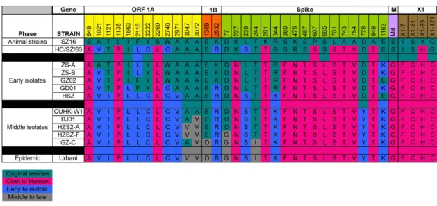
The SARS-CoV outbreak is unique in that a chronological set of sequence changes are available that span the epidemic, providing an unparalleled opportunity to identify the genetic basis for zoonotic
virus cross species transmission and human adaptation during an expanding epidemic. Molecular changes noted at with the end of the early phase and expansion into the middle phase of the epidemic include
A3047V, A3072V in the replicase and D778Y and perhaps E1163K in the Spike gene. Transition from the middle to late phase of the epidemic included an A2552V in ORF1a, E1389D in ORF1b, D77G and T244I
in the S gene, respectively (Fig 2) [40]. It has been hypothesized that these alterations were key to an expanding epidemic, yet empirical data to support these claims and functional significance of these
alterations remains unavailable. For example, it is not clear whether the ORF8 29 bp deletion is central for human adaptation as suggested, or a genetic hitchhiker amplified and maintained following a selective
sweep mediated by other beneficial mutations located elsewhere in the genome [3,40]. In addition to these changes, the SARS-CoV Spike glycoprotein was under strong positive selection, with 23 substitutions
evolving during the expanding phases of the epidemic [41]. Experimental evidence suggests both adaptation to ACE2 and antibody selection contributed to Spike changes [40,42].
Figure 1.2: Sequence changes over the SARS-CoV epidemic.
Shown here are the most significant changes important for transition of SARS from Civet to early, middle and late phases of epidemic strains. Mutations indicative of lineages that were not likely to have
1.7.Coronavirus Cross-Species Transmission: Role of Spike-Receptor Interactions in Viral Entry. Coronavirus receptor interactions are key determinants regulating host range, cross-species transmission, and tissue tropism. The various coronaviruses demonstrate broad receptor and co-receptor
usage, from proteases such as aminopeptidase N for transmissible gastroenteritis virus (TGEV), canine-CoV, feline infectious peritonitis virus (FIPV), and HCoV-229E, to cell adhesion molecules such as
CEACAM1a for MHV, to sugars as co-receptors for some alpha, beta, and gammacoronaviruses
[36,43,44]. This diverse receptor usage directly impacts host range and tissue tropism as demonstrated by
the closely related PRCoV and TGEV. PRCoV lacks the sugar-binding region of TGEV, and consequently is limited to a respiratory rather than enteric tropism [45]. The recently crystallized structure of the
Group2a coronavirus MHV complexed with its receptor, murine CEACAM1a, emphasizes again the broad diversity and flexibility of CoV Spike glycoproteins, as the core structure is hypothesized to have been
derived from a host sugar-binding protein (galectin) and subsequently modified to allow mCEACAM1a binding, thus enhancing MHV affinity for host cells [43]. Other coronaviruses encode a second putative
viral attachment protein, the hemagglutinin esterase (HE), which was likely derived from influenza C strains by recombinatory mechanisms [46]. Coronaviruses selected in vitro to broaden host range
oftentimes mutate to bind heparin sulfate for docking and entry [47]. It is notable that OC43 and BCoV have carbohydrate (sialic acid) binding capacities, as well as broader host ranges [44]. The capacity to bind
carbohydrates for docking and entry may provide an additional pathway for coronavirus host range expansion, cross-species transmission, and disease emergence, and requires further study.
The key determinant of SARS coronavirus host specificity is the Spike glycoprotein, an envelope-anchored trimeric protein responsible for binding human Angiotensin Converting Enzyme 2 (ACE2) as the
principle receptor for virus docking and entry. SARS-CoV S glycoprotein also binds C-type lectins like DC- and/or L-Sign as a co-receptor, an interaction which is blocked by mannose binding lectin [48,49].
Importantly, SARS-CoV docking and entry is also highly dependent upon transmembrane protease/serine subfamily member 2 (TMPRSS2) S and ACE2 cleavage, especially in airway and alveolar sites, and
cathepsin L cleavage and subsequent S2 fusion activation [50-52]. Several studies in the past two years have worked to clarify the plasticity of this protein, with particular emphasis on the receptor binding
the most significantly variable protein across civet and human isolates [22], and shows evidence of positive selection during both inter- and intraspecies transmission events [10,22,40,53]. The SARS Spike
can recognize and use bat, civet, mouse, and raccoon dog ACE2 receptor molecules for docking and entry, indicating that SARS trafficked along receptor orthologue networks to move between species [34,54,55]. As several alphacoronaviridae also use APN from different species, these data suggest a common theme in
coronavirus host range switching: recognizing receptor orthologues from different species [36].
Additionally, the role of different orthologue proteases for facilitating coronavirus S glycoprotein cleavage and entry processes remains undefined, and could significantly contribute to the efficiency of virus cross
species transmission processes.
SARS-CoV replicates but does not produce clinical disease in mice. Two experimental adaptations of SARS-CoV to murine hosts by serial passage independently identified a substitution in the Spike gene at
residue 436 which alone has been shown to enhance infectivity and pathogenesis in mice, and is predicted
to allow stronger binding to the murine ACE2 receptor [29,56]. However, substitutions outside of Spike are necessary for the full lethal disease phenotype in MA15, and presumably also in v2163 [57] . For example,
two other proteins, nsp9 and nsp13, contained mutations in both mouse-adapted strains, MA15 and v2163. Additionally, single substitutions in the M gene are common to MA15 and adaptation to persistent infection of human tubular kidney cells, suggesting the M protein influences tropism or pathogenesis by facilitating
the efficiency of particle egress [58]. The substitutions common to both mouse-adapted strains suggest
potential SARS-CoV virulence factors in the later stages of adaption to a novel host, and indicate potential mutation driven emergence pathways. The mouse-adapted viruses may not represent true cross-species
transmission events, as SARS could already replicate in the mouse lung, but it is notable that the most conserved change in both mouse-adapted strains enhances receptor binding at the same Spike residue. Further, serological studies indicate multiple cross-species transmissions into humans in the years before
the epidemic, suggesting that the virulence factors contributing to the later stages of adaptation to novel
hosts, in Spike or elsewhere, are critically important [23].
The receptor binding domain (RBD, aa318-510) is the strongest determinant of host range for SARS-CoV and other coronaviruses [29]. Single substitutions within the receptor binding domain can
in the RBD are sufficient to allow the virus to alter host receptor specificity [60]. Experimental adaptation of civet-Spiked SARS virus to human ACE2 receptor by Sheahan et al. demonstrated the minimal
requirements for host range expansion. In these studies, a civet-Spiked SARS-CoV was incapable of propagating in Vero cells until a human-tropic substitution was introduced at residue 479. When the Civet-Spiked virus included the K479T substitution it was capable of propagating on Vero cells and further
capable of replicating on human airway epithelial cells (HAE) and hACE2-expressing DBT cells,
demonstrating that single substitutions are capable of expanding the virus host range. Interestingly, when the K479T-civet-SARS was experimentally selected for enhanced replication on human airway epithelial
cells, the substitutions that improved replication did not exactly replicate the substitutions seen in the epidemic strains. Rather, an initial substitution at 479 was necessary for the civet-SARS to use primate ACE2 and propagate in Vero cells, but the adaptive mutations following passage on human airway
epithelial cells (HAE) selected for substitutions at two different contact interface sites at residues 442 and
472, rather than the 487 site identified in the epidemic strain [60,61]. This suggests that multiple genetic pathways exist which can improve S RBD-human ACE2 receptor interactions, providing the virus with
multiple strategies to adapt to new host species [55]. It is interesting to note that this alternative pathway for recognizing hACE2 ablated interactions with the cACE2 receptor, supporting the hypothesis that epidemic SARS-CoV strains were co-selected to efficiently recognize both civet and human ACE2 receptors.
Antibodies that neutralize SARS-CoV predominantly bind to the RBD of Spike. Rockx et al
selected and sequenced a number of different escape mutants to a panel of 23 human monoclonal
antibodies, the majority of which contained single substitutions along the RBD interface with ACE2 [62].
All but one escape site mapped within 4 angstroms of contact interface residues, and yet all viruses grew to comparable peak titers in Vero and hACE2-restricted DBT cells. However, growth on
civet-ACE2-restricted DBT cells was civet-ACE2-restricted for all escape viruses, suggesting that escape from antibody
neutralization can alter Spike-receptor binding and, consequently, host range [62]. That antibody escape
variants can stably adopt substitutions in the Spike-ACE2 receptor interface suggests that the host response to an infection may select for host range variants by a mutation-driven mechanism.
Extensive structural modeling tools are available to predict receptor binding, antibody
coronavirus Spike receptor binding domains have been complexed with receptors to date, allowing for prediction and validation of the structural determinants of binding to host and orthologous receptors (Fig
3). Application of mathematical modeling to Spike-receptor and Spike-antibody structural models allowed for the prediction of escape substitutions with a high probability of fixation in a viral population [63]. These predictions are partially in accordance with published data, predicting selection with antibody 80R
would select for a substitution at D480 of Spike, as seen in vitro following SARS-CoV escape from 80R
neutralization [63,64].
Figure 1.3: Crystal structures of coronavirus receptor binding domains (RBD) complexed with their receptors.
To date, the crystal structures of three coronavirus Spike RBD-receptor complexes have been solved: A) the RBDs of SARS complexed with human ACE2 (pdb 2AJF) [61], B) NL63 complexed with human ACE2 (3KBH) [65], and C) MHV complexed with murine CEACAM11a (3R4D) [43].
1.8.Plasticity of the Spike glycoprotein
The coronavirus Spike glycoprotein is remarkably plastic, capable of accommodating mutations
and deletions up to 479 (MHV) or 681 nucleotides (PRCoV) while retaining receptor binding and entry functions [66-68]. To date, large deletions in the SARS-CoV S glycoprotein have not been reported. The S
substitutions as well as contact interface site substitutions can be tolerated to allow escape from antibody neutralization while maintaining receptor specificity [42,59,69,70]. This flexibility allows for multiple
genetic pathways from the use of zoonotic receptors to the human ACE2 receptor [55].
Diversity and flexibility of the Spike glycoprotein is characteristic of coronaviruses beyond SARS-CoV. The lack of a clear ACE2 receptor binding motif (RBM) in the horseshoe Bat-SLCoV Spike,
and the inability to use hACE2 as a receptor, led to an early hypothesis that the human SCoV emerged from
Bat-SLCoV following a recombination event, perhaps with a NL63-like CoV, as NL63 also uses ACE2 as a receptor. Such a recombination event would have allowed direct acquisition of an ACE2 binding motif
and the resulting cross-species transmission [35]. Alternatively, SARS-CoV used batACE2 for docking and entry and introduction into human/civet populations selected for mutations that enhanced interaction with the civet or humanACE2 receptor. The recently published crystal structure of NL63-CoV complexed with
the ACE2 receptor shows no structural homology with the SARS-CoV RBM or the core RBD (Fig 3)
[65,71]. This suggests that convergent evolution, rather than recombination-mediated transfer, lead to the common use of ACE2 by NL63 and SARS-CoV [71].
Early data suggested that the RBD of SARS-CoV and perhaps HCoV-NL63 were derived by recombination processes, rather than mutation driven evolution. While these ideas remain highly speculative, these data suggested that the S glycoprotein RBDs and/or fusion cores of CoVs may be
interchangeable between distant strains. In support of this hypothesis, the consensus bat SARS-like genome
HKU3 was replication competent, but was not sufficient for sequential rounds of infection, presumably because of the lack of appropriate receptors for docking and entry. The insertion of the SARS RBD into the
HKU3 Spike allowed for the production of progeny virus that grew to high titer in ACE2-expressing DBT cells, and was capable of replicating in human airway epithelial cells and mouse lungs, although it grew with reduced efficiency in the latter [29]. Thus, under certain conditions, recombination processes can
result in bat CoV host shifting. Further, the bat-SARS-like coronavirus with the SARS RBD was capable of
replicating in mouse lungs, although with greatly reduced efficiency. It is notable that attempts to isolate CoV from bats have repeatedly failed, limiting our ability to study adaptive mechanisms or pathogenesis of CoV in host species, but that synthetic biology provides alternative sources of these viruses. The
movement of coronaviruses, specifically SARS-like coronaviruses, resides strongly in the RBD [29]. While previous studies had indicated that small changes in the Spike glycoprotein could alter host specificity of
coronaviruses, the sufficiency of a discrete RBD change in the context of a divergent 30kb genome demonstrates the RBD is a minimum determinant of species tropism. Further, it suggests a potential mechanism of host range expansion, suggesting recombination or single substitution events may allow for
infection of novel hosts. Determining receptor specificities for these novel bat coronaviruses offer
considerable opportunity to enrich our understanding of coronavirus receptor interactions, identify new receptors that coronaviruses use for docking and entry, and provide novel models for studying the ease and
mechanism of cross species transmission.
1.9.Conclusions
Fundamental insights into the molecular mechanisms and pathways that govern virus
cross species transmission is central to protecting global health. Coronaviruses readily traffic between host species and the Spike glycoprotein is the most extensively characterized viral determinant of host range
expansion. Binding of the coronavirus spike to the host receptor is the minimum determinant of infectivity and species specificity, and many recent studies have demonstrated the ability of S RBD to mutate and
engage ortholog receptors or escape antibody neutralization [60,62]. We need to know more about the breath of novel coronavirus receptors that are used in nature and the mechanisms governing ortholog
receptor recognition. Importantly, the coronavirus RBD interface is a robust iterative model for predicting structure-function relationships between mutation-driven host range expansion, virus-receptor interactions,
and antibody binding and neutralization. The SARS S-RBD model captures highly-regulated variables that recapitulate real-life biological processes critical for coronavirus cross-species transmission and host
immune response (Fig 4). The SARS RBD, receptor, neutralizing antibody interface provides considerable opportunity for predicting and studying the role of mutations in cross species transmission and immunity.
In addition, recent work has also expanded our appreciation of how intragenic recombination may influence coronavirus host range, as evidenced by targeted recombination, recombination between different bat
coronaviruses, and identifying the RBD as a minimum determinant of host-range expansion [29,39]. While the precise ancestor and route of emergence for SARS-CoV remains unidentified, extensive sampling and
directly to humans before jumping to civets. Thus, future coronavirus epidemics may be more frequent than appreciated as compared with a two step emergence model that required an intermediate host. Additionally,
while it remains unclear whether recombination or mutation of Spike mediated the emergence of SARS, both mechanisms can readily impact coronavirus host range. Future studies are needed to clarify the potential roles of host proteases or antibody mediated selection in cross-species transmission, and whether
modulation of RNA proof-reading activity could impact viral adaptation to a novel host. Further, structural
and mathematical modeling tools offer novel predictive capabilities that, when integrated with experimental studies, will assist in predicting the ease of cross species transmission and emergence and improved
Figure 1.4: Experimental Evolution at the SARS S Glycoprotein RBD-Ligand Interface.
The SARS RBD is heterogeneous and includes defined sequence variation at specific residues that engage the ACE2 receptor from different species (Part 1 and 2). Bioinformatics can be used to predict and then test the impact of targeted mutations on variant virus-receptor interactions. Iterative rounds of mutation driven selection are also possible using recombinant viruses encoding targeted mutations and variant ACE2 receptors for docking and entry. The model allows a deep structural understanding of the potential
pathways and molecular mechanisms that govern cross species transmission and pathogenesis. The
biological impact of host shifting on antigenicity can be predicted using structural models of antibody-RBD interfaces, and then studied using a panel of well characterized human and mouse monoclonal antibodies targeting the different SARS-CoV RBD domains (Part 3). In parallel, neutralizing monoclonal antibodies can be used to select for escape mutations (Part 4), allowing for iterative rounds of prediction and testing on how these mutations impact host range and ACE2 recognition.
1.10.Contributions.
CHAPTER 2: A Double-Inactivated SARS-CoV Vaccine Provides Incomplete Protection In Mice And Induces Increased Eosinophilic Pro-Inflammatory Pulmonary Response Upon Challenge1
2.1.Overview
SARS-CoV is an important emerging virus that is highly pathogenic in aged populations and is
maintained with great diversity in zoonotic reservoirs. While a variety of vaccine platforms have shown efficacy in young animal models and against homologous viral strains, vaccine efficacy has not been
thoroughly evaluated using highly pathogenic variants that replicate the acute end stage lung disease phenotypes seen during the human epidemic. Using an adjuvanted and unadjuvanted doubly-inactivated SARS-CoV vaccine (DIV), we demonstrate an eosinophilic immunopathology in aged mice comparable to
that seen in mice immunized with the SARS-nucleocapsid protein, and poor protection against a nonlethal
heterologous challenge. In young and one year aged animals, we demonstrate that adjuvanted DIV provides protection against lethal disease in young animals following homologous and heterologous challenge,
although enhanced immune pathology and eosinophilia is evident following heterologous challenge. In the absence of alum, DIV performed poorly in young animals challenged with lethal homologous or
heterologous strains. In contrast, DIV vaccines (both adjuvanted and unadjuvanted) performed poorly in
aged animal models. Importantly, aged animals displayed increased eosinophilic immune pathology in the
lungs, and were not protected against significant virus replication. These data raise significant concerns regarding DIV vaccine safety and highlight the need for additional studies into the molecular mechanisms
governing DIV induced eosinophilia and vaccine failure, especially in the more vulnerable aged animal models of human disease.
1 Meagan Bollesa, Damon Deminga, Kristin Longb, Sudhakar Agnihothram3, Alan Whitmoreb, Martin
Ferrisb, William Funkhouserd, Lisa Gralinskic, Allison Toturaa, Mark Heisea,b,e, and Ralph S. Barica,c.
Department of Microbiology and Immunologya, Carolina Vaccine Instituteb, Department of Epidemiologyc, Department of Pathologyd, and Department of Geneticse, University of North Carolina at Chapel Hill,
Chapel Hill, North Carolina.
2.2.Introduction:
Emerging in 2002 from the Guandong province of China, Severe Acute Respiratory Syndrome (SARS) presented as an atypical pneumonia with an overall mortality rate of 10-12% that exceeded 50% in
aged (>60) populations [72-74]. The etiological agent was the novel coronavirus SARS-CoV, a zoonotic virus that likely emerged from bats and spread into civets and raccoon dogs either concurrent with or prior
to the human epidemic [3,40,75]. While the epidemic strain was controlled by aggressive public health intervention strategies, the possibility of a re-emergence is fueled by the presence of SARS–like CoV
strains circulating in animal reservoirs [3,7,76]. Indeed, phylogenetic analysis of outbreak strains isolated during the late 2003/early 2004 epidemic suggest multiple independent emergences into the human
population [22,75].
SARS-CoV is a cytoplasmically replicating, positive polarity ssRNA virus with three major
membrane-bound structural proteins, Spike (S), envelope (E), and membrane (M), several unique glycoproteins, and one structural protein within the virus core, the nucleocapsid protein (N). Multiple
candidate antiviral and immunomodulatory therapeutics have been developed in response to the epidemic, and vaccines would likely be a major tool in controlling any new SARS-CoV outbreak [77]. Key to the
development of effective SARS vaccines appears to be the generation of neutralizing antibodies, targeting the S glycoprotein, which provide complete protection upon passive transfer and are consistently associated
with protection in multiple vaccine formulations [42,78-80]. SARS vaccine strategies consist of varied formulations of inactivated [81,82], live attenuated [83], recombinant subunit [84], DNA [85,86], or
subunit-vectored vaccines [87-89]. Live attenuated vaccines with deletions in nonessential proteins show some efficacy in young mice, but low antibody titers preclude sterilizing immunity and they remain
untested in more vulnerable aged animals [83]. Vectored vaccines incorporating the Spike glycoprotein alone show significant protection, but are limited by strain specificity and immunosenescence [90].
Inactivated whole virus vaccines have the advantages of relative ease of production in large quantities, stable expression of conformation-dependent antigenic epitopes, and the contribution of multiple viral
immunogens. However, disadvantages to inactivated formulations include the risk of vaccine preparations containing infectious virus, as well as the inclusion of antigenic determinants not associated with protection
have not been tested against heterologous challenges in immunosenescent models of severe end stage lung disease [90].
Effective SARS vaccines must achieve several criteria, including a) the ability to protect against heterologous viral variants that arise during independent emergence events, since many S-targeted antibodies have significantly reduced neutralization titers against heterologous spike glycoproteins
[42,88,92]; b) the ability to elicit robust immune responses in elderly populations that are difficult to
immunize and at increased risk for SARS-CoV-induced morbidity and mortality [93,94], and c) avoidance of adverse vaccine outcomes, such as the vaccine-induced immune pathology that has been demonstrated
following vaccination with the SARS-N protein [88,95]. Whole inactivated SARS-CoV vaccines have demonstrated efficacy in young animal models, generating high titers of neutralizing antibodies, yet most challenge studies have used a virus replication model devoid of clinical disease [96-98]. In humans,
inactivated SARS-CoV vaccines have been shown to induce neutralizing antibodies in healthy, young
subjects in Phase 1 clinical trials [81,84,85]. However, in neither humans nor animal models have inactivated vaccines been assessed for their ability to provide protection in aged populations or to protect
against heterologous challenges. Given the severity of disease in aged populations and the possibility that emergent SARS viruses will be antigenically distinct from the 2002 epidemic strain, animal models that capture severe age-related disease and allow assessment of heterologous SARS challenges are essential for
the preclinical validation of any vaccine or therapeutic candidate. The aged BALB/c model reproduces the
age-related susceptibility to SARS-CoV disease similar to that noted in human infections, including increased levels of SARS-CoV viral replication, more severe clinical disease, and enhanced pulmonary
histopathology [41,99,100]. When challenged with zoonotic and human chimeric SARS-CoV incorporating variant Spike glycoproteins, the aged BALB/c model reproduces severe lung damage associated with human disease including diffuse alveolar damage, hyaline membrane formation, and death, thereby also
providing a model for assessing vaccine-mediated protection against heterologous viruses [41].
To test these hypotheses, we characterize the efficacy of an inactivated whole SARS-CoV vaccine in a highly lethal homologous and heterologous challenge model that recapitulates the age-related
susceptibility and pathologic findings seen in lethal human cases. The vaccine used was the CDC strain
doubly inactivated virus (DIV) [98]. The vaccine had initially been characterized in tissue culture and young mice, where it was shown to induce neutralizing antibodies and provide protection from viral
replication. Adjuvanting with alum had minimal effect on the serum neutralizing titers or protection in these young mouse protection studies [98]. In this study, we chose to advance the protection and safety studies of DIV by assessing homologous and heterologous challenges in mice. We initially assess the
vaccine’s efficacy and potential for enhancement in a nonlethal animal model using icGD03-S. This
synthetically derived virus incorporates the Spike of a human strain isolated in 2004, providing a human virus challenge that is nonetheless divergent from the vaccine strain [41]. Extending this protection study to
the more stringent test of a lethal challenge, we utilize a mouse-adapted virus, icMA15, which is lethal in both young and old BALB/C mice, and is minimally different from the vaccine strain [57]. A chimeric virus incorporating the Spike of a civet strain (HC/SZ/61/03) onto the Urbani backbone provides a lethal
heterologous and zoonotic challenge model [41]. These three viral challenge regimens, varied adjuvants,
and an aged mouse model, help to accurately model potential challenges of vaccinating a human population against future emergences of a SARS-CoV-like zoonotic virus. Our results demonstrate a vaccine-induced
enhancement of eosinophilia and inflammatory response following challenge, as well as a failure to protect against heterologous challenge and in an aged animal model. This work highlights the challenge of vaccine design for zoonotic viruses, the need for developing broadly neutralizing therapeutics, the particular
difficulty of immunizing aged populations, and offers new routes for understanding SARS-CoV
pathogenesis.
2.3.Methods and Materials
The generation and characterization of each of the recombinant infectious clones (icUrbani,
icGD03-S, and icHC/SZ/61/03-S) have been described previously [41,101]. Briefly, all recombinant icSARS-CoV strains were propagated on Vero E6 cells in Eagle's minimal essential medium (Invitrogen,
Carlsbad, CA) supplemented with 10% fetal calf serum (HyClone, Logan, UT), kanamycin (0.25 µg/ml), and gentamicin (0.05 µg/ml) at 37°C in a humidified CO2 incubator. All work was performed in a
filters (3 M, St. Paul, MN), wore Tyvek suits (DuPont, Research Triangle Park, NC), and were double gloved.
VIRUSES AND CELLS.
The icGD03-S (AY525636) [102], icMA15 (FJ882957) and icHC/SZ/61/03-S [41] strains of SARS-CoV were propagated on Vero E6 cells in Eagle’s MEM supplemented with 10% fetal calf serum,
kanamycin (0.25 µg/ml) and gentamycin (0.05 µg/ml) at 37oC in a humidified CO2 incubator. For virus growth, cultures of Vero E6 cells were infected at an approximate MOI of 1 for 1 hr, the monolayer washed
twice with 2mls of PBS, then overlaid with complete media. At thirty hours post infection, supernatant was clarified by centrifugation at 1600 rpm for 10 minutes, aliquoted, and frozen at -70ºC. Virus stocks were
titrated by plaque assay.
MICE:
Vaccination and challenge. Due to the poor availability of aged mice, two slightly divergent
mouse strains were utilized during the course of this research. BALB/c mice (Harlan Labs, Indianapolis,
IN) were challenged with lethal viruses (icMA15 and icHC/SZ/61/03-S), while the National Institute of Aging provided BALB/cBy mice for nonlethal/epidemic strain challenges. Prior studies in our lab have
shown conserved susceptibility phenotypes in these mouse strains following SARS-CoV challenge, with slightly increased morbidity and mortality in the NIA (BALB/cBy) strain. Therefore, in the nonlethal icGD03-S challenge we expected slightly more morbidity than normally would have been predicted in
Harlan mice.
Female BALB/cAnNHsd mice (young [6-8 weeks old] and aged [12-14 months old]; Harlan Labs, Indianapolis, IN) were separated into 4 groups of 12 young and 12 aged mice. Mice within each group were
vaccinated by footpad injection with 20 µL volumes consisting of: 0.2 µg of double-inactivated SARS-CoV vaccine; 0.2 µg of double-inactivated SARS-CoV vaccine with alum; 0.2 µg of inactivated influenza; or 0.2 µg of inactivated influenza with alum. The mice were boosted with the same regimen 22-28 days later.
Aged female BALB/cBy mice (12-14 months old; NIA) were vaccinated with PBS-, alum-, or
VAP-adjuvated iFlu (n=8,10,9 respectively) or DIV (n=10,9,10) immunogens, respectively. Vaccine
formulations consisted of 0.2µg of DIV or iFlu, plus either PBS, 0.69mg/mL alum, or 105 infectious units
protective immunity in young mice [103,104]. These mice were then boosted with the same regimen 22-28 days later.
We collected blood from tail veins prior to challenge with icMA15, icHC/SZ/61/03, or icGD03-S on day 36 post vaccination. Mice were anesthetized with a ketamine (1.3 mg/mouse)-xylazine (0.38 mg/mouse) mixture administered intraperitoneally in a 50-µl volume. Mice were intranasally inoculated
with 105 PFU of icMA15, icHC/SZ/61/03, or icGD03-S in 50µl volumes and weighed daily. At two or four
days postinfection, mice were euthanized by isolfluorane, and lung and serum samples were collected for analysis. For studies involving VRP vaccinations, female BALB/C mice that were 5 weeks old were
immunized with 105 IU of VRPs expressing SARS N, Bt.CoV 279N or HA in 10µL volume by footpad injections. Three weeks later, blood was collected by tail nick method for ELISA, and the mice were boosted with 105 IU of respective VRPs . Three weeks post boost, blood was collected by tail nick for
assessing antibody responses. Mice were moved to satellite facility under BSL3 conditions, acclimatized
and were challenged with 105 pfu of rMA15 icGDO3 virus [90] by intranasal inoculation as described above.
All mice were housed under sterile conditions in individually ventilated Sealsafe cages using the SlimLine system (Tecniplast, Exton, PA). Experimental protocols were reviewed and approved by the Institutional Animal Care and Use Committee at the University of North Carolina, Chapel Hill.
PLAQUE ASSAY TITRATION OF VIRUS FROM LUNGS.
One quarter of each lung was taken for viral titer. Samples were weighed and homogenized for
60sec at 6000rpm in four equivalent volumes of PBS to generate a 20% solution. The solution was centrifuged at 13,000 rpm under aerosol containment in a table top centrifuge for 5 min, the clarified
supernatant was serially diluted in PBS, and 200-µl volumes of the dilutions were placed onto monolayers of Vero E6 cells in six-well plates. Following 1 hour of incubation at 37°C, the cells were overlaid with
0.8% agarose-containing medium. Two days later, the plates were stained with neutral red and the plaques were counted.
PLAQUE REDUCTION NEUTRALIZATION TITER ASSAYS.
icSARS-CoV to each sera dilution, incubated the virus/serum mixtures at 37°C for 30 minutes, added 200 µL of each mixture to confluent cultures of Vero E6 monolayers, and allowed them to incubate at 37°C for
hour. Following the one-hour infection, we covered each monolayer with 4 mL of 0.8% agarose melted in standard Vero E6 cell medium and resolved plaques with neutral red staining two days later. Finally, we calculated the PRNT50 values—the sera-dilution at which 50% of plaques formed relative to virus not
treated with sera.
LUNG HISTOPATHOLOGY.
One half of each lung was fixed in 4% PFA in PBS (pH 7.4) for at least seven days, imbedded in paraffin, sectioned to 5µm, and stained with H&E. Sections were blindly evaluated by Dr. Funkhouser for
extent of tissue damage and characterization of inflammation.
VISUAL ENUMERATION OF EOSINOPHILS.
Lung tissues were prepared as above and stained with H&E or Congo Red (+hematoxylin) [105].
For each slide, an initial assessment of gross lung pathology was followed by selection of a lung section
and enumeration of eosinophils within the viewing field at 400x magnification. Representative images were minimally and identically processed to enhance contrast in Adobe Photoshop CS4. For both H&E and
Congo-red stained slides, multiple 160µm2 sections proximate to airways were assessed and eosinophils counted were averaged per lung.
QUANTITATIVE REAL-TIME PCR.
One quarter of a lung from each mouse was placed into RNAlater® (Ambion) for 4 days at 4°C then frozen at -70°C. Lung samples were transferred from RNAlater® to Trizol®, homogenized for 60sec
at 6000rpm, and RNA was extracted by chloroform/isopropanol precipitation. cDNA was prepared using random hexamers and SuperScriptII Reverse Transcriptase (Invitrogen) by standard protocols. Quantitative
PCR was conducted on a Lightcycler 480II (Roche) using ABI Taqman Gene Expression Assays specific for mouse GAPD or mouse IL-4, IL-5, IFN-γ, IL-13, CCL11 (eotaxin), Cxcl1(KC). Relative quantification
was calculated as log10 fold-change (2ΔΔcT) relative to mock-vaccinated, mock-challenged controls.
FLOW CYTOMETRY.
inhalation. Lungs were perfused with 10mLs PBS by cardiac puncture, dissected, manually minced, and vigorously agitated for two hours in digestion media [RPMI, 10%FBS, 15mM HEPES, 1.7mg/mL
DNase1, (Sigma), 2.5mg/mL Collagenase A (Roche), 1x streptomycin, 1x gentamycin]. Lungs were then passed through a 75 micron cell strainer, resuspended in RPMI media [RPMI, 10%FBS, 15mM HEPES], and overlaid onto a density gradient of iodixanol dilutied to a density of 1.079 gm/cm3 with RPMI 1640
containing 10% FBS (Optiprep, Sigma-Aldrich Co., St. Louis, MO). Following centrifugation cells were
collected from the interface, washed, and viable cells counted by Countess® automated cell counter (Invitrogen). Cells were then incubated with the following panel of antibodies: APC anti-leukocyte
common antigen (LCA,CD45PE-Cy7 anti-CD11b; and PE-Cy5 anti-MHC classII antigens, all from eBioscience (San Diego, CA); FITC anti-Gr-1 and PE anti-SiglecF, from BD-Pharmingen (San Diego, CA) and PE-Texas Red anti-CD11c (Molecular Probes (InVitrogen, Carlsbad, CA). Following staining cells
were washed with FACS wash buffer [1x HBSS, 1%FBS], fixed with 2% formalin, and flow cytometry
conducted on a CyAn ADP (Beckman-Coulter) with 300,000 live cell events gathered per lung sample. Analysis was performed on the Summit software (version 5.2; Beckman-Coulter). First, we gated on LCA+
and CD11c+ cells by plotting those parameters against forward scatter and gating on positive cells. Then, Gr-1 was plotted against SigLecF as shown in figure 8A. SigLecF high, Gr-1 intermediate cells were selected and CD11b versus CD11c signal was plotted for cells in that region. CD11b +, CD11c - cells are
classified as eosinophils while alveolar macrophages are CD11c +, as shown in figure 8b. As seen in figure
8a, after gating on LCA+ and CD11c+ cells, Gr-1 positive cells are classified as neutrophils. Of the cells that remain after gating out SiglecF+ and neutrophils, we classify MHC class II negative, CD11b+ and
B220 negative cells as monocyte-derived DCs, or mDCs. Cell counts per lung was calculated as the product of the total viable lung cell population by the percentage of gated cells in live cell events. We used a two-factor ANOVA to assess the statistical significance of age and vaccine on the overall number of
eosinophils. If the ANOVA determined a factor was significant, post-hoc analyses using Tukey’s Honestly
Significant Differences (HSD) were used to further determine effects of treatment on eosinophil levels.
ENZYME-LINKED IMMUNOSORBANT ASSAY.
Antigen-specific IgG and IgG sub-isotype titers were determined by ELISA. Briefly, purified
ELISA plates (Greiner) in basic carbonate buffer (pH = 9.6). After washing with ELISA wash buffer (EWB – PBS with .016% Tween 20), diluted serum was added to the wells in EWB with 10% Blocking
Buffer (Sigma-Aldrich). After 2 hours at 4ºC, the plates were washed again and horseradish peroxidase conjugated goat anti-mouse IgG, IgG1 or IgG2a was added to the appropriate wells diluted in EWB + blocking buffer. After another 2 hours, chromogenic substrate (o-phenylene diamine in citrate buffer with
added hydrogen peroxide) was added to each well. After 30 minutes, the reaction is stopped with the
addition of 0.1M sodium flouride and read at 450 nm. A sigmoidal curve is fit to each set of optical density versus log10 of serum dilution values using the curve-fitting software of the SigmaPlot graphics package
(Systat Software, Inc.) and the inflection point (where the OD is one half of the maximum value recorded for that isotype/antigen combination) is calculated and reported as ‘half-max titer’.
2.4.Results
DIV PROVIDES PARTIAL PROTECTION AGAINST NONLETHAL HETEROLOGOUS CHALLENGE IN AGED ANIMALS
The efficacy of doubly inactivated SARS-CoV vaccines has not been evaluated in aged animals,
which show immunosenescence, increased susceptibility to clinical disease, and increased pathology, reflecting conditions in the more vulnerable SARS-CoV-infected aged populations [98]. One year old
National Institute of Aging mice were vaccinated with doubly inactivated SARS vaccine (DIV) or nonspecific immunogen (iFlu) in unadjuvanted form, adjuvanted with alum, or adjuvanted with VEE
adjuvanting particles (VAP). SARS-specific total IgG responses, as measured by ELISA, show a
significant increase in the alum adjuvanted group as compared to PBS adjuvanted group for both N-specific
(3.580 vs 2.625 log10 half-max titer) and S-specific (3.743 vs 2.781 log10 half-max titer) antibody (Fig 1A). Unexpectedly, the VAP adjuvant nearly ablated total IgG antibody compared to PBS, reducing total S-
and N-specific antibodies to titers of 0.7432 and 1.182 log10 half-max, respectively (Fig 1A). The alum-adjuvanted DIV induced a strong skew in the N- and S-specific antibodies towards IgG1, a subtype
associated with Th2 immune responses, while the non-adjuvanted DIV resulted in a more balanced or IgG2a-skewed antibody populations (Fig 1B).
To assess protection from heterologous SARS infection, the aged DIV-vaccinated mice were challenged with 105 pfu of icGD03-S, a recombinant heterologous human strain that closely resembles
though incomplete, reductions in morbidity (as measured by weight loss) by day four postinfection, while none of the nonspecific vaccination groups showed any reduction in morbidity or lung viral titer by four
days postinfection (Fig 1E,F). The VAP-adjuvanted DIV group predictably showed no reduction in morbidity or titer, consistent with the failure of DIV +VAP to induce SARS-specific antibody responses in the aged animals (data not shown). When the viral titers in the lungs were assessed, only the DIV+alum
group showed significant reductions in day four lung titers, while all other groups, including DIV
unadjuvanted, showed high levels of viral replication (Fig 1F). These results demonstrate that while DIV does provide some heterologous protection in highly susceptible aged populations, this vaccine is unable to
Figure 2.1: DIV vaccination and nonlethal heterologous challenge in aged animals. A) Log10 half-maximum ELISA titers for anti-N and anti-S total IgG antibodies following DIV immunization. One year aged NIA mice were immunized with DIV (n=10), DIV +alum (n=9), or DIV +VAP (n=9). Values were statistically compared by Mann-Whitney test. B) Log10 half-maximum ELISA titers of IgG1 vs IgG2a subtypes. Each point represents log10 IgG1 and IgG2a half-max titers for a single mouse. C) Representative images (400x magnification) of eosinophil infiltration in icGD03-S-challenged mice following DIV or iFlu vaccination regimens. Lungs taken 4 days postinfection were sectioned and stained with congo red, a reliable and specific stain for eosinophils (arrow). D) Box and whisker counts of eosinophils proximal to airways in icGD03-S-challenged aged mice, with range in whiskers. Eosinophils were counted in 4x160µm2 regions proximal to airways, 5 airways per mouse. Counts were statistically compared to adjuvanted controls by t-test with Welch’s correction. E) Mice challenged with the icGD03-S virus were weighed daily and visually assessed for morbidity. DIV and DIV +alum immunogens
titers were sporadically reduced for both DIV and DIV+alum groups, with only DIV +alum reaching statistical significance by Mann-Whitney. (*p<0.05; **<0.01; †<0.001; ‡<0.0001)
Previous work from our group and others has demonstrated that vaccination with SARS N protein
fails to protect from SARS replication while driving a vaccine-induced eosinophilic pathology. Therefore, given that none of the DIV vaccine strategies resulted in complete protection from viral replication in the
aged animals, we assessed whether any of the vaccine groups exhibited signs of eosinophilic immune pathology. Importantly, both the DIV and DIV +alum groups showed increased numbers of eosinophils in
the lungs following challenge (Fig 1C). Mice vaccinated with iFlu and iFlu +alum showed a low number of eosinophils in regions proximate to airways by congo red staining: both iFlu and iFlu+alum groups had a
median count of 1.0 eosinophils per region (Fig 1D). Both DIV and DIV+Alum vaccinated groups showed significant increases in eosinophil counts over the comparable nonspecific immune groups, at 5.0 and 35.0
eosinophils per region, respectively. Further, adjuvanting with alum significantly increased the eosinophil influx compared to DIV alone.
IMMUNIZATION OF YOUNG AND AGED ANIMALS
Few candidate vaccines have been tested in aged animals following homologous and heterologous
lethal challenge, and no whole virus vaccines have been thus tested. Therefore, we assessed whether the DIV formulations would protect against heterologous and homologous lethal challenges in young and aged animal models. As a control, vaccination with inactivated influenza virus with or without alum did not
induce detectable levels of anti-SARS (Urbani) antibody in either 12-week young or 59-week aged mice
(data not shown). When young mice were vaccinated with 0.2µg of the SARS-CoV DIV, 8/16 generated detectable levels of SARS-neutralizing antibody titers. Vaccination with DIV +Alum induced detectable
levels of neutralizing antibody in 15/15 mice and at significantly increased PRNT levels compared to DIV alone (Fig 2). Specifically, the mean (±SD) PRNT50 value for DIV alone was 221±220, which
significantly differed from the DIV+Alum group’s neutralizing titer of 2710±992 (Fig 2). Importantly, DIV
alone did not induce detectable levels of anti-SARS antibodies in aged mice, while the addition of alum
DIV vaccine formula significantly improved the induction of SARS-CoV neutralizing antibody: from moderate to high levels in young, and from immeasurable to moderate levels in aged animals.
Figure 2.2: Neutralizing antibody titers of vaccinated mice.
PRNT50 values of sera collected from young and aged mice vaccinated with DIV or DIV +alum. Neutralizing titers were significantly reduced in both aged vaccination groups compared to young groups (DIV p<0.01, DIV+alum p<0.0001; Fisher exact test). Alum adjuvant significantly increased neutralization titers for both young and aged animals (**p<0.01, ‡p<0.0001; 2-tailed Mann-Whitney test). No
neutralizing antibody was detectable for young or aged mice vaccinated with iFlu (n=15, n=16
respectively) or iFlu+alum (n=14, n=15 respectively); data not shown. PRNT50 values below the limit of detection were assigned a value of 50 and those above the upper limit of quantification a value of 3200. (LLOQ = 100 and ULOQ = 1600).
LETHAL MOUSE-ADAPTED AND ZOONOTIC CHALLENGES IN YOUNG MICE
To directly assess vaccine mediated protection from lethal disease in young and old animals, mice were challenged either with the homologous mouse adapted icMA15 virus or the heterologous zoonotic
virus icHC/SZ/61/03-S. In young animals, DIV provided partial protection, increasing survival following challenge with icMA15 from 0% to 83.3%, and significantly reducing morbidity by day 3 (p<0.01, 2-tailed Mann-Whitney test). In contrast, DIV +alum provided complete protection from morbidity and mortality by
4 days post-infection in icMA15-challenged mice (Fig 3A,B). When young mice were challenged with
icHC/SZ/61/03-S, both DIV alone and DIV +alum groups provided complete protection from mortality. The DIV +alum group showed complete protection from morbidity, while DIV-immunized groups showed
weight by day 3 (icMA15) or day 4 (icHC/SZ/61/03-S) post-infection, respectively. Furthermore, the iFlu and iFlu +alum vaccinated icMA15-infected animals showed 0% survival by day 4, while the same groups
challenged with icHC/SZ/61/03-S showed 67% and 50% survival, respectively. In short, although DIV-mediated protection was not complete, there was partial protection from homologous and heterologous lethal challenges in young animals, and this protection from weight loss and death was enhanced by the
inclusion of alum adjuvant.
When viral titers were assessed in the lungs at day 4 post infection, young mice vaccinated with DIV+alum showed no detectable viral titer following either viral challenge (Fig 3C). In contrast, for DIV
alone, lung titers following icMA15 challenge were reduced to undetectable levels in only 1 of 5 surviving mice, and the remaining 4 had titers ranging from 5.65 to 6.41 log10 pfu/g. Following icHC/SZ/61/03-S challenge, viral titers in DIV-vaccinated mice were reduced to undetectable levels for all but 1 mouse (3.74
log10pfu/g). In comparison, the iFlu-immunized groups had titers ranging from 3.65 to 4.83 log10 pfu/g
(iFlu) and 4.72 to 5.14 log10 pfu/g (iFlu +alum). Therefore, in agreement with morbidity data, DIV +alum was able to provide complete protection in young animals, while DIV alone reduced but did not eliminate