MANIPULATION OF THE HOST CELL DNA DAMAGE PATHWAYS BY HUMAN PAPILLOMAVIRUS
Daniel Carl Anacker
A dissertation submitted to the faculty of the University of North Carolina at Chapel Hill in partial fulfillment for the degree of Doctor of Philosophy in the Department of
Microbiology and Immunology in the School of Medicine.
Chapel Hill 2016
Approved by: Cary Moody Blossom Damania Nancy Raab-Traub Mark Heise
© 2016
ABSTRACT
Daniel Carl Anacker: Manipulation of the Host Cell DNA Damage Pathways by Human Papillomavirus
(Under the direction of Cary Moody)
Human papilloma virus (HPV) is thought to be the most common sexually transmitted viral infection in the United States. It poses a major public health risk since persistent infection with certain types of HPV is a major risk factor for several cancers. HPV is highly adapted for immune evasion and follows a strictly regimented life cycle in order to evade immune detection. The HPV life cycle is closely tied to host cell differentiation with late viral events, such as structural gene expression and viral genome amplification taking place in the differentiating upper layers of the epithelia, removed from immune detection. The virus accomplishes this through a complex system of host cell manipulation, and tight control of its own gene
expression and genome replication
activated in HPV positive cells and necessary for successful productive viral genome replication. We determined that Nbs1, a protein involved in the ATM DDR pathway, known to be recruited to sites of HPV replication, was required for productive viral genome replication. However, we found that Nbs1 plays a role in viral genome amplification outside of its ability to activate ATM. Our evidence suggests that Nbs1 may recruit other proteins, involved in homologous repair (HR), that may be needed for productive viral replication. We next investigated how the virus may be activating the ATR DDR in order to provide other factors necessary for viral genome synthesis. Previous research has shown that the ATR DDR is activated in HPV positive cells and that levels of the ribonucleotide reductase (RNR) small subunit M2 (RRM2) are upregulated. In this dissertation we show that levels of deoxyribonucleotide
This dissertation is dedicated to my parents who taught me the value of hard work
ACKNOWLEDGEMENTS
I am grateful to my advisor Cary Moody for her many years of encouragement and guidance. Her dedication to science and patient advising are an inspiration to always try to be the best scientist that I can be.
I would like to thank the rest of the Moody lab past and present for all of their help and support along the way. Having you guys around has made coming to work much more enjoyable.
Also, I would like to thank my committee for their help and insight over the years. I would especially like to thank members of the Moorman and Damania labs for all of their help and advice and for never getting tired of me “borrowing” their equipment and reagents.
TABLE OF CONTENTS
LIST OF FIGURES ... ix
LIST OF ABBREVIATIONS ... xi
CHAPTER 1: INTRODUCTION ... 1
OVERVIEW ... 1
INTRODUCTION ... 1
THE DIFFERENTIATION DEPENDENT VIRAL LIFE CYCLE ... 3
DNA DAMAGE REPAIR ... 7
HPV MAINIPULATION OF THE DNA DAMAGE RESPONSE ... 13
RATIONALE FOR DISSERTATION ... 17
REFERENCES ... 19
CHAPTER 2: PRODUCTIVE REPLICATION OF HUMAN PAPILOMAVIRUS 31 REQUIRES THE DNA REPAIR FACTOR NBS1 ... 28
OVERVIEW ... 28
INTRODUCTION ... 29
MATERIALS AND METHODS ... 33
RESULTS ... 40
DISCUSSION ... 68
REFERENCES ... 74
CHAPTER 3: HPV31 UTILIZES THE ATR-CHK1 PATHWAY TO MAINTAIN ELEVATED RRM2 LEVELS AND A REPLICATION-COMPETENT ENVIRONMENT IN DIFFERENTIATING KERATINOCYTES ... 83
INTRODUCTION ... 84
MATERIALS AND METHODS ... 89
RESULTS ... 95
DISCUSSION ... 111
REFERENCES ... 117
CHAPTER 4: SUMMARY, CONCLUSIONS, AND FUTURE DIRECTIONS ... 123
GENERAL SUMMARY ... 123
HOMOLOGOUS REPAIR DEPENDENT HPV REPLICATION ... 125
MODULATION OF ATR DNA DAMAGE PATHWAY TO PROVIDE REPLICATION FACTORS ... 129
DISSERTATION IMPACT ... 133
LIST OF FIGURES
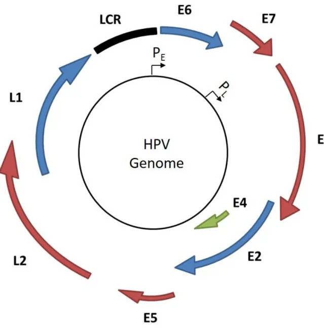
Figure 1.1 HPV viral genome organization. ... 3
Figure 1.2 The ATM dependent DNA damage signaling. ... 10
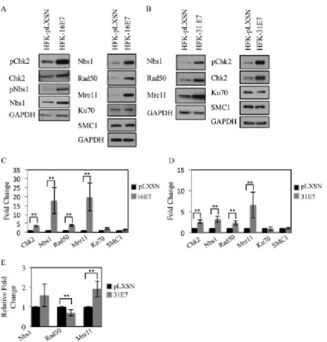
Figure 2.1. Expression of HPV E7 increases levels of proteins associated with detection and repair of DNA damage ... 42
Figure 2.2. HPV E7 interacts with Nbs1 independently of ATM. ... 44
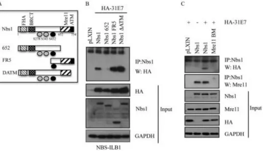
Figure 2.3. HPV 31 E7 interacts with Nbs1 through the Mre11 binding domain... 47
Figure 2.4. HPV E7 interacts with Nbs1 and Rad50, but not Mre11. ... 48
Figure 2.5. Nbs1 is not necessary for HPV31 genome maintenance. ... 50
Figure 2.6. Nbs1 is necessary for productive viral replication. ... 52
Figure 2.7. Levels of cell cycle or replication proteins are not affected by Nbs1 knockdown. ... 54
Figure 2.8. Nbs1 knockdown disrupts MRN complex formation. ... 56
Figure 2.9. Nbs1 knockdown results in decreased localization of Mre11, Rad50 and Rad51 to viral genomes. ... 59
Figure 2.10. Phosphorylated ATM and Chk2 levels are decreased variably in response to Nbs1 knockdown. ... 61
Figure 2.11. Phosphorylation of ATM and Chk2 is maintained with Nbs1 knockdown upon differentiation. ... 63
Figure 2.12. Levels of phosphorylated ATM and Chk2 do not influence viral genome amplification. ... 66
Figure 3.1. RRM2 protein and transcript levels are increased in HPV positive cells. ... 96
Figure 3.2. HPV31 positive cells exhibit elevated dNTP levels throughout the viral life cycle. ... 99
Figure 3.3. RRM2 is necessary for HPV31 replication. ... 101
Figure 3.5. E2F1 is required for the increased levels of RRM2 in HPV31
positive cells. ... 105 Figure 3.6. HPV31 increases RRM2 levels in a Chk1-dependent manner. ... 108 Figure 4.1 HPV E7 engages both the ATM and ATR DDR pathways in
order to recruit host factors and promote viral genome amplification. ... 134 Figure 4.2 DNA HR factors are recruited to HPV replication centers
LIST OF ABBREVIATIONS ATM Ataxia telangiectasia mutated
ATMIN ATM interacting
ATR Ataxia telangiectasia and Rad3 related CDC Cell division cycle
C/EBP CCAAT-enhancer-binding proteins Chk1 Checkpoint kinase 1
Chk2 Checkpoint kinase 2 CtIP CtBP interacting protein DDR DNA damage response DMSO Dimethyl sulfoxide DNA Deoxyribonucleic acid
DNA-PK DNA dependent protein kinase dNTP Deoxyribonucleotide triphosphates dNDP Deoxyribonucleotide diphosphates DSB Double strand DNA break
EBV Epstein-Barr virus
EGFR Epidermal growth factor receptor FBS Fetal bovine serum
HEK Human embryonic kidney HFK Human foreskin keratinocyte HPV Human papillomavirus HR Homologous repair
HU Hydroxyurea
IP Immunoprecipitation
KSHV Kaposi’s sarcoma-associated herpesvirus LCR Long control region
MRN Mre11-Nbs1-Rad50 mRNA Messenger RNA
NBS Nijmegen breakage syndrome NHEJ Non-homologous end joining ORF Open reading frame
PEI Polyethylenimine
PI3K Phosphoinositide 3-kinase
qPCR Quantitative Real time polymerase chain reaction Rb Retinoblastoma
RNA Ribonucleic acid
RNR Ribonucleotide reductase
RRM1 Ribonucleotide reductase subunit M1 RRM2 Ribonucleotide reductase subunit M2 SCF Skp, Cullin, F-box containing
SDS-PAGE Sodium dodecyl sulfate polyacrylamide gel electrophoresis shRNA Short hairpin RNA
SIRT1 Sirtuin 1
SMC Structural maintenance of chromosomes SV40 Simian Virus 40
CHAPTER 1: INTRODUCTION OVERVIEW
Human Papilloma Virus (HPV) is a double stranded non-enveloped DNA virus that infects epithelial cells known as keratinocytes. Specifically, alpha-HPVs target the mucosal epithelia of the anogenital tract and oropharynx. The range of HPVs can be further broken down into viral subtype with viral genome sequence and antibody recognition differing significantly between viral subtypes. Broadly they can be divided into two groups, high and low risk, based on their association with cancer. While low risk subtypes such as HPV 6 and 11 typically cause benign warts, the high risk subtypes, including HPV 16, 18, 31, and 45 are known to cause cervical, penile, anal, and head and neck cancers. Cervical cancer alone is the second leading cause of cancer deaths among women with 490,000 cases and 270,000 deaths worldwide each year (1). HPV is thought to be the most common sexually
transmitted viral infection, with an overall prevalence of 26% in US females aged 14 to 59 and a peak prevalence of over 44% in women ages 20-24 (2). Since among HPV subtypes, high risk HPVs pose the largest known threat to public health, my dissertation research specifically focused on these viruses.
INTRODUCTION
The HPV genome exists as a circular episome of 7.5 to 8kb in length depending on the specific viral subtype. The genome typically encodes eight
The early proteins are non-structural and are involved in a variety of functions ranging from viral genome replication to cell cycle control. E1 is the virally encoded helicase that also recruits host DNA replication factors to the viral origin of
Figure 1.1 HPV viral genome organization. Open reading frames are indicated with arrows and the long control region (LCR) is indicated by a black square. The early and late promoters are also indicated (PE and PL respectively).
oropharynx (12). While this specific tropism has been well described, the actual process of viral attachment and entry are not completely understood. Infection is known to require small epithelial abrasions known as microwounds (8). These micro wounds are tiny tears in the epithelia that allow virions to access the basement membrane. Once bound to the basement membrane the virion is able to come into contact with the basal layer of keratinocytes. Once in contact with the basal
keratinocyte, L1 capsid protein on the virion surface is thought to attach to heparin sulfate proteoglycans (HSPGs) on the cell surface (13). After the initial attachment event there is a conformational change in the capsid structure and the L2 protein is cleaved by host cell furin (14). At this point it is currently thought that the virion binds an unknown secondary cellular receptor which mediates viral entry into the cell. The mechanism by which the virus enters the cell is still unclear. A variety of entry
pathways have been reported, leading to the suggestion that the viral entry mechanism may be viral subtype dependent (15).
Establishment Phase: Upon Host cell entry the virus un-coats and unloads its genome into endosomes (12). Viral genomes are then released from the
proteins is mediated by the early promoter which is located directly upstream of the E6 coding region (17). The activity of the early promoter is controlled through the binding of positive and negative factors to the upstream long control region (LCR) (18-20). At this point the viral protein E2 is very important as it is the major viral transcription regulator, and regulation of viral transcription is thought to be at least partially responsible for controlling viral copy number in undifferentiated cells (21). Viral genome replication also requires E1 protein, the viral helicase (22). Acting together in complex, E1 and E2 are able to bind the viral origin of replication with high affinity (22, 23). However once the complex is bound to the viral origin of
replication, which is adjacent to the early promoter, E2 is released from the complex before DNA replication begins (22). E1 is then responsible for recruiting the cellular factors required for viral genome replication and unwinding the viral DNA (23).
degrading p53 (8). This activity prevents host cell apoptosis and inhibits cell cycle checkpoints. E7 expression during the maintenance phase is important because of its ability to promote destruction of retinoblastoma protein (Rb). The degradation of Rb deregulates the cell cycle and helps to push the cell towards a proliferative state by releasing E2F transcription factors which activate transcription of S phase genes important for DNA replication (8).
Productive phase: The virus remains in the maintenance phase while the host cell remains among the basal layer of keratinocytes. When an infected basal cell divides, one of the daughter cells remains in the actively dividing basal layer while the other begins to move upward, away from the basal layer and begins to differentiate. This activation of host cell differentiation activates the productive phase of the viral life cycle (8).
While a great deal about the link between host cell differentiation and triggering of the productive phase of HPV infection remains to be explained, some differentiation related triggers have been discovered. The viral protein E2 is
affect the late viral promoter, located downstream of the early promoter within the E7 coding region (17, 31). It has been suggested that upon differentiation the binding affinity of transcriptional repressors YY1 and Cux1 for the late promoter is decreased possibly allowing greater transcription (11). Finally, there is evidence that upon differentiation, a change in the balance of expression between transcription factor SP1 and its antagonist SP3 and the increase in transcription factor C/EBP activate the late promoter (11, 32, 33).
Aside from E2, other non-structural proteins have roles to play upon differentiation and induction of late viral events. E6 and E7 have been shown by Moody and Laimins to trigger caspase activation upon differentiation resulting in the cleavage of E1 (34). In the absence of this cleavage, a defect in viral genome replication was observed, suggesting caspase mediated cleavage activity was
necessary for successful viral amplification (34). Upon differentiation E7 continues to bind and degrade Rb family proteins, releasing E2F transcription factors. This
release of E2F factors pushes the differentiating cell back into the cell cycle and again triggers important S-phase genes necessary for DNA synthesis (19).
DNA DAMAGE REPAIR
Reliable maintenance of a stable and accurate genome is an essential function of every cell. In order to ensure faithful replication and maintenance of genomes several mechanisms exist to detect and repair damage to DNA. Depending on the type of damage that occurs, the cell has a variety of repair
mutated (ATM) and ataxia telangiectasia and Rad3-related (ATR) kinases which, along with the DNA-dependent protein kinase (DNA-PK) are members of the PI 3-Kinase related kinase family of kinases (35). Double strand DNA breaks (DSBs) can be repaired by either the high fidelity, homologous repair (HR) response regulated by ATM or the more error prone non-homologous end joining (NHEJ) response (35). Conversely damage resulting from replication stress and stalled DNA replication forks is handled by a pathway regulated through ATR (36, 37). HPV infection exploits both the ATM and ATR DDR pathways in order to successfully replicate its genome (38). It is thought that through these pathways, HPV maintains and recruits host cell factors necessary for viral genome replication, especially upon
differentiation. It is therefore important to describe these two major DDR pathways and how they are exploited by HPV infection.
ATM DNA damage repair pathway: The ATM DDR pathway is typically activated in response to DNA DSBs and primarily repairs DNA through HR. In this case, the DNA DSB is typically detected by the MRN complex which is composed of Mre11, Rad50, and Nbs1 (35). Rad50 is a member of the structural maintenance of chromosomes (SMC) family of proteins and interacts with the broken ends of DNA in the DSB (35). Mre11 facilitates MRN complex formation through binding both Rad50 and Nbs1. It also has endonuclease and exonuclease activities important for
preparing broken DNA ends for HR (39, 40). Nbs1, besides interacting with Mre11 also directly binds ATM (41, 42). Upon recognition of the DSB the MRN recruits ATM to the site of damage where it binds to Nbs1 and is activated via auto
pathway. The competing ATM interacting (ATMIN) protein has been shown to activate ATM in a MRN in a Nbs1 independent manner in response to hypotonic stress and inhibition of DNA synthesis by drugs like Hydroxyurea (HU) (46). Additionally, the DNA repair protein 53BP1 has been shown to be an activator of ATM, especially when levels of MRN are low (47, 48). Activated ATM then
coordinates repair of the DSB, phosphorylating a plethora of downstream targets including proteins involved in HR such as Nbs1, Brca1, and CtIP (49). Aside from recruiting ATM to the site of the DSB, the MRN complex stabilizes the break while MRN complex protein Mre11, along with ATM targets CtIP and Brca1 mediate resection of the broken ends to allow for recruitment of Rad51 mediated by Brca2 (50) reviewed in (35). The newly recruited Rad51 replaces phosphorylated RPA protein on the resected strands of broken DNA leading to strand invasion of the homologous template, formation of the D-loop, and ultimate repair through HR (51). Portions of this process are also dependent on ATR and its downstream effector Chk1. ATR, along with ATM, have been shown to phosphorylate RPA (48). Additionally, Chk1 has been shown to phosphorylate Rad51 (52). Both of these events are important for the formation of Rad51 filaments on the resected DNA ends and eventual stand invasion and repair of the DSB via HR (53).
recruitment of many repair factors including 53BP1, Nbs1, Brca1 and Rad51 (35) (3, 58, 59). This allows for the amplification of the repair response through the
recruitment of additional factors to the area of damage. Phosphorylation of Chk2 results in arrest in G2 phase through inactivation of CDC25 and its downstream effector CDK1 (60). This cell cycle arrest allows time for DNA repair before
continuation of the cell cycle. Activation of p53 also leads to cell cycle arrest and in the case of unrepaired damage, cell death through apoptosis (56).
Figure 1.2 The ATM dependent DNA damage signaling. A DNA double strand break (indicated by the lightning bolt) is detected by the MRN complex which then recruits ATM. ATM then phosphorylates several downstream effectors.
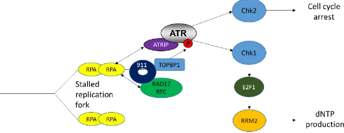
above, ATR is also known to be the central coordinator of DDR relating to replication stress and replication fork stalling (36, 37). In the case of replication stress large regions of ssDNA coated with RPA are formed due to the decoupling of the helicase and polymerase (35, 61). Polymerization of RPA1 on this ssDNA recruits ATR and its partner protein ATRIP through a direct interaction between RPA1 and ATRIP (61). In this way ATR is localized to the replication fork. At the same time the RPA-ssDNA complex also recruits the RAD17-RFC2-5 clamp loader, which in turn loads the 9-1-1 complex (RAD9-HUS1-RAD1) bound to the ATR activating TOPBP1 (36, 62-65) (reviewed in (35)). This cascade of events leads to the activation of ATR at the stressed replication fork. Activated ATR is then able to signal through its
downstream effectors Chk1 and Chk2. As discussed above Chk2 activation results in cell cycle delay. Activation of Chk1 by replication stress has recently been shown to result in high-level expression of genes related to DNA repair and nucleotide synthesis including the ribonucleotide reductase (RNR) small subunit M2 (RRM2) (66). Other studies have shown that ATR/Chk1 activation promotes RRM2
Figure 1.3 The ATR DNA damage response. ATR is recruited to sites of replication stress and stalled replication forks. ATR signaling through Chk1 can result in
increased RRM2 expression and nucleotide production to allow for DNA repair.
The ribonucleotide reductase enzyme: In proliferating cells, two RRM2 subunits together with two of the larger RRM1 subunits form the RNR tetramer (69). This enzyme responsible for the reduction of ribonucleotide diphosphates to
deoxyribonucleotide diphosphates (dNDPs) which are subsequently phosphorylated by the nucleoside diphosphate kinase to produce the balanced pools of dNTPs necessary for DNA synthesis. Outside of an actively dividing cell RRM2 can be replaced in the RNR enzyme by the p53 reactive subunit p53R2. This happens most often in G0/G1 phase when dNTP usage is typically limited and related to
S-phase (71-73). Outside of S-phase RRM2, is actively degraded through proteasome mediated degradation, facilitated by the anaphase promoting
complex/Cdh1 in G1 (74) and by SCFcyclinF in G2 (75). The increase in RRM2 levels
during S-phase coincides with an increase in RNR activity at the same time, suggesting that RRM2 is the rate limiting component of the RNR enzyme (76, 77). Confirming this, a loss of RRM2 results in decreased abundance of dNTPs available for DNA synthesis (78, 79). Appropriate RNR activity is important for maintenance of genomic integrity and cell viability. An increase or imbalance in dNTP levels can lead to mutations (80) while insufficient dNTP pools can impair DNA replication and repair (69).
HPV MAINIPULATION OF THE DNA DAMAGE RESPONSE
downstream effectors Chk2, and Brca1 in HPV positive cells. In infected cells HPV E7 forms a complex with phosphorylated ATM through its LXCXE Rb binding
domain. In addition, E7 is able to activate Chk2 phosphorylation, possibly through its interaction with ATM. This activation of ATM and Chk2 in HPV positive cells was sustained upon differentiation, where the downstream substrate Nbs1 was also found to be phosphorylated. While not necessary for viral genome maintenance in undifferentiated cells, ATM and Chk2 activation were both necessary for successful productive viral replication upon differentiation. Interestingly, Chk2 activation upon differentiation in these cells is necessary for the caspase 7 activation described above, suggesting one possible pathway connecting Chk2 activation with replication (84). Furthermore, as previously described, activation of Chk2 by ATM leads to a G2/M cell cycle arrest through the cytoplasmic sequestration of CDC25C which prevents activation of CDK1. HPV productive replication is believed to occur in a G2/M arrested state (87). Therefore, the requirement for ATM activation, specifically for productive replication suggests that HPV may be exploiting this pathway in order to hold the host cell in a state conducive for productive viral replication.
Another downstream effector of ATM thought to play a role in the HPV lifecycle is the histone variant H2AX. Phosphorylated or H2AX is found associated with HPV DNA throughout the viral life cycle and these H2AX foci expand upon productive viral replication (58). This association suggest that H2AX may play an important role in recruiting HR factors to sites of HPV genome replication.
Upregulation of SIRT1 has been observed in HPV positive cells; this upregulation has been shown to be important for the recruitment of Nbs1 and Rad51 to HPV genomes and necessary for productive viral replication (89, 90).
Some viruses, like adenovirus, actively antagonize the MRN complex and catalyze its destruction, preventing Nbs1 phosphorylation (91). In contrast, HPV infected cells maintain high levels of Mre11, Rad50, and Nbs1 both pre and post differentiation (85). This maintenance of MRN components and the phosphorylation of Nbs1 observed in differentiating infected cells suggest that Nbs1 may also be important in regulation viral replication. Further suggesting the importance of the MRN, recently our lab has shown that the HR factors Rad51 and Brca1 are necessary for productive viral replication (86). As explained above, the MRN complex plays an important role in the recruitment of Rad51 and Brca1 to DNA double strand breaks where they are necessary for the completion of HR.
were incorporated at these foci, indicating DNA synthesis and/or repair activity (3, 94).
The ATR DDR: While manipulation of the ATM DDR pathway by HPV has been studied for some time, less is known about HPV engagement of the ATR DDR pathway. However, recent studies have presented data suggesting that HPV
RATIONALE FOR DISSERTATION
Due to its small coding capacity and lifecycle being so tightly linked to host cell differentiation, HPV has evolved many ways of manipulating the host cell in order to recruit and regulate host factors necessary to replicate the viral genome. Recently, there has been a great deal of research into how HPV manipulates the host cell by targeting the ATM and ATR DDR pathways. In this dissertation I attempt to further our understanding of HPV manipulation of these pathways. Specific
proteins involved in these pathways targeted by the virus are identified along with the viral proteins responsible for the manipulation.
In the second chapter I focus on the ATM DDR and determine that Nbs1, a member of the MRN complex is necessary for productive HPV replication. However, despite the fact that Nbs1 is known to play an important role in ATM activation and ATM activity is also known to be necessary for productive viral replication, we found that depletion of Nbs1 had a minimal effect on ATM activation. Instead, our data suggests that Nbs1 is important for productive replication because it recruits HR factors such as Mre11 and Rad51 to sites of HPV replication. In fact, we show that Mre11 nuclease activity is also necessary for productive viral genome replication.
In the third chapter I examine the interaction of the ATR DDR and HPV, a considerably less explored topic. We propose a pathway in which RRM2 levels are upregulated though the release of E2F1 factors due to Rb degradation and
specifically targeting RRM2 expression through this pathway, in order to provide dNTPs for viral genome replication.
REFERENCES
1. WHO/ICO Information Centre on HPV and Cervical Cancer.. 2007. HPV and cervical cancer in the 2007 report, 2007/12/11 ed, vol 25 Suppl 3, p C1-230.
2. Dunne EF, Unger ER, Sternberg M, McQuillan G, Swan DC, Patel SS, Markowitz LE. 2007. Prevalence of HPV infection among females in the United States. JAMA 297:813-819.
3. Sakakibara N, Mitra R, McBride AA. 2011. The papillomavirus E1 helicase activates a cellular DNA damage response in viral replication foci. J Virol 85:8981-8995.
4. Gauson EJ, Donaldson MM, Dornan ES, Wang X, Bristol M, Bodily JM, Morgan IM. 2015. Evidence supporting a role for TopBP1 and Brd4 in the initiation but not continuation of human papillomavirus 16 E1/E2-mediated DNA replication. J Virol 89:4980-4991.
5. Mohr IJ, Clark R, Sun S, Androphy EJ, MacPherson P, Botchan MR. 1990. Targeting the E1 replication protein to the papillomavirus origin of replication by complex formation with the E2 transactivator. Science 250:1694-1699.
6. Yasugi T, Benson JD, Sakai H, Vidal M, Howley PM. 1997. Mapping and characterization of the interaction domains of human papillomavirus type 16 E1 and E2 proteins. J Virol 71:891-899.
7. Munger K, Werness BA, Dyson N, Phelps WC, Harlow E, Howley PM. 1989. Complex formation of human papillomavirus E7 proteins with the retinoblastoma tumor suppressor gene product. Embo j 8:4099-4105. 8. Moody CA, Laimins LA. 2010. Human papillomavirus oncoproteins:
pathways to transformation. Nat Rev Cancer 10:550-560.
9. Duensing S, Munger K. 2002. The human papillomavirus type 16 E6 and E7 oncoproteins independently induce numerical and structural chromosome instability. Cancer research 62:7075-7082.
10. Nasseri M, Hirochika R, Broker TR, Chow LT. 1987. A human papilloma virus type 11 transcript encoding an E1--E4 protein. Virology 159:433-439. 11. Kajitani N, Satsuka A, Kawate A, Sakai H. 2012. Productive Lifecycle of
Human Papillomaviruses that Depends Upon Squamous Epithelial Differentiation. Frontiers in Microbiology 3.
13. Sapp M, Day PM. 2009. Structure, attachment and entry of polyoma- and papillomaviruses. Virology 384:400-409.
14. Horvath CA, Boulet GA, Renoux VM, Delvenne PO, Bogers JP. 2010. Mechanisms of cell entry by human papillomaviruses: an overview. Virol J 7:11.
15. Letian T, Tianyu Z. 2010. Cellular receptor binding and entry of human papillomavirus. Virol J 7:2.
16. Kamper N, Day PM, Nowak T, Selinka HC, Florin L, Bolscher J, Hilbig L, Schiller JT, Sapp M. 2006. A membrane-destabilizing peptide in capsid protein L2 is required for egress of. J Virol 80:759-768.
17. Ozbun MA, Meyers C. 1998. Temporal usage of multiple promoters during the life cycle of human papillomavirus type 31b. J Virol 72:2715-2722. 18. Sichero L, Sobrinho JS, Villa LL. 2012. Identification of novel cellular
transcription factors that regulate early promoters of human papillomavirus types 18 and 16. J Infect Dis 206:867-874.
19. Carson A, Khan SA. 2006. Characterization of transcription factor binding to human papillomavirus type 16 DNA during cellular differentiation. J Virol 80:4356-4362.
20. Lambert PF. 1991. Papillomavirus DNA replication. J Virol 65:3417-3420. 21. Stubenrauch F, Lim HB, Laimins LA. 1998. Differential requirements for
conserved E2 binding sites in the life cycle of. J Virol 72:1071-1077.
22. McBride AA. 2008. Replication and partitioning of papillomavirus genomes. Adv Virus Res 72:155-205.
23. Bodily J, Laimins LA. 2011. Persistence of human papillomavirus infection: keys to malignant progression. Trends Microbiol 19:33-39.
24. Graham SV. 2010. Human papillomavirus: gene expression, regulation and prospects for novel diagnostic methods and antiviral therapies. Future Microbiol 5:1493-1506.
25. You J, Croyle JL, Nishimura A, Ozato K, Howley PM. 2004. Interaction of the bovine papillomavirus E2 protein with Brd4 tethers the viral. Cell 117:349-360.
27. Chiang CM, Dong G, Broker TR, Chow LT. 1992. Control of human papillomavirus type 11 origin of replication by the E2 family of transcription regulatory proteins. J Virol 66:5224-5231.
28. Ham J, Dostatni N, Gauthier JM, Yaniv M. 1991. The papillomavirus E2 protein: a factor with many talents. Trends Biochem Sci 16:440-444. 29. Steger G, Corbach S. 1997. Dose-dependent regulation of the early
promoter of human papillomavirus type 18. J Virol 71:50-58.
30. Hadaschik D, Hinterkeuser K, Oldak M, Pfister HJ, Smola-Hess S. 2003. The Papillomavirus E2 protein binds to and synergizes with C/EBP factors involved. J Virol 77:5253-5265.
31. Bodily JM, Meyers C. 2005. Genetic Analysis of the Human Papillomavirus Type 31 Differentiation-Dependent Late Promoter. Journal of Virology
79:3309-3321.
32. Kukimoto I, Takeuchi T, Kanda T. 2006. CCAAT/enhancer binding protein β binds to and activates the P670 promoter of human papillomavirus type 16. Virology 346:98-107.
33. Wooldridge TR, Laimins LA. 2008. Regulation of human papillomavirus type 31 gene expression during the. Virology 374:371-380.
34. Moody CA, Fradet-Turcotte A, Archambault J, Laimins LA. 2007. Human papillomaviruses activate caspases upon epithelial differentiation to. Proc Natl Acad Sci U S A 104:19541-19546.
35. Ciccia A, Elledge SJ. 2010. The DNA damage response: making it safe to play with knives. Mol Cell 40:179-204.
36. Cimprich KA, Cortez D. 2008. ATR: an essential regulator of genome integrity. Nat Rev Mol Cell Biol 9:616-627.
37. Zeman MK, Cimprich KA. 2014. Causes and consequences of replication stress. Nat Cell Biol 16:2-9.
38. McKinney CC, Hussmann KL, McBride AA. 2015. The Role of the DNA Damage Response throughout the Papillomavirus Life Cycle. Viruses 7:2450-2469.
40. Czornak K, Chughtai S, Chrzanowska KH. 2008. Mystery of DNA repair: the role of the MRN complex and ATM kinase in DNA damage repair. J Appl Genet 49:383-396.
41. Uziel T, Lerenthal Y, Moyal L, Andegeko Y, Mittelman L, Shiloh Y. 2003. Requirement of the MRN complex for ATM activation by DNA damage. Embo j 22:5612-5621.
42. Falck J, Coates J, Jackson SP. 2005. Conserved modes of recruitment of ATM, ATR and DNA-PKcs to sites of DNA damage. Nature 434:605-611. 43. Williams RS, Williams JS, Tainer JA. 2007. Mre11-Rad50-Nbs1 is a
keystone complex connecting DNA repair machinery, double-strand break signaling, and the chromatin template. Biochem Cell Biol 85:509-520. 44. Lee JH, Paull TT. 2005. ATM activation by DNA double-strand breaks
through the Mre11-Rad50-Nbs1 complex. Science 308:551-554.
45. Bakkenist CJ, Kastan MB. 2003. DNA damage activates ATM through intermolecular autophosphorylation and dimer dissociation. Nature 421:499-506.
46. Zhang T, Penicud K, Bruhn C, Loizou JI, Kanu N, Wang ZQ, Behrens A. 2012. Competition between Nbs1 and ATMIN controls ATM signaling
pathway choice. Cell Rep 2:1498-1504.
47. Mochan TA, Venere M, DiTullio RA, Jr., Halazonetis TD. 2003. 53BP1 and NFBD1/MDC1-Nbs1 function in parallel interacting pathways activating
ataxia-telangiectasia mutated (ATM) in response to DNA damage. Cancer Res 63:8586-8591.
48. Lee JH, Goodarzi AA, Jeggo PA, Paull TT. 2010. 53BP1 promotes ATM activity through direct interactions with the MRN complex. Embo j 29:574-585. 49. Matsuoka S, Ballif BA, Smogorzewska A, McDonald ER, 3rd, Hurov KE,
Luo J, Bakalarski CE, Zhao Z, Solimini N, Lerenthal Y, Shiloh Y, Gygi SP, Elledge SJ. 2007. ATM and ATR substrate analysis reveals extensive protein networks responsive to DNA damage. Science 316:1160-1166.
50. Sy SM, Huen MS, Chen J. 2009. PALB2 is an integral component of the BRCA complex required for homologous recombination repair. Proc Natl Acad Sci U S A 106:7155-7160.
52. Sorensen CS, Hansen LT, Dziegielewski J, Syljuasen RG, Lundin C, Bartek J, Helleday T. 2005. The cell-cycle checkpoint kinase Chk1 is required for mammalian homologous recombination repair. Nat Cell Biol 7:195-201.
53. West SC. 2003. Molecular views of recombination proteins and their control. Nat Rev Mol Cell Biol 4:435-445.
54. Bakkenist CJ, Kastan MB. 2015. Chromatin perturbations during the DNA damage response in higher eukaryotes. DNA Repair (Amst) 36:8-12.
55. Matsuoka S, Rotman G, Ogawa A, Shiloh Y, Tamai K, Elledge SJ. 2000. Ataxia telangiectasia-mutated phosphorylates Chk2 in vivo and in vitro. Proc Natl Acad Sci U S A 97:10389-10394.
56. Riley T, Sontag E, Chen P, Levine A. 2008. Transcriptional control of human p53-regulated genes. Nat Rev Mol Cell Biol 9:402-412.
57. Harper JW, Elledge SJ. 2007. The DNA damage response: ten years after. Mol Cell 28:739-745.
58. Gillespie KA, Mehta KP, Laimins LA, Moody CA. 2012. Human
papillomaviruses recruit cellular DNA repair and homologous recombination factors to viral replication centers. Journal of virology 86:9520-9526.
59. Fradet-Turcotte A, Bergeron-Labrecque F, Moody CA, Lehoux M, Laimins LA, Archambault J. 2011. Nuclear accumulation of the
papillomavirus E1 helicase blocks S-phase progression and triggers an ATM-dependent DNA damage response. Journal of virology 85:8996-9012.
60. Zhou BB, Elledge SJ. 2000. The DNA damage response: putting checkpoints in perspective. Nature 408:433-439.
61. Byun TS, Pacek M, Yee MC, Walter JC, Cimprich KA. 2005. Functional uncoupling of MCM helicase and DNA polymerase activities activates the ATR-dependent checkpoint. Genes Dev 19:1040-1052.
62. Zou L, Liu D, Elledge SJ. 2003. Replication protein A-mediated recruitment and activation of Rad17 complexes. Proc Natl Acad Sci U S A 100:13827-13832.
63. Ellison V, Stillman B. 2003. Biochemical characterization of DNA damage checkpoint complexes: clamp loader and clamp complexes with specificity for 5' recessed DNA. PLoS Biol 1:E33.
65. Mordes DA, Glick GG, Zhao R, Cortez D. 2008. TopBP1 activates ATR through ATRIP and a PIKK regulatory domain. Genes Dev 22:1478-1489. 66. Bertoli C, Klier S, McGowan C, Wittenberg C, de Bruin RA. 2013. Chk1
inhibits E2F6 repressor function in response to replication stress to maintain cell-cycle transcription. Curr Biol 23:1629-1637.
67. Zhang YW, Jones TL, Martin SE, Caplen NJ, Pommier Y. 2009. Implication of checkpoint kinase-dependent up-regulation of ribonucleotide reductase R2 in DNA damage response. J Biol Chem 284:18085-18095.
68. Buisson R, Boisvert JL, Benes CH, Zou L. 2015. Distinct but Concerted Roles of ATR, DNA-PK, and Chk1 in Countering Replication Stress during S Phase. Mol Cell 59:1011-1024.
69. Nordlund P, Reichard P. 2006. Ribonucleotide reductases. Annu Rev Biochem 75:681-706.
70. Tanaka H, Arakawa H, Yamaguchi T, Shiraishi K, Fukuda S, Matsui K, Takei Y, Nakamura Y. 2000. A ribonucleotide reductase gene involved in a p53-dependent cell-cycle checkpoint for DNA damage. Nature 404:42-49. 71. Chabes A, Thelander L. 2000. Controlled protein degradation regulates
ribonucleotide reductase activity in proliferating mammalian cells during the normal cell cycle and in response to DNA damage and replication blocks. J Biol Chem 275:17747-17753.
72. Mann GJ, Musgrove EA, Fox RM, Thelander L. 1988. Ribonucleotide reductase M1 subunit in cellular proliferation, quiescence, and differentiation. Cancer Res 48:5151-5156.
73. Bjorklund S, Skog S, Tribukait B, Thelander L. 1990. S-phase-specific expression of mammalian ribonucleotide reductase R1 and R2 subunit mRNAs. Biochemistry 29:5452-5458.
74. Chabes AL, Pfleger CM, Kirschner MW, Thelander L. 2003. Mouse ribonucleotide reductase R2 protein: a new target for anaphase-promoting complex-Cdh1-mediated proteolysis. Proc Natl Acad Sci U S A 100:3925-3929.
75. D'Angiolella V, Donato V, Forrester FM, Jeong YT, Pellacani C, Kudo Y, Saraf A, Florens L, Washburn MP, Pagano M. 2012. Cyclin F-mediated degradation of ribonucleotide reductase M2 controls genome integrity and DNA repair. Cell 149:1023-1034.
reductase. Differential regulation of the two subunits. J Biol Chem 260:9114-9116.
77. Eriksson S, Martin DW, Jr. 1981. Ribonucleotide reductase in cultured mouse lymphoma cells. Cell cycle-dependent variation in the activity of subunit protein M2. J Biol Chem 256:9436-9440.
78. Taricani L, Shanahan F, Malinao MC, Beaumont M, Parry D. 2014. A functional approach reveals a genetic and physical interaction between ribonucleotide reductase and Chk1 in mammalian cells. PLoS One 9:e111714.
79. Aird KM, Zhang G, Li H, Tu Z, Bitler BG, Garipov A, Wu H, Wei Z, Wagner SN, Herlyn M, Zhang R. 2013. Suppression of nucleotide metabolism
underlies the establishment and maintenance of oncogene-induced senescence. Cell Rep 3:1252-1265.
80. Hu CM, Chang ZF. 2007. Mitotic control of dTTP pool: a necessity or coincidence? J Biomed Sci 14:491-497.
81. Wilkinson DE, Weller SK. 2004. Recruitment of cellular recombination and repair proteins to sites of herpes simplex virus type 1 DNA replication is dependent on the composition of viral proteins within prereplicative sites and correlates with the induction of the DNA damage response. Journal of
virology 78:4783-4796.
82. Dheekollu J, Chen HS, Kaye KM, Lieberman PM. 2013. Timeless-dependent DNA replication-coupled recombination promotes Kaposi's Sarcoma-associated herpesvirus episome maintenance and terminal repeat stability. Journal of virology 87:3699-3709.
83. Dheekollu J, Deng Z, Wiedmer A, Weitzman MD, Lieberman PM. 2007. A role for Mre11, Nbs1, and recombination junctions in replication and stable maintenance of EBV episomes. PloS one 2:e1257.
84. Moody CA, Laimins LA. 2009. Human papillomaviruses activate the ATM DNA damage pathway for viral genome amplification upon differentiation. PLoS Pathog 5:e1000605.
85. Gillespie KA, Mehta KP, Laimins LA, Moody CA. 2012. Human
papillomaviruses recruit cellular DNA repair and homologous recombination. J Virol 86:9520-9526.
87. Banerjee NS, Wang HK, Broker TR, Chow LT. 2011. Human papillomavirus (HPV) E7 induces prolonged G2 following S phase reentry in differentiated human keratinocytes. J Biol Chem 286:15473-15482.
88. Oberdoerffer P, Michan S, McVay M, Mostoslavsky R, Vann J, Park SK, Hartlerode A, Stegmuller J, Hafner A, Loerch P, Wright SM, Mills KD, Bonni A, Yankner BA, Scully R, Prolla TA, Alt FW, Sinclair DA. 2008. SIRT1 redistribution on chromatin promotes genomic stability but alters gene expression during aging. Cell 135:907-918.
89. Langsfeld ES, Bodily JM, Laimins LA. 2015. The Deacetylase Sirtuin 1 Regulates Human Papillomavirus Replication by Modulating Histone Acetylation and Recruitment of DNA Damage Factors Nbs1 and Rad51 to Viral Genomes. PLoS Pathog 11:e1005181.
90. Allison SJ, Jiang M, Milner J. 2009. Oncogenic viral protein HPV E7 up-regulates the SIRT1 longevity protein in human cervical cancer cells. Aging 1:316-327.
91. Bridges RG, Sohn SY, Wright J, Leppard KN, Hearing P. 2016. The Adenovirus E4-ORF3 Protein Stimulates SUMOylation of General Transcription Factor TFII-I to Direct Proteasomal Degradation. MBio 7:e02184-02115.
92. Reinson T, Toots M, Kadaja M, Pipitch R, Allik M, Ustav E, Ustav M. 2013. Engagement of the ATR-dependent DNA damage response at the human papillomavirus 18 replication centers during the initial amplification. J Virol 87:951-964.
93. Sakakibara N, Chen D, McBride AA. 2013. Papillomaviruses use
recombination-dependent replication to vegetatively amplify their genomes in differentiated cells. PLoS pathogens 9:e1003321.
94. Swindle CS, Zou N, Van Tine BA, Shaw GM, Engler JA, Chow LT. 1999. Human papillomavirus DNA replication compartments in a transient DNA replication system. J Virol 73:1001-1009.
95. Hong S, Cheng S, Iovane A, Laimins LA. 2015. STAT-5 Regulates
Transcription of the Topoisomerase IIbeta-Binding Protein 1 (TopBP1) Gene To Activate the ATR Pathway and Promote Human Papillomavirus
Replication. MBio 6:e02006-02015.
96. Bester AC, Roniger M, Oren YS, Im MM, Sarni D, Chaoat M, Bensimon A, Zamir G, Shewach DS, Kerem B. 2011. Nucleotide deficiency promotes genomic instability in early stages of cancer development. Cell 145:435-446. 97. Spardy N, Duensing A, Hoskins EE, Wells SI, Duensing S. 2008. HPV-16
CHAPTER 2: PRODUCTIVE REPLICATION OF HUMAN PAPILOMAVIRUS 31 REQUIRES THE DNA REPAIR FACTOR NBS11
OVERVIEW
Activation of the ATM (Ataxia telangiectasia-mutated kinase)-dependent DNA damage response (DDR) is necessary for productive replication of Human
Papillomavirus 31 (HPV31). We previously found that DNA repair and homologous recombination (HR) factors localize to sites of HPV replication, suggesting ATM activity is required to recruit factors to viral genomes that can productively replicate viral DNA in a recombination-dependent manner. The Mre11-Rad50-Nbs1 (MRN) complex is an essential component of the DDR that is necessary for ATM-mediated HR repair, and localizes to HPV DNA foci. In this study, we demonstrate that the HPV E7 protein is sufficient to increase levels of the MRN complex, and also interacts with MRN components. We have found that Nbs1 depletion blocks productive viral replication, and results in decreased localization of Mre11, Rad50, and the principal HR factor Rad51 to HPV DNA foci upon differentiation. Nbs1 contributes to the DDR by acting as an upstream activator of ATM in response to double strand DNA breaks (DSB), and as a downstream effector of ATM activity in
1 This chapter previously appeared as an article in the Journal of Virology. The
the intra-S phase checkpoint. We have found that phosphorylation of ATM and its downstream target Chk2, as well as SMC1 (structural maintenance of chromosome 1) is maintained upon Nbs1 knockdown in differentiating cells. Given that ATM and Chk2 are required for productive replication, our results suggest that Nbs1
contributes to viral replication outside of its role as an ATM activator, potentially through ensuring localization of DNA repair factors to viral genomes that are necessary for efficient productive replication.
INTRODUCTION
Human papillomavirus are small double stranded DNA viruses that exhibit a strict tropism for epithelial cells (1). A subset of HPV types (termed high-risk) are the causative agents of cervical cancer, and are also associated with other genital malignancies, as well as an increasing number of head and neck cancers (2). The life cycle of HPV is dependent upon the differentiation of its host cell, the
keratinocyte. There are three phases of viral replication that characterize the viral life cycle (3). Upon infection of basal keratinocytes, the virus transiently amplifies to 50-100 episomal copies per cell. In undifferentiated cells, the virus is maintained at a low copy number by replicating once per cell cycle along with cellular DNA (4). In contrast, upon keratinocyte differentiation, the productive phase of the viral life cycle is activated, resulting in late gene expression, viral genome amplification to
some evidence indicating this occurs through a switch to rolling circle replication (9). Although it is well established that the viral E7 protein promotes S phase re-entry of differentiating cells to provide cellular factors necessary for productive replication (10), the mechanisms that regulate the switch to viral genome amplification in differentiating cells are not well understood.
Over the past several years, it has become evident that DNA and RNA
viruses facilitate replication by targeting the DNA damage response (11). Previously, we showed that high-risk HPV31 induces constitutive activation of an
ATM-dependent DNA damage response throughout the viral life cycle (12). ATM is a member of the phosphatidylinositol 3-kinase-like kinases (PIK) family of kinases, which along with ATR (Ataxia telangiectasia and Rad3-related protein) and DNA-PK, respond to certain types of DNA damage (13). ATM and DNA-PK are typically activated in response to double strand DNA breaks (DSB), while ATR is activated in response to single stranded DNA breaks, as well as replication stress. Our previous studies demonstrated that HPV31 requires ATM kinase activity for productive
replication upon differentiation, but not for episomal maintenance in undifferentiated cells (12). In HPV31 positive cells, the ATM response was characterized by
damage (14, 15). Recent studies by Hong and Laimins demonstrated that E7-induced STAT5 activation is necessary for ATM activation, possibly through PPAR expression (16). We, as well as others, have shown that expression of viral helicase E1 can also stimulate ATM activation (8, 17). Why HPV requires ATM activity for productive replication, as well as which ATM effectors contribute to viral DNA synthesis is not well understood.
In more recent studies, we demonstrated that multiple components of the ATM DNA damage response pathway localize to sites of HPV replication, including H2AX, Chk2, 53BP1 and components of the MRN complex (Mre11, Rad50, Nbs1) (18). In addition, we found that Rad51 and Brca1, two proteins necessary for the repair of DSBs through homologous recombination (HR) (19), are increased in expression in HPV positive cells and localize to viral DNA foci. The localization of cellular replication factors, PCNA and RPA32, to HPV DNA foci indicated these were sites of viral DNA synthesis. In addition, we observed that RPA32 is phosphorylated at sites of HPV replication, which is thought to redirect RPA’s function from DNA replication to repair synthesis, and has been linked to DSB resection (20-22). This suggests that HPV may utilize ATM signaling to recruit DNA repair machinery to viral genomes to promote replication through DNA repair. In support of this, we found that H2AX is bound to viral chromatin throughout the viral life cycle, with binding
increasing upon productive replication (18).
processing. HR is a high-fidelity repair mechanism that requires ATM activity and functions to rejoin DSBs and restart broken replication forks (19, 24). Replication-coupled recombination is thought to play role in the life cycle of several viruses, including Simian Virus 40 (SV40) (25), Herpes Simplex Virus type 1 (HSV-1) (26-28), Epstein Barr virus (EBV) (29, 30), and Kaposi’s Sarcoma Associated
Herpesvirus (KSHV) (31). The MRN complex is also essential to homology-directed repair (32). The MRN complex serves as a sensor of DNA damage that also controls the DDR through activation of ATM (33-35). ATM is recruited to sites of DSBs by directly binding Nbs1, where ATM activates the DNA damage checkpoint and regulates DNA repair by phosphorylating specific substrates (36-38). In addition to facilitating ATM activation and recruitment to DSBs, Nbs1 also acts downstream as an effector of ATM activity, and initiates HR with Mre11, a nuclease involved in resection of DNA ends (22, 39). The importance of Nbs1 is facilitating DNA repair is evident in patients with Nijmegen breakage syndrome (NBS), a disorder due to hypomorphic mutations in the Nbs1 gene, which is characterized by cellular
In this study, we investigate whether Nbs1 and maintenance of the MRN complex has an impact on the ability of HPV to efficiently replicate. We report here that HPV31 and HPV16 E7 bind to the MRN components Nbs1 and Rad50, but not Mre11. However, formation of the MRN complex is not disrupted, and rather is increased compared to uninfected keratinocytes. We have found that Nbs1 is required productive viral replication, but not episomal maintenance. Depletion of Nbs1 results in a loss of Mre11, Rad50 as well as Rad51 from sites of viral replication upon differentiation, suggesting that productive replication may occur through a mechanism dependent on recombination. Although phosphorylation of ATM and Chk2 are decreased in the absence of Nbs1, relatively high levels remain during the productive phase of the viral life cycle. Importantly, our results indicate that Nbs1 does not contribute to productive viral replication solely as an upstream regulator of ATM activity, but rather has functions downstream as well, with Nbs1 potentially acting as an effector of ATM activity and/or ensuring efficient viral DNA synthesis through a recombination-dependent mechanism.
MATERIALS AND METHODS
co-cultured with mitomycin C treated J2 3T3 fibroblasts, as described previously (45). The NBS-ILB1 fibroblast cell line was a generous gift from K. Cerosaletti, and was described previously (46). NBS-ILB1 cells were maintained in DMEM with
sodium pyruvate supplemented with 10% BGS. Generation and maintenance of cells stably expressing pLXSN or pLXSN-HPV31 E7, pLXSN-HPV16 E7 through retroviral transduction has been previously described (47). When necessary, J2 feeders were removed from HPV positive cells by incubating with Versene (PBS containing 1mM EDTA). U20S and 293T cells were grown in DMEM supplemented with 10% BGS.
Plasmids and Chemicals. The pBR322min-HPV31 plasmid has been described previously (48). The HA-tagged HPV31 E7 proteins were previously described (49) and are as follows: HA-E7 LHCYE contains an in-frame deletion of the Rb binding domain, and the HA-E7 L67R construct contains a point mutation in the HDAC binding site, converting a leucine to an arginine. The TAP-tagged HPV16 E7 construct was generously provided by J. Bodily, and was described previously (50). The retroviral plasmids pLXIN, pLXIN-Nbs1, and those expressing Nbs1 truncation mutants (Nbs1 652 and Nbs1 ATM) were kind gifts from P. Concannon, and were described previously (51, 52). The pLXIN-Nbs1 FR5 deletion construct was obtained from K. Cerosaletti and was described previously (52). The Mre11 binding mutant was also obtained from K. Cerosaletti, and was generated by site-directed mutagenesis (QuikChange, Agilent Technologies) based on the
NFKK684-687AAAA conversion. KU-55933 was obtained from Calbiochem, and MIRIN was obtained from TOCRIS.
Generation of HPV 31 positive HFKs. HFKs stably maintaining HPV31 episomes were created as previously described (45). Briefly, HPV31 genomes were excised from the pBR322 plasmid using HindIII (New England Biolabs) and religated using T4 DNA ligase (Life Technologies). Primary HFKs were transfected with 1ug of the ligated genomes and 1ug pSV2-Neo using FuGene 6 according to manufactures instructions (Promega). Stable cell lines were generated through neomycin selection (Sigma-Aldrich). After selection was complete, pooled populations were expanded for further analysis.
Induction of Keratinocyte Differentiation. For differentiation, 1.5%
methylcellulose was used as described previously (54). Cells were harvested at T0 (undifferentiated), as well as 24 and 48 hours post-suspension. High calcium medium was also used to induce differentiation as previously described (7). Cells were harvested at T0, as well as 48, 72 or 96hr post-exposure to high calcium. For both methylcellulose and calcium, at each time point DNA was harvested from one half of the cells, and protein was harvested from the other half. For every
Generation of Lentivirus. Lentivirus was produced as previously described (55). Plasmids encoding a Nbs1 shRNA (TRCN0000010393) or a scramble non-target control shRNA in the pKLO background were obtained from Open Biosystems (Pittsburg, PA). Each of these plasmids (5ug) was co-transfected with 1.6ug
vesicular stomatitis virus G plasmid DNA, and 3.37ug Gag-Pol-Tet-Rev plasmid DNA into 293T cells using polyethyleneimine (PEI) (VWR). Supernatants containing lentivirus were harvested three days posttransfection, filter sterilized, and stored at -80°C until use. CIN612 9E and HFK-31 cells were transduced with 5ml viral
supernatant consisting of Scramble or Nbs1 shRNA lentivirus particles in the
presence of 4.8ug/mL hexadimethrine bromide (Polybrene) (Sigma-Aldrich) for three days, followed by selection in puromycin to generate stable cell lines. Knockdown of Nbs1 was confirmed for each experiment by Western blot analysis.
Nuclear/Cytoplasmic Fractionation. Fractionation was carried out
of nuclear and cytoplasmic fractions was determined by Western blot analysis using antibodies to Lamin A/C (Genetex) and Tubulin (Sigma-Aldrich), respectively.
Western blot analysis/Immunoprecipitation. For Western blotting, whole cell lysates were harvested in RIPA lysis buffer supplemented with Complete Mini and PhosSTOP tablets (Roche). Total protein levels were determined via Bio-Rad protein assay. Western blot analysis was performed as described previously (49). Equal protein amounts were electrophoresed on SDS-polyacrylamide gels and transferred to polyvinylidene difluoride membranes (Immobilon-P; Millipore). For immunoprecipitations, cells were harvested in 1X cell lysis buffer (Cell Signaling), as described previously (12). The following primary antibodies were used: anti-mouse HA, rabbit HA, goat Nbs1, mouse involucrin, mouse Ku70, mouse CDC25C, mouse Cyclin A, rabbit Cyclin B, rabbit RPA32, anti-mouse GAPDH (Santa Cruz); anti-anti-mouse Cyclin E (Pharmingen); anti-anti-mouse HPV16 E7 (Life Technologies); anti-rabbit Nbs1, phospho-Nbs1 S343 (Novus Biologicals); anti-rabbit phospho-ATM S1981 and anti-mouse CDK2, anti-rabbit PCNA (Abcam); anti-rabbit phospho-Chk2 Thr68 and Chk2, anti-rabbit CDK1, and anti-rabbit SMC1 (Cell Signaling Technologies); anti-rabbit ATM, anti-rabbit
Southern Blot analysis. DNA isolation and Southern blotting were performed as previously described (54). Briefly, cells were harvested in buffer containing 400mM NaCl, 10mM Tris pH 7.5 and 10mM EDTA. Cells were lysed by the addition of 30uL 20% SDS and subsequently treated with 15ul of 10mg/mL proteinase K overnight at 37˚C. DNA was then extracted by phenol chloroform and precipitated using sodium acetate and ethanol. Resultant DNAs were digested with BamHI (which does not cut the HPV31 genome) or HindIII (which cuts the HPV31 genome once). DNAs were resolved on a 0.8% agarose gel and transferred to a positively charged nylon membrane (Genescreen plus; Perkin Elmer). Hybridization was performed using 32P-labeled HPV31 genome as a probe.
cold methanol:acetic acid (3:1) at -20C for 10 minutes followed by fixation with 4% PFA for 10 minutes at room temperature. Cover slips were then analyzed for HPV DNA by FISH using Tyramide-enhanced fluorescence (Invitrogen) as previously described(12, 57). The coverslips were mounted using Vectashield containing DAPI to counterstain the cellular DNA. Images were captured using Zeiss CLSM 710 spectral confocal laser scanning microscope. For IF, the following antibodies were used: mouse monoclonal Mre11 (1:200; GeneTex), mouse monoclonal anti-Rad50 (1:200; GeneTex), and rabbit polyclonal Rad51 (1:200; Santa Cruz). For the secondary antibody, Alexa Fluor® 568 Goat Anti-Mouse was used (Life
Technologies). IF/FISH was carried out for each repair factor on three independent experiments. The number of foci positive for both HPV DNA and each repair factor was quantified, with 25 to 40 FISH-positive cells being counted for each experiment.
gene specific primer sequences were used: Mre11 (Forward 5’-
GCCTTCCCGAAATGTCACTA -3’; Reverse 5’- TTCAAAATCAACCCCTTTCG -3’), Rad50 (Forward 5’- GGAAGAGCAGTTGTCCAGTTACG -3’; Reverse 5’-
GAGTAAACTGCTGTGGCTCCAG -3’), Nbs1 (Forward 5’- CACCTCCAAAGACAACTGCGGA -3’; Reverse 5’-
TCTGTCAGGACGGCAGGAAAGA -3’), GAPDH (Forward 3’-
CTGTTGCTGTAGCCAAATTCGT -5’; Reverse 3’- ACCCACTCCACCTTTGAC -5’). Relative transcript amounts were calculated using the CT method using GAPDH as a reference gene.
RESULTS
Expression of high-risk HPV E7 increases levels of repair factors, including the MRN complex. Previously we demonstrated that HPV31 E7 is
sufficient to induce phosphorylation of the ATM target Chk2 (12). To determine if this observation extends to HPV16 E7, we examined Chk2 phosphorylation in human foreskin keratinocytes (HFK) stably expressing pLXSN-HPV16 E7 or vector control. As shown in Figure 2.1A, we found the expression of HPV16 E7, similarly to HPV31 E7 (Figure 2.1B), is sufficient to induce Chk2, as well as Nbs1 phosphorylation, and also significantly increase total levels of Chk2 (Figure 2.1C, D). Our previous
studies also revealed that the MRN components Mre11, Rad50 and Nbs1 are expressed at increased levels in HPV positive cells compared to uninfected
Figure 2.1. Expression of HPV E7 increases levels of proteins associated with detection and repair of DNA damage. (A) Whole cell extracts were harvested from HFKs stably expressing pLXSN vector control or pLXSN-HPV16 E7. Immunoblotting was performed using antibodies to phosphorylated Chk2 (Thr68) (pChk2), total Chk2, phosphorylated Nbs1 S343 (pNbs1), total Nbs1, Rad50, Ku70 and SMC1. (B) Lysates harvested from HFK-pLXSN or HFK-pLXSN HPV31 E7 cells were analyzed by Western blot analysis using antibodies to pChk2, Chk2, Nbs1, Rad50, Mre11, Ku70 and SMC1. GAPDH served as a loading control. (C, D) Bar graphs
assayed by 2-tailed t-test. Data=mean +/- standard error. ** indicates p-value less than 0.05. (E) Quantitative real-time (qRT) PCR of gene expression analysis in pLXSN and pLXSN-31E7 lines. Expression levels are shown relative to HFK-pLXSN cells and were calculated using GAPDH serving as a reference gene. Shown is the relative fold change in gene expression over three independent experiments. The statistical analysis was assayed by 2-tailed t-test. Data= mean +/- standard error. ** indicates p-value less than 0.05.
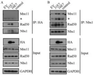
E7 binds to the MRN components Nbs1 and Rad50. In previous studies, we found that HPV31 E7 binds preferentially to the phosphorylated form of ATM (12), potentially due to the exposure of the E7 binding site upon dissociation of inactive ATM dimers to active monomers. Since Nbs1 binds ATM for recruitment to DSBs, we wanted to determine if E7 also binds Nbs1. For this, we
In order to map the Nbs1 interaction domain on HPV31 E7, we used previously characterized mutants that block either the binding of Rb (LHCYE) or histone deacetylases (HDACs) (L67R) (Figure 2.2C) (49). As shown in Figure 2.2D, both the E7 Rb binding mutant and the HDAC binding mutant were deficient in immunoprecipitating Nbs1. Since we previously found ATM also binds E7 through these two domains (12), we wanted to next determine if E7 interacts with Nbs1 indirectly through its binding to ATM. For this, we treated cells with the small molecule inhibitor of ATM, KU-55933, which inhibits ATM phosphorylation and ablates the ability of E7 to bind ATM (Figure 2.2E) (12). However, we found that Nbs1 was still able to immunoprecipitate E7, even when the E7/ATM interaction was disrupted (Figure 2.2E), indicating that E7 binds Nbs1 independently of ATM.
Figure 2.2. HPV E7 interacts with Nbs1 independently of ATM. (A) Whole cell lysates of U20S cells transiently transfected with HA-HPV31 E7 or empty vector (EV) were immunoprecipitated with antibodies to HA or Nbs1, followed by
transiently transfected with TAP-HPV16 E7 or empty vector (EV) were
immunoprecipitated using an anti-Nbs1 antibody, followed by immunoblotting with an HPV16 E7 antibody. Inputs were analyzed using antibodies to (A) Nbs1 and HA, and (B) Nbs1 and HPV16E7. (C) Structure of E7 and mutations examined in this study. Indicated are cellular targets that have been shown to interact with the Rb (LHCYE) and HDAC (L67) binding domains(97). (D) Whole cell lysates of U20S cells
transiently transfected with EV alone, HA-HPV31 E7, HA-31 E7 LHCYE, or HA-31 E7 L67R were immunoprecipitated using an anti-Nbs1 antibody and subsequently immunoblotted using an anti-HA or anti-Nbs1 antibody. Input lysates were analyzed by Western blot analysis using antibodies to HA and Nbs1. (E) Whole cell lysates of U20S cells expressing EV alone, or HA-HPV31 E7 in the presence or absence of 10uM the ATM inhibitor KU-55933 were immunoprecipitated using an anti-Nbs1 antibody and immunoblotted using an anti-HA antibody (left panel). Input lysates were immunoblotted with antibodies to Nbs1, HA, as well as phosphorylated ATM (Ser1981) and total ATM to demonstrate ATM inhibition. Lysates were also
subjected to immunoprecipitation with an antibody to HA, followed by
immunoblotting to ATM and HA. Inputs were analyzed using an antibody to HA, as well as phosphorylated and total ATM to demonstrate ATM inhibition. Ab=antibody. IP=immunoprecipitation. W=immunoblotting. ATMi=KU-55933. All results are representative of observations of three or more independent experiments.
Figure 2.3. HPV 31 E7 interacts with Nbs1 through the Mre11 binding domain. (A) Schematic of Nbs1 constructs depicting relevant binding domains and
phosphorylation sites. (B) NBS-ILB1 cells stably expressing pLXIN vector alone, or the indicted Nbs1 mutants were transiently transfected with HA-HPV31 E7.
Immunoprecipitations were performed using an antibody to Nbs1, followed by
To form the MRN complex, Nbs1 binds Mre11, which binds to Rad50 (40, 62). Since we were able to immunoprecipitate Nbs1 with E7, we next wanted to
determine if E7 interacts with other components of the MRN complex. For this, we immunoprecipitated HA-tagged HPV31 E7 or TAP-tagged HPV16 E7 from lysates harvested from U20S cells, and performed Western blot analysis for Mre11 and Rad50. Interestingly, as shown in Figure 2.4A, we found that both HPV31 E7 and HPV16 E7 could immunoprecipitate with Rad50, but neither interacted with Mre11. However, we found that the presence of E7 did not disrupt formation of the MRN complex, as Nbs1 was still able to interact with Rad50 and Mre11 (Figure 2.4B). Rather, our results indicate that the presence of E7 increases the formation of the MRN complex, which may result from increased Mre11 expression. We also found that the formation of the MRN complex is not disrupted in HPV infected cells (Figure 2.8B) or cells stably expressing HPV31 E7 (data not shown).
Figure 2.4. HPV E7 interacts with Nbs1 and Rad50, but not Mre11. (A) U20S cells were transiently transfected with vector alone (EV), HA-HPV31 E7,
HA antibody, followed by immunoblotting with Mre11, Rad50, and Nbs1 antibodies. (B) U20S cells were transiently transfected with EV alone, HA-HPV31 E7 or TAP-HPV16 E7. Immunoprecipitations were performed on lysates using an antibody to Nbs1, followed by Western blot analysis using antibodies to Mre11, Rad50, and Nbs1 antibodies. For (A) and (B), input lysates were analyzed by Western blot analysis using antibodies to HA, Mre11, Rad50 and Nbs1. GAPDH served as a loading control. IP=immunoprecipitation. * = antibody heavy chain. All results are representative of observations of three independent experiments.
Nbs1 is necessary for productive viral replication. Previously, we
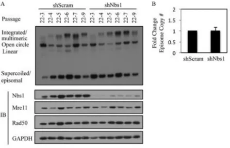
demonstrated that inhibition of ATM kinase activity has minimal effect on the ability of HPV to be maintained as an episome (12). However, in addition to being essential for ATM activation in response to DSBs, Nbs1 can also mediate ATR activation (63, 64), which could potentially be important for episomal maintenance. To examine the effect of Nbs1 depletion on episomal maintenance, we transduced HPV31 positive CIN612 9E cells with a scramble control shRNA or a previously validated Nbs1 shRNA (65), and generated stable cell lines. The cells were routinely passaged, and DNA and protein were harvested at every passage. Southern blot analysis was performed to examine the status of the episomal viral DNA. As shown in Figure 2.5 and Figure 2.12A, in two independently derived CIN612 9E lines, there was no significant difference in the ability of viral episomes to be maintained across
indicate that Nbs1 is not necessary for episomal maintenance. In addition, we observed no effect of Nbs1 knockdown on Chk1 phosphorylation (data not shown).
Figure 2.5. Nbs1 is not necessary for HPV31 genome maintenance. (A) DNA was isolated at the indicated passages from CIN612 9E cells stably maintaining a scramble shRNA (shScram) or Nbs1 shRNA (shNbs1) and analyzed by Southern blot analysis. Passage 22 (p22) represents the passage at which the CIN612 9E cells were transduced with the respective shRNAs. Each passage following transduction is represented by p22-#. Western blot analysis was performed on lysates harvested at each passage using an antibody to Nbs1 to demonstrate knockdown, as well as antibodies to Mre11 and Rad50. GAPDH served as a
loading control. IB=immunoblotting. (B) Bar graph represents episome copy number quantified and averaged across the passages of Scramble shRNA (set at 1)
performed using ImageJ software. The statistical analysis was assayed by 2-tailed t-test. Data=mean +/- standard error.
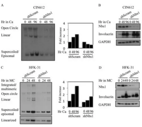
Since we previously found that ATM activity is necessary for productive viral replication, and given that Nbs1 is necessary for ATM activation in response to DSBs (32, 35, 36), we next wanted to determine the effect of Nbs1 depletion on productive replication. For this, CIN612 9E cells stably maintaining the scramble or Nbs1 shRNAs were induced to differentiate in high calcium medium, which activates the productive phase of the viral life cycle by 48 hours post-exposure (66). As shown in Figure 2.6A, Southern blot analysis for HPV DNA demonstrated that cells
CDK (CDK1), and the CDC25C phosphatase (Figure 2.7B). In addition, both RPA and PCNA were maintained at similar levels in the Nbs1 shRNA cells compared to the scramble control (Figure 2.7C). These results suggest that the block in
productive viral replication observed in response to Nbs1 knockdown was not due to alterations in cell cycle control or the lack of cellular factors directly required for viral replication. We also observed similar effects on productive viral replication following transient knockdown of Nbs1 for three days (data not shown). Overall, these results indicate that Nbs1 is specifically necessary for differentiation-dependent viral
replication.
medium. Southern blot analysis was performed to analyze viral genome
amplification. The bar graph represents quantification of the episome copy number present at each time point, relative to T0 shScramble, which was set to 1.
Figure 2.7. Levels of cell cycle or replication proteins are not affected by Nbs1 knockdown. Western blot analysis was performed on lysates harvested from
CIN612 9E cells stably maintaining the scramble (shScram) or Nbs1 shRNAs at T0, or after 72 hours of differentiation in high calcium medium using antibodies to (A) Cyclin A, Cyclin E, and CDK2; (B) Cyclin B, CDC25C, and CDK1; and (C) RPA, as well as PCNA. GAPDH served as a loading control. All results are representative observations of at least three independent experiments. Ca=calcium.
The MRN complex is disrupted upon Nbs1 knockdown. As Nbs1 functions in a complex with Mre11 and Rad50, we next wanted to determine if Nbs1 depletion affected the levels of Mre11 and Rad50. As shown in Figures 5A and 8A, Nbs1 knockdown had little effect on the levels of Mre11 or Rad50, as measured by