Published online 28 March 2017 Nucleic Acids Research, 2017, Vol. 45, No. 9 5333–5348
doi: 10.1093/nar/gkx214
Substrate preference of Gen endonucleases
highlights the importance of branched structures as
DNA damage repair intermediates
Stephanie P. Bellendir
1,†, Danielle J. Rognstad
1,†, Lydia P. Morris
2, Grzegorz Zapotoczny
1,
William G. Walton
3, Matthew R. Redinbo
3,4, Dale A. Ramsden
1,2,5, Jeff Sekelsky
1,2,4,6,*and
Dorothy A. Erie
3,4,*1Curriculum in Genetics and Molecular Biology, Chapel Hill, NC 27599, USA,2Lineberger Comprehensive Cancer
Center, Chapel Hill, NC 27599, USA,3Department of Chemistry, Chapel Hill, NC 27599, USA,4Integrative Program for Biological and Genome Sciences, Chapel Hill, NC 27599, USA,5Department of Biochemistry and Biophysics, Chapel Hill, NC 27599, USA and6Department of Biology, University of North Carolina at Chapel Hill, Chapel Hill, NC 27599, USA
Received August 22, 2016; Revised February 16, 2017; Editorial Decision March 20, 2017; Accepted March 21, 2017
ABSTRACT
Human GEN1 and yeast Yen1 are endonucleases with the ability to cleave Holliday junctions (HJs), which are proposed intermediates in recombination. In vivo, GEN1 and Yen1 function secondarily to Mus81, which has weak activity on intact HJs. We show that the genetic relationship is reversed in Drosophila, with Genmutants having more severe defects than
mus81 mutants.In vitro, DmGen, likeHsGEN1, effi-ciently cleaves HJs, 5flaps, splayed arms, and repli-cation fork structures. We find that the cleavage rates for 5flaps are significantly higher than those for HJs for both DmGen andHsGEN1, even in vast excess of enzyme over substrate. Kinetic studies suggest that the difference in cleavage rates results from a slow, rate-limiting conformational change prior to HJ cleavage: formation of a productive dimer on the HJ. Despite the stark difference in vivo that Drosophila
uses Gen over Mus81 and humans use MUS81 over GEN1, we find the in vitro activities ofDmGen and
HsGEN1 to be strikingly similar. These findings sug-gest that simpler branched structures may be more important substrates for Gen orthologs in vivo, and highlight the utility of using the Drosophila model system to further understand these enzymes.
INTRODUCTION
Recombination is an integral process for DNA damage re-pair as well as for horizontal gene transfer during
conjuga-tion or transducconjuga-tion (1). Robin Holliday proposed a molec-ular model for recombination that included a four-stranded intermediate linking two DNA helices (2). Support for the existence of this intermediate, now termed a Holliday junc-tion (HJ), came from electron microscopy of phage lambda DNA undergoing recombination inEscherichia coli(3).In vitro synthetic HJ cleavage assays using prokaryotic en-zymes, like RuvC, provided the first direct evidence for the existence of structure-selective endonucleases (SSEs) capa-ble of HJ cleavage, termed HJ resolvases (4). Prokaryotic re-solvases show preference for HJs over other DNA branched structures and cleave symmetrically about the HJ axis, cre-ating duplex DNA products with a nick that can be ligated without additional processing (5). Their in vivoresolvase function was evident in that mutations in the genes encod-ing these enzymes led to reduced recombination and sen-sitivity to DNA damaging agents (6). These properties be-came the benchmark for defining canonical resolvases.
Thein vivoand in vitroevidence for resolvase function in bacteria led to the search for similar activities and genes in eukaryotes. Electron microscopy of yeast recombination intermediates provided visual evidence that eukaryotic re-combination can involve Holliday junction intermediates (7). Extensivein vitrostudies of HJ cleavage activity identi-fied Yen1 in budding yeast and GEN1 in human cells (8,9), as well as the MUS81–EME1–SLX1–SLX4 complex (here-after called MUS–SLX complex) in humans and mice (10– 12).HsGEN1 dimerizes on HJs to coordinate symmetrical cleavage across the HJ, whereas MUS–SLX resolves HJs through the coordination of a nick by SLX1 with a counter-nick by MUS81–EME1.
*To whom correspondence should be addressed. Tel: +1 919 843 9400; Fax: +1 919 962 4574 Email: [email protected]
Correspondence may also be addressed to Dorothy A. Erie. Tel: +1 919 962 6370; Email: [email protected]
†These authors contributed equally to this work as first authors.
C
The Author(s) 2017. Published by Oxford University Press on behalf of Nucleic Acids Research.
Although GEN1 and the MUS–SLX complex display canonical resolvase activity similar to that of prokaryotic resolvases, their biochemical properties differ from those of prokaryotic resolvases in that the eukaryotic enzymes are not obligate homodimers and they cleave other branched DNA structures such as flaps, replication forks, and nicked HJs (8–12). Furthermore, mutations in the genes encoding these enzymes do not cause the same recombination defects and DNA damage sensitivities that occur when bacterial resolvases are knocked out. Loss of Mus81 results in hy-persensitivity to a broad range of DNA damaging agents (13–15), but hypersensitivities resulting from loss of Slx1 are weaker to a non-overlapping set of damaging agents (10,16). These findings suggest that many of Mus81’s re-pair functions lie outside of a MUS–SLX complex. GEN1 resolvase is genetically less complicated because it only in-volves one gene. However, mutations inS. cerevisiae YEN1
or murine Gen1 do not cause any apparent DNA repair defects on their own, but they do increase the severity of
mus81 mutant phenotypes in double mutants, suggesting that Yen1/Gen1 act primarily as backups to Mus81 (17– 20). Consequently, one of the primary challenges to char-acterizing Yen1/GEN1 has been the need to study the null effect in the background of null mutations in other endonu-cleases.
Drosophilaprovides a unique platform for understanding functions of Yen1/GEN1 because the hierarchical relation-ship between DmGen and Mus81 appears to be reversed. Unlike in other organisms,Drosophila mus81mutants are hypersensitive to only a few DNA damaging agents, and then only mildly (21). Furthermore, in the absence of the DNA repair helicase Blm, loss of DmGen causes a much more severe phenotype (death early in larval development) than loss of Mus81 (death late in pupal development) (22). Here, we confirm thatDmGen is important in DNA dam-age repair by showing that, unlike in yeast and mammalian cells,Gensingle mutants are severely hypersensitive to sev-eral different DNA damaging agents. We show that, like its human ortholog, DmGen efficiently cleaves HJs, 5 flaps, splayed arms, and replication fork structures. We find that the cleavage rate for 5flaps is significantly higher than the cleavage rate for HJs. Kinetic studies suggest that the dif-ference in cleavage rates results from a slow, rate-limiting conformational change prior to HJ cleavage: formation of a productive dimer on the HJ. We comparedDmGen to hu-man GEN1 in side-by-side experiments. While slight dif-ferences such as the propensity to dimerize do exist be-tweenDmGen andHsGEN1, we find the activities of the orthologs to be strikingly similar, including the higher cleav-age rate on flaps than on HJs. These findings suggest that simpler branched structures may be more important sub-strates for Gen orthologsin vivo, and they highlight the util-ity of using theDrosophilamodel system to further under-stand this class of enzymes.
MATERIALS AND METHODS
Drosophilastocks and genetics
All stocks were maintained at 25◦C on standard media. The following null mutations were described previously:
mus81NheI (21) and GenZ5997 (22), which was made
hem-izygous with Df(3L)6103. Sensitivity to DNA damaging agents was done as described previously (23). For nitrogen mustard (HN2), hydroxyurea (HU), and methylmethane sulfonate (MMS), 250l water containing the agent at the indicated concentration were added to each vial contain-ing feedcontain-ing larvae. Camptothecin (CPT) was dissolved in DMSO and diluted in 10% ethanol and 0.2% Tween; con-trol larvae were treated with DMSO only. For IR, vials with third instar larvae were irradiated with 20 Grays from a
137Cs source (GammaCell GG10). Progeny were scored for
5 days after eclosion began. Relative survival was calculated as the ratio of mutant to control flies per vial and was nor-malized to the ratio in untreated vials. Statistical analyses were done using Prism (GraphPad, San Diego, CA, USA).
Purification of full-length and truncated Gen (1–518) fromE. coli
DmGen cDNA was codon-optimized by GenScript. Full-length DmGen (1–726 aa) and truncated DmGen (1-518 aa) were cloned into the NdeI and XhoI sites of pET21b (Novagen, Madison, WI), which carries a C-terminal hex-ahistidine tag (His). The nuclease-dead mutations E143A and E145A, previously described by (24), were made by QuikChange site-directed mutagenesis (Agilent Technolo-gies, Santa Clara, CA, USA).DmGen-His was expressed in RDK cells (gift of Dr. Steve Matson) with 0.4 mM IPTG, andDmGen (1–518)-His was expressed in Rosetta II pLysS (Novagen) with 1.0 mM IPTG. All proteins were expressed at 18◦C for 18 h. TheDmGen (1–518)-His and
DmGen (1–518)Dead-His pellets were lysed in NiA buffer (20
mM KH2PO4 pH 7.0, 100 mM ammonium acetate, 1 mM
TCEP, 0.02% sodium azide, 500 mM NaCl, 50 mM imi-dazole), sonicated, pelleted, and the clarified supernatant was loaded onto a 5 ml HisTrap HP column (GE Health-care Life Sciences, Pittsburgh, PA) and eluted with NiB (20 mM KH2PO4 pH 7.0, 100 mM ammonium acetate, 1 mM
TCEP, 0.02% sodium azide, 500 mM NaCl, 500 mM imida-zole). Peak fractions were diluted in NiA minus salt to 50 mM NaCl and loaded onto a 6 ml Resource S column (GE Healthcare Life Sciences) pre-equilibrated with MonoSA (20 mM KH2PO4 pH 7.0, 100 mM ammonium acetate, 1
mM TCEP, 0.02% sodium azide, 50 mM NaCl) and gra-dient eluted with MonoSB (20 mM KH2PO4 pH 7.0, 100
mM ammonium acetate, 1 mM TCEP, 0.02% sodium azide, 1 M NaCl). Peak fractions were concentrated to 5 ml and loaded onto a Superdex S200 column (GE Healthcare Life Sciences) and eluted with S200 buffer (50 mM HEPES pH 7.0, 400 mM NaCl, 100 mM ammonium acetate, 1 mM TCEP, 0.02% sodium azide). Full-lengthDmGen-His andDmGenDead-His were purified over HisTrap and S200
columns. Following elution from the S200 column, purity was assessed by dynamic light scattering and SDS-PAGE.
Nucleic Acids Research, 2017, Vol. 45, No. 9 5335
Technology Corporation). The Astra V software package (Wyatt Technology Corporation) was used to determine the molar mass of the sample.
Nuclease assays
Synthetic DNA substrates were prepared by anneal-ing oligonucleotides, shown in Supplementary Table S1. Oligonucleotides (oligos) 888, 891, 892, 893, 894, 895, 897, 992 were described previously (25); 940, 994, 888+10, 990+10 were modified from these. These oligos were used to form the majority of substrates used in this study, termed ‘Bellendir substrates’. Oligos 1, 2, 3, 4 and 7 were described previously (26) and were used to form the ‘Rass substrates’ used in Figure 5. All substrates were prepared as previ-ously described (27). Briefly, one oligonucleotide was 5 end-labeled using T4 polynucleotide kinase and␥-32P ATP.
Sub-strates were annealed in annealing buffer (50 mM Tris pH 7.5, 10 mM MgCl2, 50 mM NaCl, 5 mM DTT),
PAGE-purified, and quantified byA260.
For nuclease assays, tr-DmGen-His (1–518-His) or
tr-HsGEN1-His (1–527) (gift of Dr. Steve West) was incu-bated with the32P-labeled structures in a 10l reaction mix-ture containing 50 mM Tris pH 8, 100g/ml BSA, 1 mM DTT, 10% glycerol, 50 mM KCl, and 5 mM MgCl2
(Bel-lendir buffer––used in all kinetic experiments unless other-wise stated) or 60 mM sodium phosphate pH 7.4, 100g/ml BSA, 1 mM DTT, and 5 mM Mg(OAc)2 (Rass buffer) at
either room temperature for reactions containing DmGen or 37◦C for reactions containingHsGEN1. For fixed end-point assays, unless otherwise indicated, 20 nM protein was incubated with 1 nM substrate. For ligation of products, 1 U of T4 DNA Ligase (NEB) was incubated at 22◦C for 30 min. The reaction was stopped by adding an equal vol-ume of formamide loading dye (85% formamide, 50 mM ethylenediaminetetraacetic acid (EDTA), 1% bromophenol blue, 1% xylene cyanol), heated at 95◦C for 5 min, and a fraction was loaded onto a polyacrylamide gel. After elec-trophoresis, gels were dried and imaged on a Typhoon Trio+ (GE Healthcare Life Sciences). Bands were quantified using ImageQuant (GE HealthCare Life Sciences).
For the kinetic analysis, the experiments were conducted by two methods: (i) simultaneous addition, in which the reaction was initiated by the simultaneous addition of
DmGen orHsGEN1, Mg2+and DNA, and (ii) prebinding
analysis, in which,DmGen and either 5flap or HJ0 were in-cubated together for a few minutes before the reactions were initialized with the addition of MgCl2. For time points, 1
l aliquots were removed and quenched in 2.5 mg/ml Pro-teinase K, 2.5% SDS, and 125 mM EDTA. The amounts of protein and substrate used in the kinetics assays are given in the figure legend. To determine the percentage substrate cleaved, the amount of product was calculated as a frac-tion of the total radioactivity per lane. For the HJ0, only half the cleavage products (those in which the labeled strand is cut) are detected. Conversely, the HJ-Rass structure dis-played propensity to adopt a specific orientation that leads to biased cleavage orientation (28). To account for this, rates were determined from native gels. For the 5 flap, a frtion of the substrate was unproductive or degraded. To ac-count for this non-functional substrate, data were
normal-ized to the expected amount of detectable product. The ap-parent pseudo first-order rate constant,kapp, for each
con-centration was determined by fitting the full reaction curves to a single-exponential function [(y =A*exp(kappt) + C]
using KaleidaGraph software (Synergy Software, Reading, PA, USA). To examine the concentration dependence of the rates of cleavage of the flap an HJ0, kapp was plotted as
a function ofDmGen concentration, and these plots were fit to hyperbolic binding curves to determine the apparent binding affinity,K1/2, ofDmGen to the flap or HJ0, using
KaleidaGraph.
DNA-binding assays
DmGen (1–518)-His was incubated in a 10l reaction with 50 pM32P-labeled DNA in binding buffer (10 mM HEPES
pH 7.5, 100g/ml BSA, 1 mM DTT, 5% glycerol, 60 mM KCl) containing 5 mM EDTA. Incubation was at RT for 30 min. Reactions were immediately analyzed by 4% neutral PAGE at 4◦C. After running, gels were either dried or ex-posed overnight at –80◦C and imaged on a Typhoon Trio+ (GE Healthcare Life Sciences). Bands corresponding to un-bound and un-bound DNA were quantified using ImageQuant (GE HealthCare Life Sciences). TheK1/2of binding and the
Hill co-efficient for the binding of the HJ0 or the 5flap was determined by fitting the data using KaleidaGraph software (Synergy Software, Reading, PA) and applying the follow-ing equation:
θ=(θmax∗pn)/
(K1/2)n+pn
whereθis the fraction of total substrate that is bound,θmax
is the maximum fraction of substrate bound,pis the protein concentration, andnis the Hill coefficient.K1/2is the
con-centration at which half of the substrate is bound by protein andKD=(K1/2)n.
Expression of Gen inS. pombeand sensitivity analysis
Strains, RusA plasmids, and pREP41 plasmids are listed in Supplementary Table S2. Transformations were performed using the lithium acetate-based method described previ-ously (29). For spot tests, strains containing plasmids were grown to saturation in EMM2−leucine dropout medium, washed twice with water, diluted to OD600=1, and 10-fold
serially diluted to 10−4 cells/ml. Ten microliters aliquots from each dilution were spotted onto minimal medium plates containing MMS, CPT, HU or BLEO, then incu-bated at 32◦C for 4 days before being photographed.
Immunofluorescence microscopy
Polyclonal antibodies were raised to a peptide spanning residues 236–335 of DmGen and affinity-purified by Ge-nomic Antibody Technology (SDIX, Newark, DE, USA). All imaging was done with a laser-scanning confocal mi-croscope (710, Carl Zeiss) and analyzed with ImageJ.
Technologies). DAPI (1:1000) staining was done for 2 min at room temperature.
ForDrosophilaS2 cells,DmGen cDNA was cloned into the pMT-V5-HisA vector (Life Technologies), which con-tains the CuSO4-inducible metallothionein promoter and
a C-terminal His tag. The construct was stably trans-fected into S2 cells. Cells were plated at 1×10⁶ cells/ml
on poly-L-lysine-treated coverslips.DmGen-His expression was induced for 3 days before staining. Staining was per-formed as in (30). The primary antibodies were rabbit
anti-DmGen-N (1:10 000) and mouse anti-His (1:500). The pri-mary antibodies were visualized with goat anti-rabbit IgG (H+L)-Alexa Fluor 488 (1:10,000) and goat anti-mouse IgG (H+L)-Alexa Fluor 555 (1:10 000, Life Technologies). DNA was detected by staining with DAPI (1:5000, Thermo Fisher Scientific, Carlsbad, CA, USA) for 1 min at room temperature.
Atomic force microscopy
50MDmGen (1–518)-His was diluted to 2M in stor-age buffer (50 mM HEPES pH 7.0, 400 mM NaCl, 100 mM ammonium acetate, 1 mM TCEP, 10% glycerol) and then to 20 and 37 nM in high salt buffer (25 mM HEPES pH 7.5, 100 mM sodium acetate, 10 mM magnesium ac-etate, 5% glycerol, 1 mM DTT) and 20l was immediately deposited onto freshly-cleaved mica. The mica surface was then immediately washed with water, and a stream of nitro-gen gas was used to dry the surface. Images were acquired with a Nanoscope IIIA atomic force microscope (Veeco, Santa Barbara, CA, USA) in tapping mode with a resolu-tion of 512×512 pixels at a scan rate of 1.97 Hz and over a 1×1 m scan size. AFM tips were from NanoSensors (Neuchatel, Switzerland) with a spring constant between 21 and 98 N/m and resonance frequencies between 146 and 236 kHz. AFM images for the samples were consistent over two depositions and multiple tips (at least two for each deposition). Poor images resulting from blunted tips were excluded from analysis. At least seven representative im-ages of each sample were second order plane-fitted and flat-tened, and three-dimensional images were generated using NanoScope Analysis version 1.53r1 (Bruker Corporation, Billerica, MA, USA). Volume analysis of protein peaks was conducted with Image SXM 195-1 (Steve Barrett, Univer-sity of Liverpool, UK) as described in (31). Volumes corre-sponding to protein aggregates were excluded from analy-sis. Volume plots were generated using KaleidaGraph 4.1.3 (Synergy Software). Protein molecular mass was converted into predicted AFM volume using the following equation:
V=1.2∗M−14.7
whereVis AFM volume in nm3, andMis molecular mass
in kDa (32).
Sequence alignments
Sequence alignments were performed using ClustalX 2.1 (33) and edited in GeneDoc version 2.7.000 (34).
RESULTS
DmGen mutants are more sensitive to DNA damage than mus81mutants
In yeast and humans, Yen1 and GEN1 seem to act in DNA damage repair secondarily to Mus81 (17,19,20,35). This relationship seems to be switched in Drosophila as previous studies show that flies mutant in Gen and Blm, which encodes a helicase that can participate in dissolu-tion of double-HJs, die earlier in development thanMus81 Blmdouble mutants (22). To more thoroughly assess the relationship between DmGen and Mus81, we examined the sensitivity of single and double mutants to a variety of DNA damaging agents (Figure 1). To investigate ef-fects on replication-associated damage, we used (a) camp-tothecin (CPT), a topoisomerase I poison that results in replication-associated DSBs (36); (b) methyl methanesul-fonate (MMS), an alkylating agent that induces lesions that can block replication forks (37) and (c) hydroxyurea (HU), which inhibits ribonucleotide reductase, leading to decreased dNTP pools and fork slowing and stalling (38). Wild-type flies are not sensitive to these agents at the doses tested (Figure 1). Gen mutants show significant sensitiv-ity to each agent, indicating an important role in respond-ing to replication-associated damage (Figure1A–C). Con-versely,mus81mutants do not show sensitivity to CPT or MMS; however,mus81 Gendouble mutants have more se-vere sensitivity thanGensingle mutants, indicating a sec-ondary role for Mus81 in repairing damage during replica-tion (Figure1A and B). Interestingly, mus81mutants are healthier than wild-type flies following HU treatment (Fig-ure1C) (21), and themus81 Gendouble mutant does not show increased sensitivity compared to theGensingle mu-tant. These data indicate that in flies, DmGen facilitates repair of HU-induced stalled or slowed replication forks, whereas Mus81 may exacerbate problems caused by HU.
We treated larvae with mechlorethamine (HN2) and ion-izing radiation (IR) to investigate sensitivity to interstand crosslinks and DSBs, respectively.Genmutants were signif-icantly more sensitive to both agents, withmus81mutants showing sensitivity only to HN2 (Figure1D and E); how-evermus81 Gendouble mutants are significantly more sen-sitive to IR (Gensingle mutants were already completely in-viable at the tested dose of HN2), suggesting that Mus81 may play a backup role toDmGen in DSB repair (Figure
1D and E).
In summary,Genmutants are more sensitive thanmus81
mutants to all of the DNA damaging agents tested (Fig-ure1). The increased sensitivity ofmus81 Gendouble mu-tants indicates that in flies, Mus81 plays a secondary role to DmGen. These findings contrast with data from yeast and human cells that show that Yen1/GEN1 is secondary to MUS81 (17,19,20,35).
DmGen rescues the DNA-damage sensitivity of S. pombe mus81mutants
Nucleic Acids Research, 2017, Vol. 45, No. 9 5337
Figure 1. Drosophila Genmutants are more sensitive to DNA damaging agents thanmus81mutants. Graphs show survival of mutants relative to control siblings (see Materials and Methods). (A) 0.025 mM camptothecin (CPT); (B) 0.04% methyl-methane sulfonate (MMS); (C) 70 mM hydroxyurea (HU); (D) 0.004% nitrogen mustard (HN2); (E) 2000 rads ionizing radiation (IR). Each point corresponds to one vial; means and 95% confidence intervals are shown. Dotted lines indicate 100% relative survival (note that Y axes differ between treatments). Pairedt-tests between mutant and control individuals were done to evaluate sensitivity of mutants to each treatment; statistical significance of sensitivity is indicated below each genotype. Differences between genotypes were assessed by one-way ANOVA and are indicated above each graph. n.s.=not significant (P>0.05); **P<0.01; ***P<0.001; ****P<
0.0001.
has amus81ortholog but no Yen1/GEN1 ortholog (13,14). TruncatedHsGEN1 (residues 1–527) expressed inS. pombe mus81mutants rescues sensitivity to DNA damaging agents (39). Similarly, we find that expression of a truncated form ofDmGen (residues 1–518, similar to truncatedHsGEN1 (39)) rescues sensitivity ofmus81Δmutants to MMS, CPT, HU and the radiomimetic drug bleomycin (BLEO) (Supple-mentary Figure S1A). This rescue is dependent onDmGen nuclease activity, as expression of nuclease-dead DmGen has a dominant-negative effect, resulting in less growth than seen in the negative control (Supplementary Figure S1A). This effect, which is also seen with nuclease-dead
HsGEN1 or budding yeast Yen1 (17,39), strongly suggests that the catalytically inactive enzymes bind repair interme-diates and block alternative repair pathways. We conclude that DmGen (1–518) is functionalin vivo in the repair of DNA damage inmus81Δmutants, suggesting that despite their different genetic phenotypes, HsGEN1 andDmGen share one or more critical activities that can compensate for loss of Mus81 activity.
DmGen localizes to the cytoplasm of early embryos and S2 cells
The reversed roles ofDmGen and Mus81 inDrosophila rel-ative to humans and yeast could indicate differences in ac-cess to the damaged DNA as a result of different cellular localizations. The activity of human GEN1 and yeast Yen1 is limited to cells undergoing mitosis by protein localiza-tion and/or activation. Specifically,HsGEN1 is sequestered in the cytoplasm until nuclear membrane breakdown, and
yeast Yen1’s activity and access to the nucleus are controlled by dephosphorylation (40–42). A previous study using a polyclonal antibody toDmGen suggested that it localizes to the nucleus of 0–3 h old embryos (24). To further assess
DmGen localization, we generated a polyclonal antibody to a different epitope (residues 236–335). Immunofluorescence using this antibody reveals thatDmGen localizes largely or exclusively to the cytoplasm in wild-type embryos (Supple-mentary Figure S2A). Because this result contrasts with the previous study, we further confirmed cytoplasmic localiza-tion in cultured cells by overexpression ofDmGen carrying a C-terminal hexahistadine (His) tag under control of an inducible promoter (Supplementary Figure S2B). A small fraction of uninduced cells show leaky expression of His-taggedDmGen, as evidenced from the strong staining by the His antibody; most of the uninduced cells showed only background staining with the His antibody but show sig-nificant staining throughout the cytoplasm with our poly-colonal antibody toDmGen, suggesting that this cytoplas-mic protein is endogenousDmGen (Supplementary Figure S2B, top panels). After induction, both anti-DmGen and anti-His antibodies detect high levels of a cytoplasmic pro-tein, with no detectable signal in interphase nuclei (Supple-mentary Figure S2B, bottom panels). These results strongly suggest that our antibody binds toDmGen in cells and that
be-tweenDmGen and its orthologs are not simply due to dif-ferences in gross protein localization.
DmGen cuts 5flaps, replication forks, splayed arms and Hol-liday junctions
Thein vivodata strongly suggest thatDmGen, like its or-thologs, is a structure-specific endonuclease and might have Holliday junction resolvase activity. In previous in vitro
experiments, N-terminal 6xHis-tagged DmGen exhibited weak activity on 5flaps, double flaps, and replication forks, but no activity on HJs (24). These results contrast with the strong activity seen with recombinantHsGEN1 and Yen1 which were tagged on the C-terminus, and the recent crys-tal structures of GEN1 from the thermophilic yeastC. ther-mophilum(CtGEN1) and humans (HsGEN1) implicate the N-terminal region in cleavage (8,9,43–46). We considered the possibility that a tag near the N-terminal nuclease do-main might impact cleavage activity ofDmGen and sought to compare N-terminally and C-terminally tagged proteins. We expressed and purified both N- and C-terminal taggedDmGen in full-length and truncated (1–518) forms (Supplementary Figure S3A and B). To examine substrate specificity, we incubated DmGen with radiolabeled DNA substrates. The nuclease activity of the N-terminal-tagged proteins on the 5 flap is weak (∼3% cut), but the C-terminal-tagged versions of DmGen show high activity (92% cut), with no evidence of contaminating nuclease ac-tivity (Supplementary Figure S3C and S4C). Both full-length and truncated C-terminal His-tagged DmGen ex-hibit robust cleavage of 5flaps, replication fork-like struc-tures (RFs), splayed arms (SAs), and fixed, mobile, and nicked HJs (Figure2A; Supplementary Figure S4). We did not detect cleavage of unbranched dsDNA, nicked duplex DNA, or 3 flaps (Figure2A; Supplementary Figure S4). Yen1 and HsGEN1 have not been shown to cleave SAs (8,40). To determine whetherDmGen’s cleavage of the SA is a novel substrate specificity, we tested whether our sub-strates were cleaved differently by DmGen than by Hs-GEN1 (Supplementary Figure S5). We see that like Dm-Gen, HsGEN1 cleaves SAs, and cuts our 5flap and HJ sub-strates at the same sites (Supplementary Figure S5).
Analysis of the cleavage products by denaturing polyacry-lamide gel electrophoresis (PAGE) reveals that the predom-inant cut sites are approximately at or one nucleotide (nt) 3 of the junction branch point on the 5flap, RF and SA (Fig-ure2A; Supplementary Figures S4 and S5). On the mobile HJ12 substrate, which contains a 12 bp homologous core within which the junction can migrate, we observe multiple cut sites, all within the 12 bp core (Figure2A; Supplemen-tary Figure S4). To map HJ cleavage sites on all four strands of the HJ0, we alternately labeled each strand of the immo-bile HJ0 structure (Supplementary Figure S6). The major cut sites appear to be at the junction and/or one to two nt 3to the junction branch point (Supplementary Figure S6). To further analyze the cleavage of the HJ0, we created a derivative of the HJ0 termed HJ0+10ntby annealing one
la-beled strand of∼50 nt, one unlabeled strand of∼50 nt, and two unlabeled strands of ∼60 nt (Figure2B and C). This junction allows us to determine if DmGen cuts symmetri-cally about the axis to produce nicked duplex products that
can be ligated. If the HJ cleavage products can be ligated, a new band corresponding to a∼60 nt DNA will appear on the gel in the presence of ligase but not its absence (Figure
2B). Analysis of the cleavage and ligation products of the HJ0+10nt substrate reveals ∼55% detectable product upon cleavage alone. Incubation of cleavage products with T4 lig-ase shows a new 60 nt band (30% of total) and a reduction of the cleaved product (25% of total) (Figure2C). These data show thatDmGen, like HsGEN1, exhibits canonical resolvase activity in addition to robust endonuclease activ-ity on 5flaps, RFs and SAs.
Because full-length and truncated C-terminally-tagged
DmGen have similar substrate specificities and activities, and the truncated protein shows activity inS. pombe (Fig-ure2; Supplementary Figures S1–S4), we used the more sta-ble truncated protein in subsequentin vitroexperiments; for simplicity, we refer to this truncated protein asDmGen.
DmGen can dimerize on DNA substrates
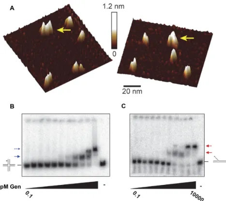
HsGEN1 and CtGEN1 are monomers in solution and dimerize on HJs (8,9,43). We analyzed DmGen by size-exclusion chromatography with multi-angle light scattering (SEC-MALS) after affinity and ion-exchange chromatogra-phy steps and compared it to a BSA standard (Supplemen-tary Figure S7). BSA eluted from the column in three dis-tinct peaks, with the monomer (66 kDa) eluting between 14 and 15 min. The majority ofDmGen elutes as a monomer, exhibiting an average molecular weight of∼58 kDa (pre-dicted 60 kDa), however there is a small amount of dimer present. (Supplementary Figure S7B). To further explore possible dimerization ofDmGen, we used atomic force mi-croscopy (AFM), which allows us to directly observe the oligomerization state of the protein (Figure3A). Previous studies showed that there is a linear relationship between the molecular mass of a protein and its observed volume in AFM images, allowing oligomerization state and asso-ciation constants of protein–protein complexes to be de-termined (31,32). Plots of the distribution of volumes of
DmGen deposited at 20 and 37 nM DmGen show a ma-jor peak consistent with the volume expected for aDmGen monomer and a smaller peak consistent with the volume expected for aDmGen dimer (Figure3A and Supplemental Figure S8). Due to the low population of dimer and crowd-ing of the protein on the surface, accurate determination of the protein dimerization constant is not possible; how-ever, based on the population of protein contained within the monomer and dimer peaks, we estimate the protein dis-sociation constant to be in the hundred nanomolar range (Supplementary Figure S7).
To determine the affinity and stoichiometry ofDmGen binding to DNA, we used electrophoretic mobility shift as-says (EMSAs) (Figure3B and C). We incubated increasing concentrations ofDmGen (0.1 pM–10 nM) with 50 pM of either 5 flap or HJ0 in the presence of EDTA to chelate the metal ions to prevent cleavage of the substrate. On the HJ0, two slower-migrating bands appear with increasing
Nucleic Acids Research, 2017, Vol. 45, No. 9 5339
Figure 2.DmGen is a structure-specific endonuclease and a resolvase. (A) Substrates radiolabelled at the 5end of one strand (asterisks) were incubated with full-length (FL)DmGen or truncated (518)DmGen. Arrows indicate sites of cleavage determined by denaturing PAGE, as shown below. The bracket indicates the expected size range of the cleavage products for the HJ12 substrate. (B) Schematic illustrating the cleavage and ligation experiment of the HJ0+10ntsubstrate formed by annealing one labeled strand of∼50nt, one unlabeled strand of∼50nt, and two unlabeled strands of∼60nt. The addition
of∼10nt on one arm are indicated in green and the asterisk indicates the location of the radiolabel. Cleavage with DmGen creates nicked duplex DNA that can be ligated to create duplex DNA. When visualized on a denaturing PAGE, the presence of a newly ligated longer strand can be observed. (C) Denaturing PAGE of HJ0+10ntcleavage and ligation experiment depicted in (B). (0) indicates substrate only and (+) indicate the addition ofDmGen and/
or T4 ligase. Cartoons on the right of gel indicate the various products formed. Analysis of the cleavage and ligation products of the HJ0+10ntsubstrate
Figure 3. DmGen can dimerize. (A) Topographical AFM images of truncatedDmGen showing monomers and dimers. 20 nM truncatedDmGen was deposited onto naked mica and imaged with tapping mode AFM in air. The gradient bar represents 0–1.2 nm height above the mica surface. Yellow arrows denoteDmGen dimers. 1m x 1m images and volume analyses can be found in Supplementary Figure S8. (B) EMSA analysis ofDmGen with HJ0. TruncatedDmGen (0.1, 0.5, 1, 5, 10, 50, 100, 250, 500, 1000, 10 000 pM) was incubated at room temperature with 50 pM radiolabeled DNA in the presence of EDTA and bound products were separated using 4% native PAGE at 4◦C. The HJ0 cartoon indicates the position of the DNA alone, whereas the positions of the HJ0 bound by monomer and dimer are indicated by a solid arrow and a dashed arrow, respectively. (C) The same as in (B), except with the 5flap. Plots graphing the fraction of DNA bound as a function ofDmGen concentration for three to four independent EMSAs can be found in Supplementary Figure S9.
withDmGen binding to HJ0 with high affinity, similar to those seen withCtGEN1, which binds to HJs with a high affinity (∼10 nM) . We determined theDmGen concentra-tion at which half of the substrate is bound (K1/2,EMSA,HJ) by
plotting the fraction of DNA bound (both shifted bands) as a function of DmGen concentration for four indepen-dent EMSAs and fitting them to the Hill equation (Supple-mentary Figure S9A). This analysis yields aK1/2,EMSA,HJfor
binding ofDmGen to the HJ0 equal to 0.19±0.24 nM. In addition, at 10 nMDmGen, all of the DNA is in the super-shifted band, consistent with 100% of the HJ0 being bound by a dimer or two monomers ofDmGen (Figure3B).
Interestingly,DmGen also forms two shifted species on a 5flap substrate (Figure3C), with the first shifted species occurring at concentrations of DmGen between 100 and 500 pM, followed by complete conversion to a supershifted species by 1 nMDmGen (Figure3C). TheK1/2,EMSA,5’FLAP
for the 5flap substrate determined from three independent experiments is 0.18±.08 nM. By 1 nM DmGen, all of the DNA is in the supershifted band (Figure 4B and Supple-mentary Figure S9B). The supershifted (dimer) band is un-expected because forHsGEN1 a monomer is sufficient for 5flap cleavage (9). No band shifts were observed when the same experiment was performed with linear dsDNA (Sup-plementary Figure S9C), indicating that the supershifted
bands do not result from nonspecific binding ofDmGen to the DNA.
DmGen cleaves 5flaps faster than Holliday junctions
We investigated the kinetics of flap and HJ0 cleavage by
DmGen as a function of both substrate and enzyme concen-tration in multiple-turnover assays (i.e. excess substrate rela-tive to protein) (Figure4A and B). 5 nM 5flap is completely cleaved within 1–4 min usingDmGen concentrations rang-ing from 0.5 to 3 nM (10- to 1.7-fold excess DNA), with the rate of cleavage increasing with increasing concentration. In contrast, the rates of HJ0 cleavage are∼10-fold slower, with reactions taking 10–30 min to plateau (Figure4B). The re-action containing 3 nMDmGen goes to completion, but re-actions with the lower concentrations (0.5–1 nM) plateau at 20–60% of the substrate being cleaved, respectively (Figure
concentra-Nucleic Acids Research, 2017, Vol. 45, No. 9 5341
Figure 4. DmGen cleaves 5flaps faster than HJs. Graphs show time courses of nuclease progression under conditions of excess (A) 5flap or (B) HJ0. For each time course, aliquots were taken at various time points (note that the time scales differ in each panel). The intensity of each cleavage product was quantified by ImageQuant, and the data were normalized to the expected amount of detectable product (see Experimental Procedures). Each point represents the mean of three experiments, except in (A), which is the mean of two experiments. Error bars indicate standard error of the mean. The curves drawn though the data are best fits to single exponentials (C) The rate of cleavage as a function ofDmGen concentration under conditions of excess
DmGen. The data from each individual replicate in (A and B and Supplementary Figure S10) were fit to a single exponential curve given by the equation
y=m1*x/(m2+x), wherem1=maximum rate (kapp,max) at saturating protein concentrations andm2=the apparent dissociation constant (K1/2). Note
that the first point (3 nM protein) was performed with 5 nM 5flap or HJ0, whereas the rest of the experiments (20, 60, 100 and 200 nM protein) were performed with 2 nM DNA. From these fitsK1/2andkapp,maxare 62±3 nM and 46±9 s−1for the flap and 660±500 nM and 31±19 s−1for the 5flap
and HJ0, respectively.
tion increases above the concentration of DmGen, the ex-cess HJ0 acts as a trap, binding monomers ofDmGen and removing them from solution, thereby reducing the con-centration of active substrate-dimer complexes. HsGEN1 shows similar inhibition of cleavage when the HJ substrate is in excess (9,43). We do not observe such substrate inhibi-tion with the flap, likely because flap cleavage requires only a monomer ofDmGen.
Classic steady-state enzyme kinetics (multiple turn-over assays) allow for the determination of thekcat andKm of
an enzyme by plotting the rate of reactions as a function of substrate concentration. Because substrate inhibition oc-curs if the concentration ofDmGen is below the concentra-tion of HJ0 substrate, such assays could not be performed. To circumvent the problem of substrate inhibition, we mea-sured rates of cleavage of the flap and HJ0 as a function of DmGen concentration, maintaining DmGen in excess over substrate (Figure4C and Supplementary Figure S10). The rates measured in these reactions are the rate at which
DmGen cleaves a single substrate molecule and therefore represent steps at or before cleavage. To determine the de-pendence of the cleavage rate onDmGen concentration, we first determined the pseudo-first-order rate constants,kapp,
by fitting the rate curves for each DmGen concentration to single exponentials and then plottedkappas a function
ofDmGen concentration. The rate of cleavage (orkapp) of
both the flap and HJ0 as a function ofDmGen concentra-tion are fit well by simple hyperbolic binding curves, allow-ing us to determine theK1/2,KINETIC(DmGen concentration
necessary to achieve half maximal cleavage rate or simply ‘apparent affinity’) and the apparent maximal cleavage rate (Figure4C). TheK1/2,KINETICand apparent maximal
cleav-age rates are 62±3 nM and 46±9 s−1for the flap and 660 ±500 nM and 31±19 s−1for the HJ0, respectively. In all concentrations ofDmGen tested, the rate of 5flap cleavage is least∼7-fold greater than the rate of HJ0 cleavage (Figure
ex-cess protein found thatHsGEN1cleaves a HJ more rapidly than a 5flap, suggesting that HJs are preferred substrates over 5flaps forHsGEN1 (9). In contrast, our data suggest that 5flaps may be the preferred substrates forDmGen.
HsGEN1 cleaves 5flaps faster than HJs
We next determined whether the difference in substrate specificity ofDmGen andHsGEN1 may reveal abona fide
difference in enzyme activity between the orthologs or may result from dissimilar assay conditions (buffer and DNA se-quence of substrates). The published study onHsGEN1 (9) employed a low-salt phosphate buffer (referred to here as Rass buffer), whereas we used a medium-salt Tris buffer (Bellendir buffer); in addition, the sequences of the sub-strates were different in the two studies (Figure5A). Con-sequently, we compared HsGEN1 with DmGen side-by-side. The human andDrosophilaproteins cleave each sub-strate (5 flap and HJ created from Bellendir or Rass oli-gos) at the same site (Supplementary Figure S5). DmGen andHsGEN1 have very similar rates of cleavage on both HJ substrates in both buffers (Figure 5B, dark blue and light blue bars); however, rates of 5flap cleavage vary with sequence and with buffer. In the Bellendir buffer, the two proteins have similar cleavage rates on both 5 flap sub-strates, and 5 flap cleavage is significantly faster than HJ cleavage (Figure5B). In the Rass buffer, cleavage rates fer for the two sequences, with a 34-fold and a 6-fold dif-ference between the highest and lowest rates of flap cleav-age forHsGEN1 andDmGen, respectively (Figure5B and Supplementary Table S3). In summary, DmGen cleaves 5 flaps faster than HJs in all conditions tested, andHsGEN1 cleaves 5 flaps faster in all but one of the conditions we tested (Figure5B and Supplementary Table S3). Thus, vary-ing the buffer conditions and oligo sequences used in ki-netic assays reveals additional properties of HsGEN1 be-yond those described previously. These results suggest that simpler branched substrates, such as flaps, are preferred by bothDmGen andHsGEN1 and that both enzymes utilize similar mechanisms of recognition and cleavage.
The rate-limiting step of HJ0 cleavage is assembly of a pro-ductive dimer complex on the substrate
Examination of theDmGen kinetic data together with the EMSAs can shed light onto possible mechanisms of 5flap and HJ cleavage. Given that the EMSAs show very tight binding of a DmGen monomer (K1/2,EMSA ∼0.2 nM) to
both the flap and the HJ0 (Figure3B–C and Supplementary Figure S9), it is likely that the concentration dependence of the cleavage rate that we observe (Figure4C) results from a second monomer ofDmGen binding to the substrate prior to cleavage. From our EMSAs, we estimate that the con-centration for a second monomer binding to either the HJ0 or the 5 flap is 0.5–10 nM. TheK1/2,KINETIC,5’FLAP for the
flap from our kinetic data is slightly larger than would be predicted for dimerization on the 5flap from the EMSAs, but this difference may be due to the different temperatures at which the two experiments were performed (EMSAs at 4◦C and kinetic assays at 25◦C). TheK1/2,KINETIC,HJfor the
HJ0 (660 nM), however, is significantly larger than apparent
Nucleic Acids Research, 2017, Vol. 45, No. 9 5343
binding affinities for the dimer seen in the EMSAs, in which we observe 100% dimer at 10 nMDmGen. These differences indicate that the K1/2,KINETIC,HJ value for the HJ0,
deter-mined from kinetic measurements represents a step other than the binding seen in the EMSAs.
If the rate-limiting step to HJ0 cleavage is a confor-mational change after binding, the weak K1/2,KINETIC,HJ
(660 nM) we observe for cleavage of the HJ0 could rep-resent nonspecific of binding of the second monomer of
DmGen prior to a conformational change that leads to the stableDmGen2:HJ0 complexes seen in the EMSAs. To
test this possibility, instead of adding DNA,DmGen, and Mg2+simultaneously as done above (Figure4A–C), we
pre-incubatedDmGen with HJ0 in the absence of Mg2+, allow-ing time for the dimer to assemble on the HJ0, then ini-tiated cleavage by the addition of Mg2+ (Figure6). If the
rate-limiting step is conformational change after the sec-ond monomer binds the HJ0 (i.e. productive assembly of the dimer on HJ0) that results in a stable DmGen2:HJ0
complex that is slow to dissociate, a burst of rapid cleav-age with an amplitude equal to the concentration of pre-formedDmGen2:HJ0 complexes will be observed in the
pre-incubation experiment (47). Using 3 nMDmGen with of 5 or 10 nM HJ0, we observe a burst of cleavage before the first time point (5 s), followed by a slow rate of cleavage similar to that seen in the simultaneous addition experiment (Fig-ure6A and B). This observation strongly suggests that given sufficient time, DmGen can cooperatively assemble into a productive complex on the HJ0. We used aDmGen con-centration of 3 nM, corresponding to 1.5 nM dimer with 5 or 10 nM HJ0. If all theDmGen molecules were active and pre-bound as dimers to HJ0 and poised to undergo cleav-age, we would expect burst heights of 30% for 5 nM HJ0 and 15% for 10 nM HJ0. We observed burst heights of 28% and 10%, respectively (Figure6A), suggesting that the majority ofDmGen is active and bound in a productive dimer com-plex prior to the addition of Mg2+. These results are consis-tent with our equilibrium binding EMSAs, which suggest that the binding affinity of a dimer ofDmGen for the HJ0 is between∼3 and 10 nM (Figure3B). Taken together, our results strongly suggest that the rate-limiting step to HJ0 cleavage is a conformational change after binding of the second monomer ofDmGen to the HJ. We conducted simi-lar pre-incubation experiments with the flap (Figure6C). In contrast to the results of pre-binding with the HJ0, no initial burst of cleavage is discernable, suggesting that the interac-tion ofDmGen with the flap is dynamic and that binding of a monomer or dimer to the flap does not result in a complex that is slow to dissociate (Figure6C and6D).
DISCUSSION
In this work, we show that DmGen functions as a key structure-selective endonuclease (SSE) during repair of DNA damage. Our genetic data reveals a fundamental dif-ference betweenDmGen and the well-characterized human and fungal orthologs. In Drosophila, Gen single mutants have extreme hypersensitivity to DNA damaging agents but
mus81single mutants do not (Figure1); as in other organ-isms,mus81 Gendouble mutants have even more severe hy-persensitivities. These data are in stark contrast to genetic
results seen in other species, where Mus81 is the predom-inant SSE and Gen orthologs play a secondary role in re-pair (17,19,20,35). Interphase protein localization does not appear to account for the reversed dominance observed in
Drosophila, asDmGen, like Yen1 and HsGEN1, seems to be primarily or exclusively cytoplasmic (Supplementary Fig-ure S2) (40,48,49); however, it is possible that the order of activation during mitosis is switched inDrosophilarelative to yeast and human cells.
DmGen, like HsGEN1, cleaves a variety of branched structures including 5flaps, replication forks, splayed arms and HJs (Figure 2 and Supplementary Figure S5). The mechanism of HJ cleavage appears to be similar between
DmGen andHsGEN1, as the two proteins exhibit similar rates of cleavage across various conditions (Figures5–7). BothDmGen and HsGEN1 are robust 5flap endonucle-ases.HsGEN1’s cleavage rate on flaps is more sensitive to reaction conditions than that ofDmGen, though even this difference might be due to different physiological temper-atures (Figure5). This combination of genetic differences and biochemical similarities observed betweenDmGen and
HsGEN1 highlights the unique platform thatDrosophila
provides to the DNA repair/structure selective endonucle-ase fields to further our understanding of the functions of these enzymesin vivo. Below, we discuss insights into the mechanism of cleavage of HJs and flaps by DmGen or-thologs, structural considerations that may underlie novel-ties acrossDmGen orthologs, and the importance of HJs versus other substrates.
The mechanism of 5flap and HJ cleavage byDmGen
Comparison of our biochemical data withDmGen to those with the yeast and human orthologs suggests that they fol-low similar mechanisms of cleavage of 5 flaps and HJs. Taking both our kinetic and EMSA data into account al-lows us to elucidate important features of the mechanism of HJ and 5flap cleavage. HJ cleavage requires the assem-bly of a dimer of DmGen (or orthologs) on the HJ prior to cutting (9,43,45,46). Our prebinding experiments (Fig-ure 6A), which reveal a burst of cleavage followed by a slow turnover, indicate that after the second monomer binds the complex undergoes a conformational change that stabi-lizes theDmGen2:HJ0 complex and leads to rapid cleavage.
These data support previous models for the activity of or-thologs on HJs, which suggest that a rate-limiting confor-mational change occurs after binding of a second monomer (9,43,45,46). Our EMSAs show tight binding of both the monomer and dimer ofDmGen to the HJ (Figure4A and Supplementary Figure S9). Our observation of HJ0 sub-strate inhibition further supports tight monomer binding, and the near-stoichiometric burst amplitudes in prebinding experiments support tight dimer binding; however, in ex-periments in whichDmGen, DNA, and Mg2+were mixed
simultaneously, we observe a very weak concentration de-pendence of DmGen for HJ cleavage, with a K1/2,KINETIC
consistent with nonspecific DNA binding (Figures4B and
6A). Accordingly, we propose that the first monomer binds tightly to the HJ (K1/2,EMSA∼0.2 nM), followed by a second
monomer binding with a weak affinity (K1/2,KINETIC∼700
Figure 6. Production of an active complex is the rate-limiting step in HJ0 cleavage. (A) To determine whether the rate-limiting step of the HJ0 reaction is a slow conformational change that results in a stable Gen2:HJ0 complex that can rapidly cleave the DNA, 3 nMDmGen was pre-incubated with 5 or
10 nM HJ0 for several minutes before starting the time course experiment with MgCl2. Quantification and analyses were done as in (Figure4A–C). (B)
Data from the corresponding simultaneous addition experiments in Figure4B (3 nMDmGen with 5nM HJ0,pink) is replotted on a 1-minute time scale for comparison to (A). (C) The same as in (A), except with the 5flap. (D) Data from the corresponding simultaneous addition experiments in Figure4A (3nMDmGen with 5nM 5flap,pink) was replotted on 1-minute time scale for comparison to (C). Note that the data in this figure are not normalized, as in Figure4.
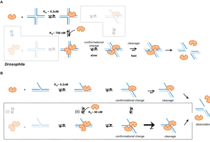
Subsequently, this complex undergoes a rate-limiting con-formational change that positions opposing DNA strands in theDmGen active sites to form a stable productive dimer-HJ0 complex that is slow to convert back to the nonspecif-ically bound form. After formation of a productive dimer– HJ complex, the dimer rapidly nicks the two opposing strands of the junction in a cooperative and symmetric man-ner, yielding two nicked duplexes. It is also possible that a
DmGen dimer can form in solution prior to binding the HJ (Figure7A, bottom left box); however, our results in-dicate that the HJ must be in the proper conformation for the secondDmGen to bind tightly. The importance of the HJ conformation is also supported by the crystal structure ofCtGEN1 bound to an HJ after cleavage. In the presence of cation, unbound HJs are found in the stacked X confor-mation (50). In the HJ model based on the crystal structure of CtGEN1 in the product complex, the HJ is bent into
a non-planar conformation (46). Given the affinity of the monomer for DNA, binding of preformed dimers to the HJ may promote dissociation of one monomer. It is also pos-sible that a DNA conformational change occurs prior to a secondDmGen monomer binding the HJ (Figure7A, top right box).
Consistent with data on HsGEN1 (9,43), our EMSA data taken together with our kinetic data on the 5 flap under excess substrate conditions suggest that a monomer of DmGen can cleave a 5 flap. This result is not unex-pected, as flap cleavage requires only a single nick. Our ki-netic data using excess protein further suggest that a second monomer ofDmGen can bind to the flap (with an apparent affinity/K1/2,KINETIC,5’FLAPof 60 nM) and increase the
Nucleic Acids Research, 2017, Vol. 45, No. 9 5345
Figure 7. Mechanism ofDmGen cleavage of HJs and 5flaps. (A) Mechanism of Gen function on HJs.DmGen monomer binds the HJ with aKDof∼0.2 nM (K1/2,EMSA,HJfrom EMSAs), followed by weak, nonspecific binding of a second monomer with aKDof∼660 nM (K1/2,KINETIC,HJfrom Kinetics).
Formation of a productive dimer complex, exhibiting the correct DNA conformation required to position opposite DNA strands in theDmGen active sites, is slow. Once a productive dimer–HJ0 complex is formed, the dimer cooperatively nicks across the junction. (bottom left box) It is unlikely that a pre-formed dimer will encounter a HJ exhibiting the proper conformation required for cleavage. If the dimer–DNA complex is not productive, one monomer likely dissociates from the HJ, allowing otherDmGen proteins access to the junction. (top right box) It is also possible that a DNA conformational change occurs prior to the second monomer binding; however, given our observation that production of a productive dimer–DNA complex is the rate-limiting step, this is unlikely to represent a main pathway. See discussion for additional details (B) Mechanism ofDmGen function on 5flaps. (top)DmGen monomer binds the 5flap, triggering a DNA conformational change and rapid cleavage of the flap strand one nt 3of the junction branch point. (bottom) ADrosophila -specific pathway is depicted in the box. (i) A pre-formed dimer can bind the 5flap, (ii) Alternatively, two monomers can sequentially bind the 5flap, with the first monomer having a very high affinity (∼0.2 nM from EMSAs) and the second monomer having a weaker affinity of∼60 nM (based on the
K1/2,KINETIC, 5’ flapdetermined from kinetic data). The additional DNA contacts provided by the second monomer may constrain the 5flap conformation
to facilitate cleavage.
for cleavage and that the dimer facilitates this change. Based on these observations we propose the mechanism shown in Figure7B. ADmGen monomer binds the 5flap with high affinity (K1/2,EMSA ∼0.2 nM, based on EMSAs), followed
by a conformational change that positions the DNA in the active site and the subsequent rapid cleavage of the 5flap one nucleotide 3of the branch point (Figure7B, top). At lowDmGen concentrations or in excess substrate, this path dominates; however as the concentration of DmGen in-creases, a second monomer or a preformed dimer ofDmGen can bind the 5flap and increase the rate of cleavage∼5–10 fold. This proposed mechanism is supported by our obser-vation thatDmGen can form dimers in solution and that the rate of flap cleavage increases withDmGen concentration, with aK1/2,KINETIC=60 nM (Figures3,4,5and
Supplemen-tary Figures S7-10). We suggest that thisK1/2,KINETIC
repre-sents the binding affinity of the second monomer ofDmGen to the flap (Figure7B, (ii)). The additionalDmGen–DNA binding interactions provided by the second monomer may
Protein structural comparisons across Gen orthologs high-light differences leading to substrate specificity and dimeriza-tion
Studies ofDmGen orthologs across species show cleavage of a variety of branched DNA structures and different propen-sities for dimerization. Insight into possible structural rea-sons for these differences in biochemistry can be gleaned from analysis of the protein primary sequences and crys-tal structures of theDmGen orthologs and the 5flap cut-ter FEN1, which is a monomeric family member. Sequence alignment of theDmGen and FEN1 orthologs reveals sig-nificant differences among the orthologs in the region of
DmGen 76–125 (Supplementary Figure S11). This region is adjacent to the active site and forms part of the dimer in-terface in the crystal structure ofCtGEN1and the proposed dimer interface ofHsGEN1 (45,46). In FEN1, this region forms a helical arch with a cap through which the single-stranded 5flap is threaded. It has been suggested that this cap would prevent DNA structures without free ends from threading into the catalytic site, thereby limiting the activity of FEN1 to substrates containing a single-stranded 5end (46,52). InCtGEN1, both the arch and cap regions are ab-sent, and the protein is predominately a monomer in solu-tion and cuts HJs but not flaps, replicasolu-tion forks or splayed arms . InHsGEN1, only the cap region is absent; the pro-tein is a monomer in solution, but it cleaves 5flaps, repli-cation forks, HJs, and splayed arms (Supplementary Fig-ure S5). Flap cleavage byHsGEN1 shows greater sensitivity thanDmGen to the buffer and to the sequence at the junc-tion (8,45,53).DmGen has both the helical arch and cap (al-though the cap sequence is not conserved with FEN1); this protein can dimerize on substrates and can cut 5flaps, repli-cation forks, HJs, and splayed arms. Interestingly, Yen1 also contains both the helical arch and cap region, and it cuts 5 flaps, replication forks, and HJs, but not splayed arms, and appears to dimerize on flaps (Figure 6E from (40)). Taken together, these observations suggest that the presence and/or the sequence of helical arch and cap govern not only substrate specificity but also the propensity toDmGen and its orthologs to dimerize. The ability of DmGen to dimerize on substrates as well as its broad substrate specificity may contribute to its preferential usage over Mus81in vivo.
Substrate specificities ofDmGen andHsGEN1 suggest that HJs are uncommon repair intermediates
Although it may seem unusual for the roles of Gen and Mus81 to have switched in Drosophila relative to other species, comparison of thein vitroandin vivoactivities of
DmGen in flies and Mus81 in other species sheds light on the commonalities of these two enzymes that allow them to be interchangeable within cells. The predominant enzymes in flies (DmGen) and in yeast and human cells (Mus81) cut a variety of branched structures, with substantially higher activities on simpler structures, such as flaps, over the more physically restrained HJ. WhileDmGen and its orthologs cut 5to a branch point and Mus81 cuts 3, this polarity dif-ference may be less relevantin vivoand may simply dictate the orientation of binding of the endonuclease to the sub-strate. This suggestion is supported byin vitrodata that indi-cate that both Mus81 andDmGen orthologs cleave nicked
HJs and replication forks to form nick duplexed products (9,54,55). In addition,in vivostudies show that overexpres-sion of Yen1 can rescuemus81 mutants in budding yeast (17), and expression of HsGEN1 or DmGen can rescue
mus81mutants in fission yeast, which lacks a Yen1 ortholog ((40) and Supplementary Figure S1). We propose that these simpler structures may be more importantin vivo, perhaps because they are more frequently encountered. Consistent with this hypothesis, recent studies show that replicative stress in yeast results in nicked and gapped HJs, while an-other study highlights non-HJ intermediates of replication repair (56,57).
In repair, DNA intermediates arising from strand ex-change that generates a displacement-loop (D-loop) resem-ble flaps or nicked HJs, and these intermediates have many potential advantages over a canonical HJ as substrates for SSEs. First, flaps or nHJs would presumably be an earlier intermediate in repair, and further synthesis and ligation would require additional energy. Second, flaps or nHJs only require one nuclease domain for cleavage and could explain why eukaryotic ‘resolvases’ are predominantly monomers or heterodimers with only a single catalytic subunit. These points suggest that there may be mechanisms to avoid formation of HJs in favor of less complex intermediates. Nonetheless, intact HJs may accrue if repair intermediates are not processed fast enough such that further synthesis and ligation occurs, or if blocked replication forks are re-gressed (58). Even so, the biological presentation of these intact HJ may be very different from the syntheticin vitro
HJs used for testing cleavage activity.In vivo,intact HJs may be opened by helicases or ssDNA binding proteins, and con-sequently resemble the splayed arms or bubbles studiedin vitro. Such intermediates have been proposed to explain the requirement for the Blm helicase to make meiotic crossovers inC. elegansmeiosis (59). Accordingly, we propose that dur-ing damage repair, canonical resolvase activity of Mus81 andDmGen orthologs on intact HJs may be a backup or failsafe to resolve intact HJs if they accumulate, but the pri-mary role of these endonucleases is to cleave other struc-tures that arise.
SUPPLEMENTARY DATA
Supplementary Data are available at NAR Online.
ACKNOWLEDGEMENTS
We thank Steve West for the kind gift of HsGEN1 protein. We thank Steve West and Anton Gartner for suggesting we move the tag to the C-terminus of DmGen; Gerry Smith for providingS. pombestrains and plasmids; Wolf-Dietrich Heyer for comments on an early manuscript; Ashutosh Tri-pathy and the UNC MacInFac core facility for performing SEC-MALS; and Tony Perdue and the UNC Microscopy core facility for assistance.
FUNDING
Nucleic Acids Research, 2017, Vol. 45, No. 9 5347
M.R.R.]; National Cancer Institute (NCI) [R01 CA084442 to D.A.R]; NIGMS [5T32GM007092 to S.P.B.] (in part); National Science Foundation Graduate Research Fellow-ship and The Graduate School at The University of North Carolina at Chapel Hill (to D.J.R.). Funding for open ac-cess charge: National Institute of General Medical Sciences [1R35GM118127].
Conflict of interest statement.None declared.
REFERENCES
1. Lloyd,R.G. and Sharples,G.J. (1992) Genetic analysis of
recombination in prokaryotes.Curr. Opin. Genet. Dev.,2, 683–690. 2. Holliday,R. (1964) A mechanism for gene conversion in fungi.
Genet.Res.,78, 282–304.
3. Valenzuela,M.S. and Inman,R.B. (1975) Visualization of a novel junction in bacteriophage lambda DNA.Proc. Natl. Acad. Sci. U.S.A.,72, 3024–3028.
4. Mizuuchi,K., Kemper,B., Hays,J. and Weisberg,R.A. (1982) T4 endonuclease VII cleaves holliday structures.Cell,29, 357–365. 5. Bennett,R.J., Dunderdale,H.J. and West,S.C. (1993) Resolution of
Holliday junctions by RuvC resolvase: cleavage specificity and DNA distortion.Cell,74, 1021–1031.
6. Sharples,G.J., Benson,F.E., Illing,G.T. and Lloyd,R.G. (1990) Molecular and functional analysis of theruvregion ofEscherichia coli
K-12 reveals three genes involved in DNA repair and recombination.
Mol. Gen. Genet.,221, 219–226.
7. Bell,L. and Byers,B. (1979) Occurrence of crossed strand-exchange forms in yeast DNA during meiosis.Proc. Natl. Acad. Sci. U.S.A.,76, 3445–3449.
8. Ip,S.C., Rass,U., Blanco,M.G., Flynn,H.R., Skehel,J.M. and West,S.C. (2008) Identification of Holliday junction resolvases from humans and yeast.Nature,456, 357–361.
9. Rass,U., Compton,S.A., Matos,J., Singleton,M.R., Ip,S.C., Blanco,M.G., Griffith,J.D. and West,S.C. (2010) Mechanism of Holliday junction resolution by the human GEN1 protein.Genes Dev.,24, 1559–1569.
10. Castor,D., Nair,N., Declais,A.C., Lachaud,C., Toth,R., Macartney,T.J., Lilley,D.M., Arthur,J.S. and Rouse,J. (2013) Cooperative control of Holliday junction resolution and DNA repair by the SLX1 and MUS81-EME1 nucleases.Mol. Cell,52, 221–233. 11. Garner,E., Kim,Y., Lach,F.P., Kottemann,M.C. and
Smogorzewska,A. (2013) Human GEN1 and the SLX4-associated nucleases MUS81 and SLX1 are essential for the resolution of replication-induced Holliday junctions.Cell Rep.,5, 207–215. 12. Wyatt,H.D., Sarbajna,S., Matos,J. and West,S.C. (2013) Coordinated
actions of SLX1-SLX4 and MUS81-EME1 for Holliday junction resolution in human cells.Mol. Cell,52, 234–247.
13. Boddy,M.N., Lopez-Girona,A., Shanahan,P., Interthal,H., Heyer,W.D. and Russell,P. (2000) Damage tolerance protein Mus81 associates with the FHA1 domain of checkpoint kinase Cds1.Mol. Cell. Biol.,20, 8758–8766.
14. Interthal,H. and Heyer,W.D. (2000) MUS81 encodes a novel helix-hairpin-helix protein involved in the response to UV- and methylation-induced DNA damage inSaccharomyces cerevisiae.Mol. Gen. Genet.,263, 812–827.
15. Dendouga,N., Gao,H., Moechars,D., Janicot,M., Vialard,J. and McGowan,C.H. (2005) Disruption of murine mus81 increases genomic instability and DNA damage sensitivity but does not promote tumorigenesis.Mol. Cell. Biol.,25, 7569–7579. 16. Fricke,W.M. and Brill,S.J. (2003) Slx1-Slx4 is a second
structure-specific endonuclease functionally redundant with Sgs1-Top3.Genes Dev.,17, 1768–1778.
17. Blanco,M.G., Matos,J., Rass,U., Ip,S.C. and West,S.C. (2010) Functional overlap between the structure-specific nucleases Yen1 and Mus81-Mms4 for DNA-damage repair inS. cerevisiae.DNA Repair (Amst.),9, 394–402.
18. Ho,C.K., Mazon,G., Lam,A.F. and Symington,L.S. (2010) Mus81 and Yen1 promote reciprocal exchange during mitotic recombination to maintain genome integrity in budding yeast.Mol. Cell,40, 988–1000.
19. Tay,Y.D. and Wu,L. (2010) Overlapping roles for Yen1 and Mus81 in cellular Holliday junction processing.J. Biol. Chem.,285,
11427–11432.
20. Mu ˜noz-Galvan,S., Tous,C., Blanco,M.G., Schwartz,E.K.,
Ehmsen,K.T., West,S.C., Heyer,W.D. and Aguilera,A. (2012) Distinct roles of Mus81, Yen1, Slx1-Slx4, and Rad1 nucleases in the repair of replication-born double-strand breaks by sister chromatid exchange.
Mol. Cell. Biol.,32, 1592–1603.
21. Trowbridge,K., McKim,K.S., Brill,S. and Sekelsky,J. (2007) Synthetic lethality in the absence of the Drosophila MUS81 endonuclease and the DmBlm helicase is associated with elevated apoptosis.Genetics,
176, 1993–2001.
22. Andersen,S.L., Kuo,H.K., Savukoski,D., Brodsky,M.H. and Sekelsky,J. (2011) Three structure-selective endonucleases are essential in the absence of BLM helicase inDrosophila.PLoS Genet.,
7, e1002315.
23. Yıldız, ¨O., Majumder,S., Kramer,B.C. and Sekelsky,J. (2002) Drosophila MUS312 interacts with the nucleotide excision repair endonuclease MEI-9 to generate meiotic crossovers.Mol. Cell,10, 1503–1509.
24. Kanai,Y., Ishikawa,G., Takeuchi,R., Ruike,T., Nakamura,R., Ihara,A., Ohashi,T., Takata,K., Kimura,S. and Sakaguchi,K. (2007) DmGEN shows a flap endonuclease activity, cleaving the blocked-flap structure and model replication fork.FEBS J.,274, 3914–3927. 25. Kaliraman,V., Mullen,J.R., Fricke,W.M., Bastin-Shanower,S.A. and
Brill,S.J. (2001) Functional overlap between Sgs1-Top3 and the Mms4-Mus81 endonuclease.Genes Dev.,15, 2730–2740. 26. Benson,F.E. and West,S.C. (1994) Substrate specificity of the
Escherichia coliRuvC protein. Resolution of three- and four-stranded recombination intermediates.J. Biol. Chem.,269, 5195–5201. 27. Wright,W.D., Ehmsen,K.T. and Heyer,W.D. (2011) Assays for
structure-selective DNA endonucleases.Methods Mol. Biol.,745, 345–362.
28. Bennett,R.J. and West,S.C. (1995) RuvC protein resolves Holliday junctions via cleavage of the continuous (noncrossover) strands.Proc. Natl. Acad. Sci. U.S.A.,92, 5635–5639.
29. Okazaki,K., Okazaki,N., Kume,K., Jinno,S., Tanaka,K. and Okayama,H. (1990) High-frequency transformation method and library transducing vectors for cloning mammalian cDNAs by trans-complementation ofSchizosaccharomyces pombe.Nucleic Acids Res.,18, 6485–6489.
30. Lake,C.M., Holsclaw,J.K., Bellendir,S.P., Sekelsky,J. and Hawley,R.S. (2013) The development of a monoclonal antibody recognizing theDrosophila melanogasterphosphorylated histone H2A variant (gamma-H2AV).G3 (Bethesda),3, 1539–1543. 31. Ratcliff,G.C. and Erie,D.A. (2001) A novel single-molecule study to
determine protein–protein association constants.J. Am. Chem. Soc.,
123, 5632–5635.
32. Yang,Y., Wang,H. and Erie,D.A. (2003) Quantitative characterization of biomolecular assemblies and interactions using atomic force microscopy.Methods,29, 175–187.
33. Larkin,M.A., Blackshields,G., Brown,N.P., Chenna,R., McGettigan,P.A., McWilliam,H., Valentin,F., Wallace,I.M., Wilm,A., Lopez,R.et al.(2007) Clustal W and Clustal X version 2.0.
Bioinformatics,23, 2947–2948.
34. Nicholas,K.B., Nicholas,H.B. Jr and Deerfield,D.W.I. (1997) GeneDoc: analysis and visualization of genetic variation.
EMBNEW.NEWS,4, 14.
35. Sarbajna,S., Davies,D. and West,S.C. (2014) Roles of SLX1-SLX4, MUS81-EME1, and GEN1 in avoiding genome instability and mitotic catastrophe.Genes Dev.,28, 1124–1136.
36. Liu,L.F., Desai,S.D., Li,T.K., Mao,Y., Sun,M. and Sim,S.P. (2000) Mechanism of action of camptothecin.Ann. N.Y. Acad. Sci.,922, 1–10.
37. Groth,P., Auslander,S., Majumder,M.M., Schultz,N., Johansson,F., Petermann,E. and Helleday,T. (2010) Methylated DNA causes a physical block to replication forks independently of damage signalling, O(6)-methylguanine or DNA single-strand breaks and results in DNA damage.J. Mol. Biol.,402, 70–82.
38. Alvino,G.M., Collingwood,D., Murphy,J.M., Delrow,J., Brewer,B.J. and Raghuraman,M.K. (2007) Replication in hydroxyurea: it’s a matter of time.Mol. Cell. Biol.,27, 6396–6406.