FUNCTIONAL MOVEMENT DEFICITS IN RELATION TO SPORT-RELATED CONCUSSION
Robert C. Lynall
A dissertation submitted to the faculty of the University of North Carolina at Chapel Hill in partial fulfillment of the requirements for the degree of Doctor of Philosophy in the Human
Movement Science Curriculum in the School of Medicine.
Chapel Hill 2016
ABSTRACT
Robert C. Lynall: Functional Movement Deficits In Relation to Sport-Related Concussion
(Under the direction of Jason P. Mihalik)
The objective of this dissertation was to identify tandem gait dynamic balance deficits and assess dynamic functional movement in recreational athletes with and without a history of concussion within the past 18 months. We recruited a convenience sample of 30 college-aged recreational athletes. There were two groups (15 participants per group): 1) Recent concussion group (median time since concussion 126 days, range 28-432 days), and 2) Matched control group with no recent concussions. Control participants were matched to injured participants based on sex, age (±1 year), mass (±10%), and height (±5%). We measured center of pressure outcomes under 4 tandem gait (heel-to-toe walking) conditions: 1) Tandem gait (eyes open, no cognitive distraction), 2) Tandem gait, eyes closed (no cognitive distraction), 3) Tandem gait, eyes open, cognitive distraction (Brooks Visuospatial Task), and 4) Tandem gait, eyes closed, cognitive distraction (Brooks
Visuospatial Task). We investigated joint kinematics and reaction time during 3 movement tasks: 1) Jump-landing, 2) Anticipated-cut, and 3) Unanticipated-cut. The recently
concussed group demonstrated slower velocity during tandem gait compared to the control group (4.0 cm/s difference; F1,27=4.26; p=0.049; ES=0.38). Greater dual-task cost was
observed for center of pressure speed (F3,26=5.13; p=0.032) such that the concussion group
cutting tasks, but the control group displayed better reaction time cost (-10.7%) than the concussed group (-0.8%) during anticipated cutting (F2, 25=5.26; p=0.030). The concussed
group displayed greater trunk flexion compared to the control group during anticipated cut towards the non-dominant side (5.1° difference; F2, 27=5.89; p=0.022; ES=0.63). There may
be subtle movement differences that are detectable more than a month after return-to-activity following concussion, but the clinical meaning of these findings is unclear.
TABLE OF CONTENTS
LIST OF TABLES ... vii
LIST OF FIGURES ... viii
LIST OF ABBREVIATIONS ... ix
CHAPTER I: INTRODUCTION ... 1
Specific Aims & Research Hypotheses ... 4
CHAPTER II: LITERATURE REVIEW ... 7
Introduction ... 7
Concussion Assessment Battery ... 8
Impairments in Gait Following Concussion ... 11
Functional Movement Following Concussion ... 19
CHAPTER III: METHODOLOGY ... 31
Specific Aim 1 ... 31
Specific Aim 2 ... 35
CHAPTER IV: MANUSCRIPT 1 ... 44
Introduction ... 44
Methods ... 46
Results ... 49
Discussion ... 51
Conclusion ... 55
CHAPTER V: MANUSCRIPT 2 ... 61
Methods ... 62
Results ... 67
Discussion ... 68
Conclusion ... 72
CHAPTER VI: SUPPLEMENTARY RESULTS AND DISCUSSION ... 78
Results ... 78
Discussion ... 79
CHAPTER VII: DISSERTATION SUMMARY AND CONCLUSIONS ... 83
Results Summary ... 83
Methodologic Limitations ... 83
Interpretation of Findings ... 85
Future Directions ... 88
Significance ... 91
LIST OF TABLES
Table 1. Common spatiotemporal gait variables in the concussion gait
literature ... 27
Table 2. Common dynamic balance variables in the concussion gait literature ... 28
Table 3. Summary of literature reporting musculoskeletal injury risk following concussion. ... 29
Table 4. Tandem gait outcome variables. ... 42
Table 5. Outcome variables associated with Aim 2. ... 43
Table 6. Group demographics and statistical comparisons. ... 57
Table 7. Tandem gait outcomes between groups for each condition adjusted for average days since ... 58
Table 8. Reaction time and reaction time cost (RTC) during jump landing, anticipated cut, and unanticipated cut, adjusted for days since concussion. ... 74
Table 9. Trunk, hip, and knee angles (°) at initial ground contact for each group during jump landing, adjusted for days since concussion. ... 75
Table 10. Trunk, hip, and knee angles (°) at initial ground contact for each group during anticipated cut, adjusted for days since concussion. ... 76
Table 11. Trunk, hip, and knee angles (°) at initial ground contact for each group during unanticipated cut, adjusted for days since concussion. ... 77
LIST OF FIGURES
Figure 1. Effect of condition on velocity (m/s) during tandem gait. ... 30
Figure 2. Example of a Brooks Visuospatial Task card. ... 41
Figure 3. Dual-task cost during eyes open tandem gait conditions. ... 59
Figure 4. Dual-task cost during eyes closed tandem gait conditions. ... 60
Figure 5. Time to stabilization (s) during single leg hop on the dominant and non-dominant legs between groups. ... 81
Figure 6. Observed effect sizes during anticipated cut. ... 92
Figure 7. Center of pressure path (cm) main effects for condition. ... 93
LIST OF ABBREVIATIONS
ACL Anterior Cruciate Ligament
ANCOVA Analysis of covariance
BESS Balance Error Scoring System
CI Confidence Interval
cm Centimeters
cm/s Centimeters per second
COP Center of Pressure
DT Dual-task
DTC Dual-task Cost
EC Eyes Closed
ES Effect Size
Hz Hertz
ICC Intraclass Correlation Coefficient
kg Kilograms
L5 Lumbar spine level 5
m Meters
m/s Meters per second
n Frequency
N Newtons
PKMAS Protokinetics Movement Analysis Software
RTC Reaction Time Cost
s Seconds
SCAT3 Sport Concussion Assessment Tool 3rd Edition
SOT Sensory Organization Test
ST Single-task
TG Tandem Gait
TGEC Tandem Gait Eyes Closed
TGDT Tandem Gait Dual-task
TGDTEC Tandem Gait Dual-task with Eyes Closed
CHAPTER I: INTRODUCTION
Failure to properly diagnose and manage concussion can result in catastrophic secondary events of severe brain injury or death after a second injury is sustained when symptoms from the initial injury have yet to fully resolve.1 A large proportion of brain-related
fatalities in football players under the age of 21 were associated with a recent history of symptomatic sport-related concussion.2 Athletes failing to report symptoms during activity is
common, and further complicates clinical management of sport-related concussion.3 This
underscores the need for more objective, clinician-friendly tests.
To that end, several validated post-injury assessments identify symptoms4 and
deficits in mental status,5 neurocognition,6 and static balance7 following concussion. Over 85% of patients demonstrate full recovery within 7 days of injury as measured by symptom reports and traditional measures of neurocognition and static balance deficits following concussion.8 At this point in the patient’s recovery, athletes are often guided back to full return-to-participation over the course of several days. When accounting for this gradual return-to-participation structure, the majority of athletes will return to full participation within 10-15 days of the brain injury.
Several investigations have called into question the validity of labeling an athlete as ‘recovered’ based solely upon symptom, neurocognitive, and static balance measures. Athletes who have suffered an initial concussion are 3 to 6 times more likely to suffer a subsequent concussion.9, 10 Additionally, of 12 same-season repeat concussions that
and static balance.9 Further, research has demonstrated athletes are at an increased risk for
musculoskeletal injury following return-to-participation after concussion.11-14 These findings
suggest there may be lingering motor control deficits remaining well after the clinical recovery one can measure with current assessment tools, which may increase the risk of subsequent neurological and musculoskeletal injury. While several studies have reported similar outcomes in regards to musculoskeletal injury risk following concussion, limitations to these works exist. Importantly, none of these investigations observed other factors that may influence injury risk following concussion, such as overall exposure to injury and behavioral risk and care taking profiles of individual athletes.
The increased risk for musculoskeletal injury may not be surprising given the existing literature detailing deficits in standard gait following concussion, both acutely15, 16 and
persisting beyond return-to-play.17, 18 Considering these documented gait deficits, such as loss of dynamic balance control75 and conservative adaptions,56 and the increased
musculoskeletal injury risk, it is surprising that functional movement assessments are not performed following suspected concussion. This may be due to the small dynamic balance differences observed in laboratory settings, which may not translate to clinically observable outcomes, or to the large movement variance observed between individuals. A functional movement assessment may have clinical utility as both a diagnostic tool and a mechanism by which return-to-participation can be safely evaluated and monitored.
A tandem gait task with a recorded time component has been suggested as a valid, cost-effective, and clinician friendly means of objectively identifying post-concussive
deficits.19 Unfortunately, the tandem gait task included in the Sport Concussion Assessment
Tool-3rd Edition (SCAT3)19 was created without any scientific validity or evidence of how
task completion exceeds 14 seconds, the patient is said to have failed the tandem gait task. No consideration is given to trials in which the patient is unable to maintain balance
throughout the trial. In fact, the SCAT3 recommends re-starting the trial in cases where the patient is unable to maintain balance while walking along a straight line. Based on this single pass/fail criterion, 80% of healthy high school athletes fail the tandem gait task.20 This lack
of clinical specificity is unacceptable. More information is needed so an effective test of functional movement can be incorporated into clinical athletic training. Additionally,
increasing neuromechanical constraints by adding a cognitive distractor task during tandem gait (dual-task) may further challenge the ability of the patient to maintain neuromuscular control. This dual-task paradigm, used previously to investigate standard gait deficits following concussion,15, 16, 18 seeks to challenge the patient in the same way athletic
participation will challenge them as they return to sport activity.
Functional deficits noted post-concussion may be related to disrupted cortical pathways. Using transcranial magnetic stimulation to assess cortical hypoexcitability
following concussion, researchers have demonstrated lower intra-cortical facilitation,21 lower maximal voluntary muscle activation,21 increased motor evoked potential latency, and decreased motor evoked potential amplitude.22, 23 These results suggest the brain’s ability to control movement may be impaired, both acutely and after return-to-participation, following concussion. Further, small changes in cortical response to external stimuli may be
exacerbated in highly dynamic environments. For this reason, it is important to explore potential movement differences between concussed and healthy individuals in a dynamic, sport-like setting.
post-concussion. It is unlikely a standard gait task alone will be in-depth enough to understand all biomechanical maladaptations that may occur after brain injury. Identifying lower extremity biomechanics that may increase injury risk and potential functional reaction time deficits present during sport-specific activities will inform the mechanisms underlying the increased risk of musculoskeletal injury following concussion. This may enhance rehabilitation
protocols following concussion to combat the biomechanical maladaptations leading to lower extremity musculoskeletal injuries.
The overall objective of this dissertation was to identify tandem gait dynamic balance deficits and assess dynamic functional movement in recently concussed recreational
athletes (within the past 18 months), relative to comparison subjects with no concussion within the past 18 months. Our central hypothesis was that sport-related concussion would result in dynamic balance deficits during tandem gait that are still identifiable after the
athlete has returned to play due to lingering motor control and dynamic functional movement deficits from the injury that are not assessed using conventional concussion assessment tools. Identifying lingering concussion deficits may lead to safer and more effective return-to-participation strategies, which could help decrease potential long-term deficits associated with concussion.
Specific Aims & Research Hypotheses
Specific Aim 1. Determine differences in tandem gait dynamic balance between concussed recreational athletes and non-concussed control participants.
during dual-task conditions during standard gait. Diminished attentional capacity may negatively affect previously concussed individuals’ ability to perform simultaneous gait and cognitive tasks. Thus, we hypothesize recently concussed recreational athletes will demonstrate greater dual-task cost (center of pressure path, speed, velocity, and cognitive component) as compared to controls who were not recently concussed during the tandem gait dual-task conditions.
Hypothesis 1C. Due to lingering neuromuscular control maladaptations from concussion, we hypothesize dynamic balance outcomes (center of pressure path, speed, and velocity) will worsen as condition difficulty increases (in order: tandem gait, tandem gait with eyes closed, tandem gait with Brooks Visuospatial Task, tandem gait with eyes closed and Brooks Visuospatial Task). Additionally, we hypothesize there will be a significant group by condition interaction, such that the recently concussed group will perform significantly worse than the control group as the conditions increase in
difficulty.
Specific Aim 2. Identify functional movement and dynamic balance differences that present following traditional return to full participation after concussion in
recreational athletes and non-concussed control participants.
Hypothesis 2A: Reaction time is affected after concussion and is only assessed in a static environment. These reaction time deficits may take longer to resolve when assessed during dynamic movement tasks such as those athletes experience during sport. We hypothesize recently concussed recreational athletes will demonstrate slower movement reaction times and greater reaction time cost during anticipated and unanticipated cutting tasks as compared to healthy matched controls when reaction time is assessed in a more dynamic, sport-like environment.
slight joint kinematic alterations after traditional recovery from the brain injury, possibly contributing to increased risk of musculoskeletal injury. Thus, we hypothesize recently concussed recreational athletes will demonstrate biomechanical risk factors for lower extremity injury (increased knee adduction angle and trunk flexion angle at initial ground contact, decreased knee flexion angle at initial ground contact) as compared to healthy matched controls.
Hypothesis 2C: Measures of static balance recover within 3-5 days following concussion, but reports of dynamic balance during gait suggest balance differences between concussed and healthy participants, even after static balance has recovered. If
dynamic balance assessments are more sensitive to lingering balance control deficits, we hypothesize recently concussed recreational athletes will demonstrate decreased dynamic balance control (increased center of pressure speed and path and increased time to stabilization) as compared to controls who were not recently concussed.
Hypothesis 2D: Beyond movement, joint proprioception may play an important role in musculoskeletal injury risk. Persistent motor cortex alterations following concussion may decrease joint proprioception. Thus, we hypothesize recently concussed
CHAPTER II: LITERATURE REVIEW
Introduction
Many effective concussion assessment and management tools have been
developed and described in the literature.5, 7, 19, 24 Despite widespread clinical use of various
tools,25 current return-to-participation assessment lacks investigation of functional
movement. This may be an important missing component to concussion evaluation, as increased rates of musculoskeletal injury12-14, 26 along with dynamic balance and
spatiotemporal deficits during gait following concussion have been identified.15, 27, 28 Despite this knowledge, there is no agreed upon assessment of functional movement after
concussion. Standard gait dynamic balance deficits such as increased medial-lateral center of mass displacement and reduced velocity have been identified under sophisticated laboratory conditions. Unfortunately, very few clinicians have access to this technology. This, among other factors, is likely a key reason why functional movement assessments have not been recommended for clinical practice.
Tandem gait has been proposed as a method to identify dynamic balance deficits following concussion.19 However, based on a single pass/fail criterion (time to task
completion of 14 seconds), 80% of healthy high school athletes fail the tandem gait task.20
athletes move after concussion in a dynamic, sport-like environment will help inform our understanding of the increased rates of lower extremity musculoskeletal injury.14 Concussion Assessment Battery
Due to the complex nature of concussion, which has the potential to result in a wide range of post-injury deficits, multiple position and consensus statements call for a multi-faceted approach to concussion diagnosis and management.19, 29 At a minimum, this
assessment battery should include objective tests of balance, neurocognition or neurological status, and symptoms. It is important to include assessments in each of these domains because recovery in one domain does not always translate to recovery in one or more of the other domains.30 Over the last 2 decades, several clinician-friendly (easy to apply, accurate,
and low cost) objective measures have been developed and validated for use in concussion diagnosis and management. These assessments have been created in response to known impairments following concussion.
Static Balance Deficits Following Concussion
The maintenance of postural control is a complex process involving brain integration and control of afferent and efferent nervous system pathways. Essential afferent information is provided to the brain from cutaneous receptors, in addition to somatosensory, visual, and vestibular inputs. Under normal conditions, inputs from cutaneous receptors in the foot along with visual and somatosensory information are adequate to maintain the center of gravity within the body’s base of support.31 The role of the vestibular system is emphasized in cases where there is disruption or conflicting information from one or more of the aforementioned systems. Deficits in postural control following concussion have been mainly attributed to sensory integration issues.31 In cases where input from one of the postural control systems
effectively identify and ignore distracting input from altered environmental conditions. For example, under false visual conditions (visual reference moves in phase with subject’s center of pressure), a healthy individual is able to adapt by identifying and ignoring this false visual input and instead rely on somatosensory and/or vestibular information to maintain balance. Therefore, it is hypothesized concussed individuals suffer from an inability to ignore altered environmental conditions, causing them to select a motor response based on these altered cues.31-33
Postural control deficits following concussion have been demonstrated using both sophisticated laboratory measures and less expensive clinician-friendly methods.7 The
Sensory Organization Test (SOT), performed on a force plate system called the NeuroCom Smart Balance Master (Clackamas, OR), allows for the manipulation of visual and/or
somatosensory input during quiet stance. Investigations of post-concussion postural stability deficits utilizing the SOT have demonstrated increased postural sway for up to 3 days following the injury.32-34 These findings have been repeated with a more clinically feasible test known as the Balance Error Scoring System (BESS).4, 32, 34 The BESS consists of 6 total trials utilizing 3 stances (feet together, single leg, and tandem stance) and 2 surface
conditions (stable and unstable). Importantly, this test employs no expensive force plate systems while still delivering an objective outcome score based on an error scoring system that has been validated against force plate measures.7, 32, 34
Neurocognitive Deficits Following Concussion
Neurocognitive testing following concussion, once labeled as “one of the cornerstones of concussion management,”35 can yield valuable objective information.
Although cognitive recovery may overlap with symptom recovery, it is possible the 2 may be independent of each other.36-38 Neurocognitive deficits have been identified following
concussion utilizing both traditional paper and pencil tests39 as well as computerized testing
Concussed participants have demonstrated deficits in several neurocognitive domains, including verbal and visual memory42, 43 and reaction time.44 Importantly, these
deficits have been shown to linger beyond the usual recovery of static balance and symptom deficits.37, 45 This underscores the need to investigate all possible deficits following
concussion.
Symptom Deficits Following Concussion
Self-reported symptom assessments are one of the most common means of diagnosing concussion.25 While specific symptoms of concussion vary widely after each
injury, common symptoms include headache, dizziness, loss of memory, visual
disturbances, and feeling “in a fog.” The number of symptoms and the severity of each are generally recorded following injury. Symptom recovery appears to differ between athletes of different ages. For college-aged athletes, concussion symptoms commonly recover between 5 and 10 days for the majority of injured subjects.4, 39, 46 Evidence suggests younger athletes
may take longer to recover from concussion and this recovery time may be lengthier in those suffering from a previous history of concussion. Amongst a cohort of high-school aged subjects suffering from their first concussion, the median time to symptom recovery was 12 days. Amongst those who had suffered more than 1 previous concussion, median time to symptom resolution was 28 days.47 These differences in concussion recovery, among others, underscore the need for a complete and thorough assessment battery that accounts for all possible post-injury deficits.
Tandem Gait Assessment Following Concussion
In response to known static balance deficits and increasing evidence of functional movement deficits following concussion, a consensus panel of concussion experts from around the world have advocated for the addition of a tandem gait task to sideline
concussion management protocols.19 This tandem gait task consists of 4 trials of heel-to-toe
throughout the trial without being given direction as to the placement of the hands. Each trial is timed and the shortest time to complete the task is recorded and considered to be the scored trial. Trials lasting longer than 14 seconds are deemed failed trials. If at any time the subject is unable to maintain balance while walking, the trial is stopped and re-started. This tandem gait task was based on 2 published articles investigating normative values for a wide age range of participants (16-37 years)48 and surface/footwear interactions that may
affect test outcomes.49 Importantly, no studies to date have investigated the effect of
concussion on tandem gait outcomes. One study observed 80% of healthy high school aged athletes take longer than 14 seconds to complete the tandem gait task and, thus, are
considered to have failed the tandem gait test in a healthy state.20 In order for a more
sensitive and specific tandem gait task to be developed, much more understanding is needed of how concussion affects tandem gait dynamic balance outcome variables. Impairments in Gait Following Concussion
Variables of Interest in Gait Studies
Many gait variables have been explored to study deficits following concussion. These variables can be group into 2 categories: spatiotemporal variables and dynamic balance variables. Spatiotemporal variables are those outcome measures that are defined by time and/or distance. Dynamic balance variables are those outcome measures that attempt to quantify sway through various means during gait. Although these variables are not standard across all published research, in most cases between-study differences in defining these variables are negligible. Tables 1 and 2 display the most common calculations of some variables of interest in the concussion gait literature.
reported in the same studies, it is worth noting several of them are interrelated. For example, a person displaying more conservative gait is likely to reduce stride time and decrease stride length. These reductions will likely result in a decrease in any number of anterior/posterior dynamic balance variables during standard gait, including velocity, range of motion, and displacement. While this is the most intuitive association between the variables, spatiotemporal variables will not always change in the same manner. Stride time could decrease while stride length increases, resulting in no detectable change in the dynamic balance variables. For this reason, dynamic balance variables in the sagittal plane are usually thought to be conservative adaptions in gait as opposed to balance loss.50
Dynamic balance measures in the frontal plane, along with several frontal plane
spatiotemporal variables, likely give the best insights into postural control during gait. Center of mass range of motion and maximum separation of the center of mass and center of pressure (both in the frontal plane) are correlated with stride width.50 Frontal plane center of
mass measures of velocity do not necessarily relate to any spatiotemporal variables, but have been shown to be an important variable to describe imbalance in the frontal plane during gait following concussion.51 Many measures have been studied and are found throughout the concussion gait literature. Due to the disparity in outcomes reported in the concussion literature, there is no way to identify one or some of these variables as superior. Further, no investigations have explored a concussed sample during tandem gait.
The Single-Task Paradigm in Gait Studies Following Concussion
The study of gait following concussion under single-task conditions during level walking has only revealed minimal group differences (concussed vs. healthy controls). From a spatiotemporal perspective, gait velocity appears to be the most sensitive variable,16, 17, 28
although increased stride time in concussed individuals has been noted as well.16
of pressure,17 and sagittal plane center of mass velocity.28 These differences, however, are
only present within 48 hours of injury. When the concussed individual moves to the 5th day
post-injury and beyond, differences in single-task performance on both spatiotemporal and dynamic balance variables disappear. Together, these acute differences likely point to the adoption of a more conservative gait strategy following concussion.
The Dual-Task Paradigm in Gait Studies Following Concussion
The classic version of the dual-task paradigm explores 2 tasks simultaneously, with one of the tasks designated as primary.52 These tasks, one of which is typically cognitive
based while the other motor based, compete for the subject’s attentional demands. Baseline measures are taken of both tasks performed in isolation (single-task) to establish normal values for that participant. If under dual-task conditions the secondary task performance drops, it is suggested the participant had insufficient attentional reserve to maintain the secondary task at the baseline level.53
In this classic dual-task paradigm, it is imperative primary task performance is maintained. Because this is difficult in a laboratory setting, researchers have found
additional ways to assess performance under dual-task conditions. Instead of designating a primary and secondary task, participants perform both tasks simultaneously without being instructed to direct their focus to either. This method of dual-task investigation is very clinically applicable as most real-world scenarios call for some degree of split attention (dual-task) throughout the day. Multiple outcomes are possible when dual-task conditions are investigated. Performance in the motor task could increase or decrease with or without a corresponding change in cognitive performance.52, 54
DTC = [(DT – ST) / ST] x 100
This cost, represented as a percentage, can certainly be influenced by task priority.
Generally speaking, the task with the greatest dual-task cost is thought to be the secondary task, although this can also be influenced by task difficulty.52
Several different cognitive tasks have been used in conjunction with a motor task in the concussion gait literature. The most commonly employed cognitive task is the Modified Mental Status Examination, which consists of a series of simple mental tasks.16, 17, 50 These
tasks include spelling common 5-letter words in reverse, counting backwards by sevens, or reciting the months of the year in reverse order. Other cognitive tasks have been reported as well, including a simple reaction time task,16 auditory or visual Stroop Task,55-57 and the
Brooks Visuospatial Task.18 Howell et al. investigated the effect of dual-task complexity on
gait stability following concussion in adolescents.27 The authors reported increased cognitive task complexity has a greater effect on gait balance control. This is important, especially when attempting to identify lingering gait deficits after concussed individuals have returned to full sport participation. Employing the most challenging dual-task protocol may allow researchers and clinicians to better identify lingering deficits during functional movement.
To that end, we have used the Brooks Visuospatial Task during pilot testing. The Brooks Visuospatial Task is designed to tax the visuospatial component of cognition.18 Previous research has established an important connection between cognition and motor function.12, 13 It has been speculated these associations exist because cognition and motor function rely on the same neural network.57 Sosnoff et al. explored this association in
concussed individuals.58 They reported cognitive/motor function associations were present
score. This indicates these deficits are not necessarily due to a decreased pool of attentional resources, but may be due to deficits within a shared process, such as
visuospatial attention.58 Thus, because the Brooks Visuospatial Task taxes the visuospatial
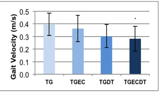
component of cognition, it has the potential to exacerbate cognitive and motor deficits observed after concussion. Additionally, the Brooks Visuospatial Task is very challenging. For example, Figure 1 illustrates the effect of various tandem gait conditions on gait velocity in a small cohort (n=9) of healthy college-aged individuals walking across a 14-foot gait mat. Differences in gait velocity during tandem gait with eyes closed and the Brooks Visuospatial Task as compared to tandem gait alone and tandem gait with eyes closed were observed, suggesting increasing task complexity with the Brooks Visuospatial Task. These pilot data helped inform hypothesis 1C, that there would be an interaction between concussion (group) and tandem gait condition. This paradigm with increasing difficulty is ideal, as we
investigated concussed individuals after they were cleared to fully return to sport participation.
Both spatiotemporal and dynamic balance deficits have been observed during gait under dual-task conditions following concussion. In contrast to the acute single task deficits, dual-task deficits are present at both acute time points15-17, 28, 50, 56, 59 and beyond athlete return-to-participation.17, 18, 50, 59 Not only do these deficits suggest dual-task gait paradigms are more sensitive to concussion, it also implies deficits persist long after the injured athlete is believed to have fully recovered based on common clinical measures of concussion. When group comparisons are considered acutely under dual-task conditions, concussed subjects appear to adopt a more conservative gait strategy by decreasing stride length15, 17
and velocity16, 17, 28 along with decreasing sagittal plane center of mass velocity.16, 28, 50, 56
between center of mass and center of pressure,17 and frontal plane center of mass
velocity.16
At time points beyond the average return-to-participation timeframe, group
differences persist when the dual-task paradigm is employed. It appears concussed subjects continue to display a more conservative gait strategy as evidenced by decreased stride length at the 14 day post-injury time point and decreased separation between center of mass and center of pressure in the sagittal plane at both 14 and 28 days following injury.17
Interestingly, there is evidence of gait deficits persisting in a cohort of previously concussed individuals who were, on average, over 6 years post-injury. This cohort demonstrated increased double leg stance time along with decreased single leg stance time and velocity.18
While all of the findings discussed thus far have emerged from studies employing similar college-aged cohort methodology, it is worth discussing that several other studies have employed different methodologies to achieve the same end. Dual-task cost has been discussed as a potentially important variable of interest in the study of gait following concussion. Dual-task cost related to gait speed has been shown to be sensitive to group differences in a college-aged cohort. These differences were present in multiple dual-task conditions as well as obstacle avoidance conditions in which the participants had to step over an obstacle.60 Group differences during obstacle avoidance tasks are intriguing findings. Avoiding an obstacle may increase postural control demands on the
neuromuscular system, not unlike athletic participation. Further exploration of dynamic balance variables under obstacle avoidance conditions is warranted and may further inform deficits following concussion, especially as it relates to the body’s ability to dynamically control posture.
High-school aged cohort studies have also revealed group differences. One study reported dual-task cost outcomes in a cohort of high school aged participants.61 Across the
cost for average walking speed, sagittal plane center of mass velocity, and frontal plane separation between center of mass and center of pressure. Importantly, the authors of this study also report concussed participants were significantly less accurate on the concurrent cognitive task as compared to matched controls. Dual-task group differences in dynamic balance variables along with a decline in cognitive task performance have also been noted in a high-school aged cohort for up to 2 months following injury.27 Additionally, dynamic
balance outcome variables have been shown to regress between pre- and post-return-to-participation following concussion in high-school aged athletes during dual-task gait conditions.62
It is important to recognize limitations to the study of gait following concussion. None of the reported studies have collected baseline (pre-concussion) data. Thus, it is possible reported dynamic balance group differences were present prior to the concussion.
It is clear dual-task paradigms are more sensitive to gait deficits following
concussion, but the underlying reason for this is not entirely understood. Generally, there are several theories that attempt to identify the underlying mechanism for dual-task
interference. The bottleneck theory suggests only a single processing operation can happen at any given time.63 If two tasks compete for the same processing mechanism, a bottleneck
results and one or both of the tasks will be affected. A separate but related hypothesis suggests a capacity sharing of information processing.64 As opposed to the bottleneck
theory, capacity sharing suggests capacity for a given task is reduced when another task is attempted simultaneously.63, 64 In this situation, both tasks may be performed, but each task
may be affected by the other. Quantifying the effect of one task on another is an important consideration in dual-task paradigms.54 It is imperative dual-task paradigms include both
cognitive and motor task outcome measures in order to appropriately explain the observed dual-task interference.65 In the capacity sharing model, insufficient attentional capacity to
efficiently divide attention between the cognitive and motor task may affect outcomes.66 For example, if a given participant has a fear of falling during a gait or balance task, he or she may devote the majority of their attentional capacity to the motor task. Thus, cognitive performance may suffer while motor performance remains steady. In this case, we could interpret this interference as motor-related cognitive interference, meaning motor task performance remained steady with a subsequent drop in cognitive performance.54 Without
appropriate measure of both cognitive and motor task performance, this effect may be missed completely or misinterpreted.
Further work with concussed individuals has sought to explain the mechanism underlying dual-task interference, specifically the ability of concussed individuals to maintain and switch attention between tasks. Using the Attentional Network Task, researchers have demonstrated specific attention deficits following concussion.67 It appears the alerting
extent, the orienting components of attention are affected. Briefly, the alerting component of attention is associated with the ability to maintain vigilance during continuous task
performance. The executive component allows for appropriate conflict resolution while the orienting component allows for efficient selection of information based on sensory input.68
These findings are interesting when taken in context with dual-task gait deficits following concussion. The deficits in the orienting component of attention suggest an impaired ability to move attention from a central focus point. Additionally, the executive component deficits suggest those who have suffered a concussion are less able to appropriately ignore irrelevant or contradictory information. Adding a cognitive task to a relatively simple motor task, such as standard gait, affects both the orienting and executive components of attention, suggesting a potential mechanism behind the noted standard gait deficits in dual-task conditions.
Functional Movement Following Concussion
As discussed previously, athletes undergo a concussion assessment battery prior to full return-to-participation following concussion. While this battery is effective in identifying lingering static balance, neurocognitive, and symptom deficits, functional movement deficits go completely ignored. This is problematic given the presence of long-term gait deficits under dual-task conditions.17, 18, 50, 59 Additionally, increased risk of subsequent concussion9,
10 along with increased risk of musculoskeletal injury following the initial concussion11-14
suggests an incomplete recovery. It is also possible that athletes are fully recovered from concussion, but other factors such as inherent behavioral differences (i.e. risk taking) and injury exposure may be driving reported musculoskeletal injury group differences. Table 3 details the current literature reporting musculoskeletal injury risk after concussion. Although evidence of an association between concussion and musculoskeletal injury is growing in the literature, it is important to acknowledge alternative confounders that may affect this
was different between cohorts. Playing style, injury reporting and care-seeking behaviors, and playing position are all additional confounders to the association between concussion and musculoskeletal injury. Understanding how athletes move during sport-related activities after concussion may further inform clinical best practice in patient care and lead to an increased understanding of lingering effects that contribute to increased injury rates. Further, examining reaction time in a dynamic environment, just as the athlete will experience during competition, will lead to an increased understanding of attentional contributions to movement deficits.
Reaction Time Deficits Following Concussion
Reaction time is a commonly assessed cognitive domain following concussion that has been shown to be sensitive to injury in numerous investigations.36, 41, 69-71 In addition to
computerized and paper-and-pencil reaction time tests, a more functional reaction time test has been developed for efficient sideline use.24 This clinical reaction time measure involves
inexpensive components that are easily administered and highly portable and has been shown to be valid, reliable, and sensitive to concussion.24, 72, 73 Briefly, this clinical reaction time measure involves a stick marked every centimeter attached to a heavy cylinder, such as a hockey puck. The clinician holds the stick such that the hockey puck is level with the subject’s hand. The clinician then drops the stick at various time intervals while the subject attempts to catch it as quickly as possible. Eight total trials are performed, and the distance scores are input into a formula that converts the subject’s score to a reaction time measure. As reaction time is essential not only to sport performance but to injury prevention as well, the correlation between clinical reaction time and protective reaction time (moving the hands to protect the head from an incoming ball) has been explored. Clinical reaction time
demonstrated a strong correlation to protective reaction time.74 This finding is important as
neurocognitive testing and simple clinical testing is clearly affected by concussion. Unfortunately, the current methods to assess reaction time are far removed and much simpler than the reaction time required to perform at a high level and protect oneself during sport. Therefore, further examination of reaction time in more dynamic and demanding situations is warranted.
At-Risk Movement Patterns
Beyond reaction time assessment, understanding biomechanical movement patterns that increase musculoskeletal injury risk following concussion may lead to a better
understanding of interventional methods to reduce this injury risk. While many lower extremity injuries are possible, research into the mechanism associated with anterior cruciate ligament (ACL) injury is prevalent. Although various combinations of movement may contribute to ACL injury, increased anterior shear force at the proximal tibia appears to be the major contributor to increased ACL loading as demonstrated by several
investigations utilizing cadaveric models.75, 76 The quadriceps muscles are a major contributor to anterior shear force on the proximal end of the tibia.77, 78 For a given
quadriceps force, anterior shear force on the proximal tibia increases as knee flexion angle decreases.79 Therefore, landing from a jump or cut with a decreased knee flexion angle may lead to increased risk of ACL injury.
Although it has not received as much attention in published literature, hip adduction may be an important contributing risk factor for ACL injury. In yet unpublished findings, Marshall et al. describe the largest prospective cohort study investigating risk factors for ACL injury.80 Military academy cadets who demonstrated hip adduction (less than 0° hip
an ipsilateral hip shift, possibly due to gluteus medius weakness, but it is clear diminished hip control during a jump landing is prospectively associated with ACL injury.
Core stability and trunk control may be other factors related to increased risk of injury. Movement of the trunk in the frontal plane may lead to increased valgus stress on the knee. For instance, landing from a jump with a lateral trunk shift to the right side of the body increases the overall valgus moment placed upon the right knee. Along these lines,
research has indicated that increased lateral trunk displacement following a sudden force applied to the trunk may be the best predictor of knee injury.81 Sagittal plane trunk
biomechanics may also play a role in mediating ACL injury risk. Blackburn and Padua reported increased trunk flexion is associated with increases in knee and hip flexion angles as well as a decreased risk of ACL injury.82 In contrast, the study by Marshall et al.
referenced above found increased trunk flexion (greater than 40° vs. less than 25°)to be associated with a higher prospective rate of ACL injury.80 The authors suggest this finding
reflects a landing strategy in which the participant lands with too much trunk flexion. Taken together, these results suggest there is a desired amount of trunk flexion that has the potential to mitigate ACL injury risk. These findings are important as rehabilitation and athlete-training programs may be modified to emphasize movements that are associated with decreased risk of injury.
It should be noted that many factors may play a role in mediating ACL injury risk such as sex,83 bony anatomy,84 and genetic predisposition.85 These factors, for the most part, are unchangeable in a given athlete. Therefore, we will focus on potential
biomechanical deficits in athletes following concussion that may be modifiable. This may allow for future interventions to affect some or all of the causes of increased rates of lower extremity musculoskeletal injury following concussion.
deficits lingering beyond athlete return to play18, 57, 62 along with the increased risk of
musculoskeletal injury following concussion.12, 14, 26 We studied outcomes that have been
associated with ACL injury risk, but these outcomes do not represent all potential
maladaptations that may occur after concussion. This investigation was intended to be a preliminary attempt to quantify lower extremity functional biomechanics after concussion. Future research protocols should build on our methodology in order fully explore
biomechanical outcomes after concussion.
Additional Functional Movement Assessments
While the above biomechanical variables are related to ACL injury risk, many more adaptations could occur following brain injury that may contribute to increased risk of musculoskeletal injury. Ankle sprains are the most common orthopedic injury associated with sports participation, accounting for about 25% of all injuries.86 Measures of static
postural stability are often used to diagnose and manage those with chronic ankle instability. These static measures, however, may not be sensitive enough to distinguish between functionally stable and unstable ankles. Several reports have suggested dynamic measures may be more sensitive to differences between groups. Specifically, time to stabilization appears to be a better measure of ankle stability than traditional measures of static balance.58, 87 Briefly, time to stabilization is measured following a jump from a
pre-determined height. The participant lands on a single leg and is instructed to maintain their best balance as quickly as possible following landing. The total time it takes to reach a pre-determined stable posture is described as the time to stabilization.
While the underlying mechanism for deficits in dynamic stabilization for those who display functional ankle instability has largely been attributed to afferent pathway
sensorimotor deficits associated with the initial ligament injury,88 functional deficits noted
have demonstrated lower intra-cortical facilitation,21 lower maximal voluntary muscle
activation,21 increased motor evoked potential latency, and decreased motor evoked
potential amplitude.22, 23 These results suggest the brain’s ability to control movement may
be impaired, both acutely and after return-to-participation, following concussion. Further, small changes in cortical response to external stimuli may be exacerbated in highly dynamic environments. For this reason, it is important to explore potential movement differences in a dynamic, sport-like setting. Beyond investigating gross biomechanical differences as
proposed here, further investigation will need to be done to explore the direct effect of disrupted cortical pathways on movement. Future methodology that incorporates movement outcomes and cortical pathway outcomes will greatly inform the hypotheses proposed here.
Further, investigation of lower extremity proprioception following concussion may give additional insight into potential cortical deficits leading to increased risk of
musculoskeletal injury. Joint position sense (awareness of and ability to recreate the joints position in space) is an important component to proper functional movement. Effectively repeating movements during repetitive movements, such as gait or cutting during athletics, is essential to avoid injury. Following injury to a joint, local sensory receptors in the skin, tendon/muscle, ligaments, or joint capsule that provide proprioceptive information to the central nervous system may become damaged, leading to deficits in overall joint position sense.89, 90 Concussion is interesting in that these peripheral sensory receptors are not
damaged, making it unlikely they contribute to any loss of proprioception. Importantly, the brain is essential to the organization and storage of information related to proprioception. Thus, brain injury may affect motor control that is dependent on appropriate joint position sense. To our knowledge, no one has directly measured joint position sense in a dynamic environment following concussion. Including closed kinetic chain joint position sense
contribution of proprioception to increased risk of acute lower extremity injury during athletics.
Measures of static balance following concussion are an important part of a
comprehensive test battery. As noted, previous research into static balance deficits following concussion have shown deficits persisting for approximately 3 days following injury.32-34
More functional assessments of balance, such as those discussed above involving gait tasks, have shown deficits persisting well beyond this 3-day time frame.17, 18, 50, 59
Additionally, investigators have sought to determine the relationship between static and dynamic balance measures. Several published articles reveal significant, but relatively weak, relationships between the static and dynamic measures.91-94 Despite the statistically
significant correlations, clinical conclusions drawn from these data suggest static and dynamic measures of balance are not reflective of the same phenomenon. These results only underscore the need for more functional assessments of balance following concussion.
It is important to consider that the hypotheses discussed to this point may only represent one mechanism affecting movement following concussion. Other psychosocial factors such as care-seeking and risk-taking may influence lower extremity musculoskeletal injury risk following concussion. Additionally, accounting for lower extremity injury exposure and head impact exposure may be important. Future work should seek to account for these and other confounders in the context of functional movement following concussion.
The Clinical Significance of Assessing Functional Movement Following Concussion
While one prominent concussion consensus statement, the Consensus Statement on Concussion in Sport, advocates for a brief tandem gait assessment,19 no work has been
done to understand deficits that may be present under tandem gait conditions following concussion. Before an appropriate clinical test can be designed, we must explore the dynamic balance deficits that may be present in tandem gait following concussion.
present in tandem gait following concussion, beyond athlete recovery and return-to-participation.
While understanding these tandem gait deficits will greatly inform our clinical management of concussion, investigation of tandem gait alone is not enough to fully understand functional movement deficits. Athletes are at an increased risk of sustaining subsequent concussions9, 10 and lower extremity musculoskeletal injuries11-14 following an
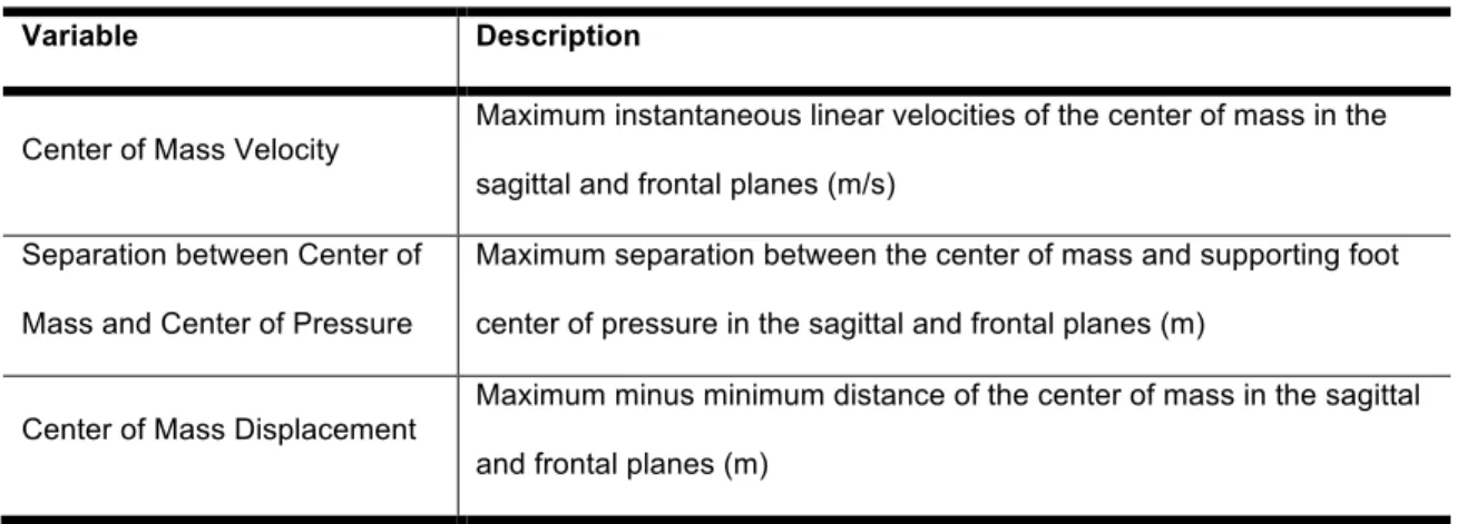
Table 1. Common spatiotemporal gait variables in the concussion gait literature Variable Description
Velocity Divide the sum of all stride lengths by the sum of all stride times (m/s)
Stride Length Distance between 2 successive heel strikes in the sagittal plane (m)
Stride Width Distance between 2 successive heel strikes in the frontal plane (m)
Stride Time Time between 2 successive heel strikes (s)
Double Support Time Percentage of total trial spent with both feet in contact with the ground
Single Support Time Percentage of total trial spent with one foot in contact with the ground
Total Time The total time it takes to complete a given trial (s)
Table 2. Common dynamic balance variables in the concussion gait literature
Variable Description
Center of Mass Velocity Maximum instantaneous linear velocities of the center of mass in the sagittal and frontal planes (m/s)
Separation between Center of
Mass and Center of Pressure
Maximum separation between the center of mass and supporting foot
center of pressure in the sagittal and frontal planes (m)
Center of Mass Displacement
Maximum minus minimum distance of the center of mass in the sagittal
Table 3. Summary of literature reporting musculoskeletal injury risk following concussion.
Study Cohort
Post-Concussion
Timeframe
Outcome
(95% Confidence Interval)
Makdissi et al.95 Professional Australian Football
1 competitive match
Injury Rate Ratio
- 2.23 (0.93, 5.04) Brooks et al.12 Mixed College 90 days Odds Ratio - 2.48 (1.04, 5.91) Cross et al.13 Professional Rugby
Union 24 months
Injury Rate Ratio
- 1.6 (1.4, 1.9)
Lynall et al.14 Mixed College 12 months
Injury Rate Ratio
- Pre-conc vs. post-conca = 1.97 (1.19, 3.28)
- Conc vs. control = 1.64 (1.07, 2.51)
Nordstrom et
al.26 Professional Soccer 12 months
Hazard Ratiob
- 1.47 (1.05, 2.05)
Burman et al.96 Mixed Athlete 24 months
Odds Ratio
- Pre Concussionc = 1.98 (1.45, 2.72)
- Post Concussiond = 1.72 (1.26, 2.37) Pietrosimone et al.97 Retired Professional Football Reported history
Odds Ratioe
- 1 conc vs. 0 conc = 1.59 (1.30, 1.94)
- 2 conc vs. 0 conc = 2.29 (1.85, 2.83)
- 3+ conc vs. 0 conc = 2.86 (2.36, 3.48)
All studies compared a concussed group to a control group, unless otherwise noted. All outcome ratios are reported as concussion/control.
a Compared injury rates in year post-concussion to year pre-concussion in concussion group only. b Reported results are when controlling for number of injuries in the year preceding concussion. c Analyzed injuries prior to concussion.
d Analyzed injuries after concussion.
e Reported odds ratios for those who had a history of 1, 2, or 3+ concussions compared to those
Figure 1. Effect of condition on velocity (m/s) during tandem gait.
Nine healthy college-aged subjects completed tandem gait under 4 conditions. * Indicates slower velocity as compared to TG and TGEC. TG = tandem gait; TGEC = tandem gait eyes closed; TGDT = tandem gait dual-task; TGDTEC = tandem gait dual-task with eyes closed.
CHAPTER III: METHODOLOGY
Specific Aim 1
Design and Setting
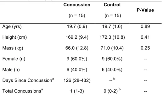
We recruited a convenience sample of 30 college-aged recreational athletes (no varsity inter-collegiate athletes were included). There were two groups (15 participants in each group): 1) Recent concussion group (median time since concussion of 126 days, range 28-432 days), and 2) Matched control group with no recent concussions. Control
participants were matched to each injured participant based on sex, age (± 1 year), mass (± 10%), and height (± 5%). Participants must have reported being moderately physically active for at least 30 minutes 3 times a week. We excluded participants for any of the following conditions: attention deficit hyperactivity disorder, seizure disorders, lower extremity injury resulting in physical activity time loss of ≥3 days within the last 6 months, any history of lower extremity or low back surgery, concussion requiring admittance to the hospital, any current concussion symptoms, or a previous history of >3 concussions. For inclusion in the concussed group, participants must have sustained a concussion diagnosed by a medical professional within the last 1.5 years. Participants in the matched control group must have been without diagnosed concussion for at least 3 years. We recorded the number of days since the most recent concussion in the concussed group. The Institutional Review Board at the University of North Carolina at Chapel Hill approved our study and all participants signed an informed consent document prior to testing.
Instrumentation
static and dynamic balance and gait assessment. The Zeno Walkway contains a 16-level pressure sensing pad and circuitry inside a low profile and portable housing. The walkway is 16 feet long by 2 feet wide and allows for analysis in a single pass or multiple passes. ProtoKinetics Movement Analysis Software (PKMAS) allows for recording and analysis of spatiotemporal and dynamic balance variables. The technology employed by the Zeno Walkway has been shown to have strong concurrent validity and good to excellent test-retest reliability for the assessment of spatiotemporal gait variables.98-100 Our own internal
testing revealed excellent center of pressure outcome reliability (ICC2,k >0.963) and strong
correlations to force plate center of pressure outcomes (r>0.75).
Data Collection Procedures
The cognitive task used during the dual-task conditions was the Brooks Visuospatial Task,101 which has been previously used to investigate dual-task effects on gait following
concussion.18 The Brooks Visuospatial Task was chosen as our pilot data indicated
significantly slower tandem gait velocities during dual-task trials using this cognitive task. Additionally, the Brooks Visuospatial Task challenges the visuospatial component of cognition. This may be important post-concussion, as an increased cognitive-motor
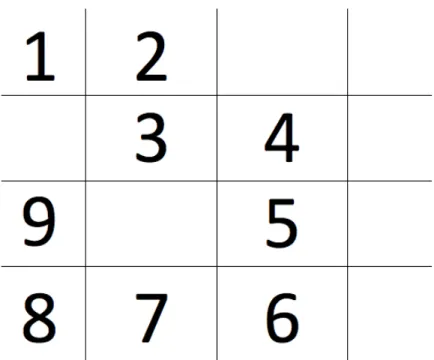
association has been observed, suggesting damage to a shared cognitive-motor component such as visuospatial attention.102 Each participant had one minute to memorize the order of
digits 1-8 on a 4x4 grid (Figure 2). After the minute-long period, the participant identified the position of the next consecutive digit without looking at the grid. For example, participants presented with the grid in Figure 2 would say, “1st row, 1st column, 1, right 2, down 3, right 4,
down 5, down 6, left 7, left 8.” During dual-task conditions, specific directions were given that instructed the participant to focus on maintaining fast and balanced tandem gait while trying their best to accurately complete the cognitive task. Error frequency and time taken to complete the Brooks Visuospatial Task were recorded by the primary investigator using a stopwatch during all trials involving the Brooks task. Prior to any tandem gait trials, each participant completed 3 baseline Brooks Visuospatial Tasks while seated to ensure task familiarization.
Data Reduction and Analysis
reduce the data and calculate all outcomes listed in Table 4. The first 140 cm of each trial were analyzed. This was necessary because trials were stopped when the participant finished the Brooks Visuospatial Task during conditions 3 and 4. Several cut-points were explored, and the 140 cm cut-point resulted in the least amount of discarded trials (n=1) while still capturing multiple footfalls during a given trial. This distance allowed for at least 4 footfalls per trial per participant, which is more than has been analyzed and previously reported.56, 62, 103
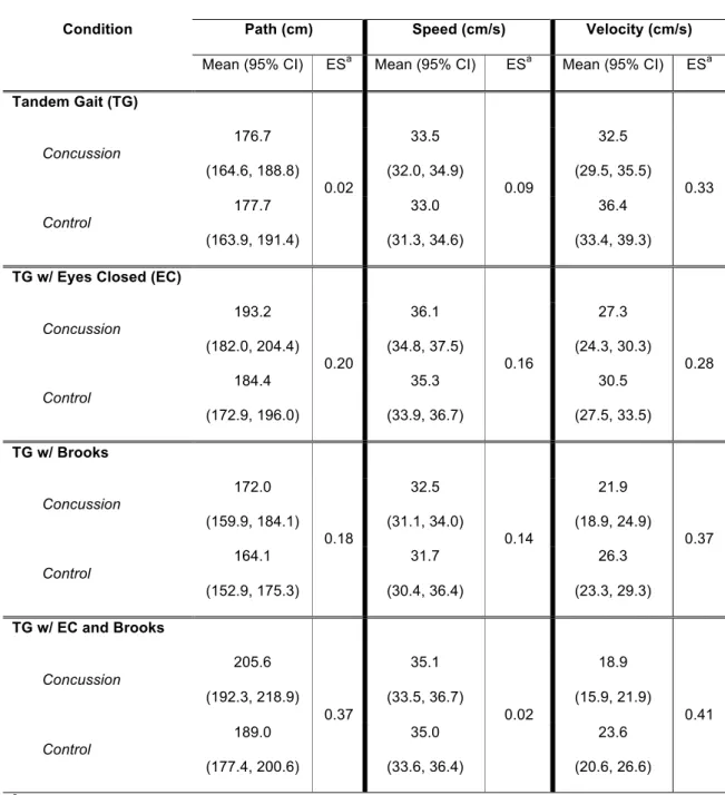
Data were averaged across all trials for each condition, and these average values were used for all statistical analyses. To explore tandem gait velocity, speed, and center of pressure path, we utilized a 4 (condition) x 2 (group) mixed-model analysis of covariance (ANCOVA). Brooks Visuospatial Task time to task completion was analyzed using a 3 (condition, including Brooks Visuospatial Task baseline, dual-task eyes open, and dual-task eyes closed) x 2 (group) mixed-model ANCOVA. Bonferroni corrected t tests were used to analyze any significant interactions or main effects. Dual-task cost was analyzed utilizing a between subjects ANCOVA. Because velocity was statistically different between groups, it was used a covariate when investigating center of pressure speed and path as well as all center of pressure dual-task outcomes. Additionally, we covaried for the number of days between the last concussion and the testing session in all statistical models. The number of days post-injury for each concussion group participant was subtracted from the group mean days since concussion (177 days). Control participants were assigned a value of zero. This created a mean centered days since concussion value, which was used as a covariate in all statistical models. An a priori alpha value of 0.05 was established.
Dual-task cost was calculated separately for eyes open and eyes closed conditions. When interpreting DTC, positive values indicate worse performance during dual-task
combined with time to complete the Brooks Visuospatial Task to form a single combined dual-task cost outcome for the cognitive task.
Specific Aim 2
Design and Setting
The study participants and exclusion criteria for Aim 2 were identical to Aim 1.
Instrumentation
The Vicon System (Vicon Motion Systems, Centennial, CO) consists of 10 infrared
video cameras in conjunction with two piezoelectric non-conductive force platforms (Model
#4060-NC Bertec Co., Columbus, OH) embedded in the floor. Each participant was outfitted
with 20 individual retro-reflective markers affixed to the skin or spandex over the jugular
notch, the tip of each shoulder, the L5 area of the low back, bilaterally on the
anterior-superior iliac spine of the pelvis, greater trochanter, medial epicondyle of the femur, lateral epicondyle of the femur, medial malleolus, lateral malleolus, first metatarsal head, and fifth metatarsal head. Cluster markers were affixed over the sacrum and bilaterally on each thigh, shank, and foot. Left side clusters consisted of 4 retro-reflective markers while the right side and sacral clusters consisted of 3 markers. Following an initial static trial, all individual markers except those at the tip of each shoulder and jugular notch were removed and the participant completed the testing with the clusters. Kinematic data were collected at 150 Hz and calibrated for a 4m long x 3m wide x 2.5m high volume while kinetic data were collected at 1500 Hz. All video data will be time synchronized with the analog force plate data. The world axis system was established as positive anteriorly in the sagittal plane, left in the frontal plane, and superior in the transverse plane.
Data Collection Procedures
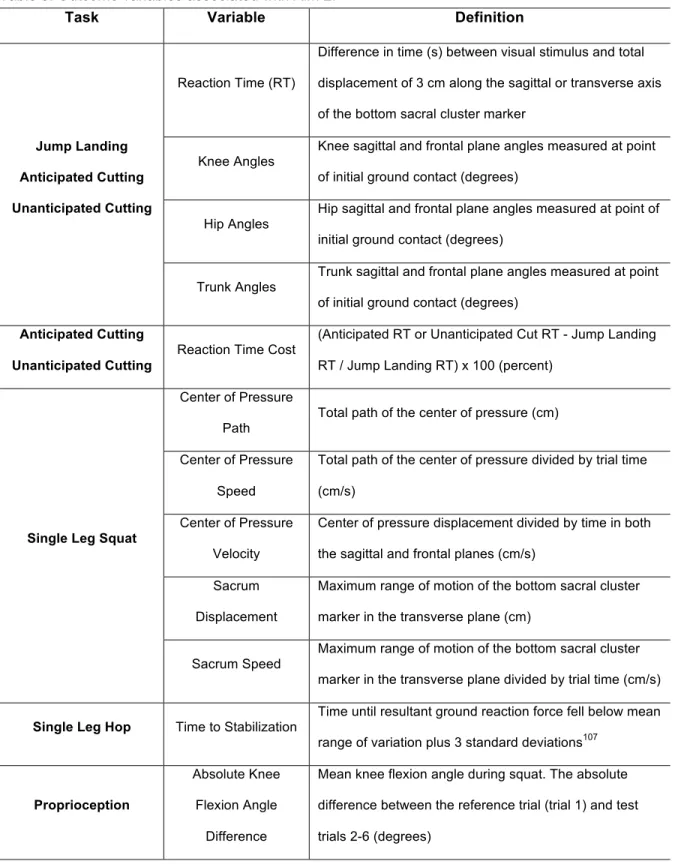
confident with the task. All functional tasks are described below and all outcome variables are described in Table 5.
Jump Landing. Participants stood atop a 30 cm box placed a horizontal distance equal to 50% of their height behind the force plates. The participants were instructed to “get set,” meaning they were to take an athletic stance upon the box and await a stimulus to signal the beginning of the trial. A visual stimulus (green light) placed approximately 3 m in front of the participant was triggered randomly within 5 seconds by the investigator,
indicating the start of the trial. The participant jumped forward off the box (told to “jump out, not up”) and performed a double-leg landing with the right foot in contact with one force plate and the left foot in contact with the other force plate before jumping vertically for maximal height. The participant was instructed to initiate the movement as quickly as possible following the visual stimulus. Each participant completed 5 jump landings.
Anticipated Cut. Participants stood atop a 30 cm box placed a horizontal distance equal to 50% of their height behind the force plates. The participants were instructed to “get set,” meaning they were to take an athletic stance upon the box and await a stimulus to signal the beginning of the trial. A visual stimulus (green light) placed approximately 3 m in front of the participant was triggered randomly within 5 seconds by the investigator,
indicating the start of the trial. The participant jumped forward off the box (told to “jump out, not up”) and landed on a single leg. Immediately upon landing, the participant cut at a 45° angle in the direction provided by the investigator prior to the trial (cut towards dominant = land on non-dominant foot, cut towards non-dominant = land on dominant foot). Each participant completed 5 trials cutting in each direction (10 total trials).
front of the participant was triggered randomly within 5 seconds by the investigator,
indicating the start of the trial. The participant jumped forward off the box (told to “jump out, not up”) and landed on a single leg. Immediately upon landing, the participant cut at a 45° angle. For the unanticipated cut, the participant was not informed which direction to cut. As the participant jumped from the box, they triggered a timing gate set at 0.76 m behind the force plates. This distance was chosen to maximize the time each participant would have to react to the directional stimulus, but be in a position where shorter participants would not be excluded. This timing gate triggered a visual stimulus (set of blue and green lights) to the participants left or right. Participants were instructed to cut towards the light in the same manner as the anticipated cutting task above (cut towards dominant = land on non-dominant foot, cut towards non-dominant = land on dominant foot). Each participant completed 10 total trials, regardless of whether or not they were performed correctly. Trials were discarded if the participant did not land appropriately on a single leg or cut in the wrong direction.
Single Leg Squat. Participants were asked to stand on a single leg, with their toes facing forward. The non-weight bearing leg was flexed to 90° at the knee approximately 75° at the hip, the hands placed on the hips, and the head and eyes facing forward. Participants flexed their weight-bearing knee to a squat, to maximal comfort, and then returned to the upright posture. Participants performed 5 single leg squat trials on each leg. Each trial consisted of 5 squat repetitions completed without pause at a self-selected pace.
Single Leg Hop. Participants stood atop a 30 cm box placed a horizontal distance equal to 50% of their height behind a force plate. Participants placed both hands on their hips and jumped off the box with both feet and landed on a single leg. Participants were instructed to come to a stable position as quickly as possible upon landing. The total trial time was 10 seconds, with participant holding their best single leg posture for as much of the trial as possible. Participants performed 5 single leg hop trials on each leg (10 total trials).