EXAMINATION OF SELECTIVE BROTH CULTURE MEDIA FOR THE DETECTION AND QUANTIFICATION OF VIBRIO CHOLERAE IN DRINKING WATER USING AN
ADAPTED COMPARTMENT BAG TEST
Megan Lott
A thesis submitted to the faculty at the University of North Carolina at Chapel Hill in partial fulfillment of the requirements for the degree of Master of Science in the Department of Environmental Sciences and Engineering in the Gillings School of Global Public Health.
Chapel Hill 2018
Approved by:
Mark Sobsey
Jill Stewart
ii © 2018 Megan Lott
iii ABSTRACT
Megan Lott: Examination of selective broth culture media for the detection and quantification of Vibrio cholerae in drinking water using an adapted Compartment Bag Test
(Under the direction of Mark Sobsey)
Cholera outbreaks may be mitigated by monitoring drinking waters for the bacterial disease agent, Vibrio cholerae. Present microbial methods for V. cholerae require advanced training and specialized equipment; these methods may not be appropriate for low-resource settings most vulnerable to cholera outbreaks. Our team hypothesized that the Aquagenx
iv
TABLE OF CONTENTS
LIST OF TABLES ... viii
LIST OF FIGURES ... xi
LIST OF ABBREVIATIONS ... xii
CHAPTER 1: INTRODUCTION ... 1
Objectives ... 3
Experimental Design ... 3
CHAPTER 2: REVIEW OF LITERATURE ... 6
The Genus Vibrio ... 6
Vibrio cholerae ... 7
Vibrio cholerae in the Aquatic Environment ... 9
The Epidemiology of Cholera ... 9
Methods for Cultivation and Enumeration V. cholerae ... 11
Field-Based Methods for Detection and Enumeration of V. cholerae ... 13
The Compartment Bag Test ... 15
Selective Culture Media for Vibrio and V. cholerae ... 16
Basal Media ... 17
Selective Agents... 17
Selective Culture Conditions ... 20
Growth Indicators and Specific Substrates ... 20
v
CHAPTER 3: METHODS ... 25
Sample Collection and Preparation ... 25
1% Primary Effluent-Surface Water Sample ... 25
Natural Surface Waters ... 25
Media Preparation ... 26
Examining the Exclusivity of Media for Culturing V. cholerae ... 28
Enumeration of Non-Target Organisms from 1% Primary Effluent-Surface Water Matrix on Solid Agar Media ... 28
Quantification of Non-Target Organisms from 1% Primary Effluent-Surface Water Matrix in Broth Media ... 29
Quantification of Non-Target Organisms from Surface Waters ... 30
Identification of Non-Target Organisms in Taurocholate Tellurite Peptone ... 31
Examining the Efficiency of Media for Culturing V. cholerae ... 32
Quantification and Enumeration of V. cholerae (Multiple Tube Format) ... 32
Quantification and Enumeration of V. cholerae (Multiple Well Format) ... 32
Examining the Efficiency of Basal Media for Culturing V. cholerae ... 33
Screening Candidate Basal Media for Growth of V. cholerae ... 33
Efficiency of Basal Media for Quantification of V. cholerae ... 34
Antibiotic Susceptibility Testing of V. cholerae and Non-V. cholerae Bacteria ... 35
Preparation of Antibiotics ... 35
Preparation of Inoculum ... 36
vi
Examining the Exclusivity of Newly-Proposed Culture Media for V. cholerae ... 37
Data Analysis ... 40
CHAPTER 4: RESULTS ... 41
Examining the Exclusivity of Media for Culturing V. cholerae ... 41
Enumeration of Non-Target Organisms from 1% Primary Effluent-Surface Water Matrix on Solid Agar Media ... 41
Quantification of Non-Target Organisms from 1% Primary Effluent-Surface Water Matrix in Broth Media ... 43
Quantification of Non-Target Organisms from Surface Waters ... 45
Identification of Non-Target Organisms in Taurocholate Tellurite Peptone ... 48
Examining the Plating Efficiency of Media for Culturing V. cholerae ... 49
Quantification and Enumeration of V. cholerae (Multiple Tube Format) ... 49
Quantification and Enumeration of V. cholerae (Multiple Well Format) ... 54
Examining the Plating Efficiency of Basal Media for Culturing V. cholerae... 63
Screening Basal Media for Growth of V. cholerae ... 63
Plating Efficiency of Basal Media for Quantification of V. cholerae ... 64
Antibiotic Susceptibility Testing of V. cholerae and Non-V. cholerae Bacteria ... 67
Examining the Exclusivity of Newly-Proposed Culture Media for Culturing V. cholerae ... 71
CHAPTER 5: DISCUSSION ... 74
Limitations ... 82
Future Directions ... 84
vii
APPENDIX A: Composition of Newly Proposed Media for Selective Culture
of V. cholerae ... 87
APPENDIX B: Raw Data for Enumeration of Non-Target Organisms from 1% Primary Effluent ... 89
APPENDIX C: Raw Data for Quantification of Non-Target Organisms from 1% Primary Effluent ... 90
APPENDIX D: Raw Data for Quantification of Non-Target Organisms in Surface Water ... 91
APPENDIX E: Raw Data for Quantification and Enumeration of V. cholerae (Multiple Tube Test)... 92
APPENDIX F: Raw Data for Quantification and Enumeration of V. cholerae (Multiple Well Test) ... 93
APPENDIX G: Raw Data for Efficiency of Basal Media ... 95
APPENDIX H: Raw Data Antimicrobial Susceptibility Testing – MIC Determination ... 96
APPENDIX I: Examining the Exclusivity of Newly-Proposed Selective Culture Media... 98
viii
LIST OF TABLES
Table 1. Composition of selective culture media described for V. cholerae ... 23
Table 2. Vibrio-specific media examined ... 27
Table 3. Criteria for enumerating colonies appearing the same as V. cholerae. ... 29
Table 4. Criteria for scoring wells for growth of all non-target organisms ... 30
Table 5. Criteria for scoring tubes for growth of all non-target organisms. ... 31
Table 6. Constituents of basal media. ... 33
Table 7. Organisms included in antibiotic susceptibility testing ... 35
Table 8. Composition of novel culture media for the selective culture of V. cholerae... 38
Table 9. Test organisms to examine the exclusivity of culture media for Vibrio spp. and V. cholerae. ... 39
Table 10. Criteria for scoring wells for growth of known, relevant test organisms. ... 40
Table 11. Frequency of observing non-target growth appearing the same as V. cholerae on selective agar media for across three replicate trials ... 43
Table 12. Frequency of observing any non-target growth in selective broth media for Vibrio spp. across three replicate trials... 45
Table 13. Results of the Wilcoxon Signed-Rank Test comparing median concentrations for V. cholerae O139 as determined by multiple tube test with TTGB and by spread plate on LBA, TCBS, and TTGA ... 51
Table 14. Results of the Wilcoxon Signed-Rank Test comparing median concentrations for V. cholerae O1 El Tor Ogawa as determined by multiple tube test with TTGB and by spread plate on LBA, TCBS, and TTGA ... 52
Table 15. Results of the Wilcoxon Signed-Rank Test comparing median concentrations for V. cholerae Non-O1 as determined by multiple tube test with TTGB and by spread plate on LBA, TCBS, and TTGA ... 53
ix
Table 17. Results of the Wilcoxon Signed-Rank Test to compare pooled median concentrations of V. cholerae by medium type ... 54 Table 18. Results of the Kruskal-Wallis test comparing median concentrations of V. cholerae O139 by medium type, incubation temperature, and medium format ... 56 Table 19. Results of the Wilcoxon Signed-Rank Test comparing median concentrations of V. cholerae O139 by medium type ... 56 Table 20. Results of the Kruskal-Wallis test comparing median concentration of V. cholerae O1 El Tor Ogawa by medium type, temperature, and medium format ... 57
Table 21. Results of the Wilcoxon Signed-Rank Test comparing median concentrations of V. cholerae O1 El Tor Ogawa as determined by LB, TTGB, TCBS and CV ... 57
Table 22. Results of the Kruskal-Wallis test comparing median concentration of V. cholerae Non-O1 by medium type, temperature, and medium format ... 58
Table 23. Results of the Wilcoxon Signed-Rank Test comparing median
concentrations of V. cholerae Non-O1 as determined by LB, TTGB, TCBS and CV ... 58 Table 24. Results of the Kruskal-Wallis test comparing pooled estimates of the median concentration of V. cholerae by strain type, medium type, temperature, and medium format ... 59 Table 25. Results of the Wilcoxon Signed-Rank Test comparing median concentrations of V. cholerae as determined by LB, TTGB, TCBS and CV ... 60 Table 26. Results of the ANOVA test comparing the productivity ratio of each medium by strain type, medium type, temperature, and medium format ... 62 Table 27. Results of the Paired T-Test comparing productivity ratios of TTGB, TCBS and CV ... 62 Table 28. Screening of basal media for overnight growth of V. cholerae ... 63
Table 29. Results of the Kruskal-Wallis test comparing median concentrations of V. cholerae by strain type, basal medium type, added NaCl, and complete
x
xi
LIST OF FIGURES
Figure 1. Experimental design ... 5
Figure 2. Classifications of V. cholerae by toxin-production, biovar, and serogroup ... 8
Figure 3. EPA Standard Analytical Protocol for V. cholerae O1 and O139 in Drinking and Surface Water ... 11
Figure 4. CBT Kit Instructions ... 15
Figure 5. Enumeration of non-target organisms with Vibrio appearance cultured from 1% primary effluent in surface water ... 42
Figure 6. Quantification of all non-target organisms from 1% primary effluent in surface water ... 44
Figure 7. Quantification of all non-target organisms from natural surface waters ... 47
Figure 8. Plating efficiency of TTGB for the quantification of V. cholerae by multiple tube test ... 50
Figure 9. Plating efficiency of selective culture media for the quantification of V. cholerae by multiple well test and agar media enumeration ... 55
Figure 10. Estimated concentrations of V. cholerae as determined by LBA/LB, TTGA/TTGB, TCBS/TCBS Broth, and CV/CV Broth ... 60
Figure 11. Average productivity ratio of each selective medium ... 61
Figure 12. Efficiency of basal broth media for the quantification of V. cholerae. ... 64
xii
LIST OF ABBREVIATIONS
APW Alkaline Peptone Water
BE Beef Extract
BHI Brain Heart Infusion
CBT Compartment Bag Test
CFU Colony Forming Units
CPC Cellobiose, Polymyxin, Colistin Agar
CV CHROMagar Vibrio Agar
EUCAST European Committee on Antimicrobial Susceptibility Testing GSLS Gelatin Salt Lauryl Sulfate
GTFCC Global Task Force on Cholera Control
LB Luria Broth
LBA Luria Broth Agar
MALDI-TOF MS Matrix-Assisted Laser Desorption/Ionization Time of Flight Mass Spectrometry
MHB Mueller-Hinton Broth
MIC Minimum Inhibitory Concentration
MPN Most Probable Number
MTT Multiple Tube Test
SDS Sodium Dodecyl Sulfate
SLS Sodium Lauryl Sulfate
STT Sucrose Teepol Tellurite Agar
xiii
TCI Thiosulphate Chloride Iodide Agar TTGA Taurocholate Tellurite Gelatin Agar TTGA Taurocholate Tellurite Gelatin Broth
US EPA United States Environmental Protection Agency
VBNC Viable but Non-Culturable
VP Vibrio parahaemolyticus Agar
WASH Water, Sanitation and Hygiene
WHO World Health Organization
1
CHAPTER 1: INTRODUCTION
Vibiro cholerae is the etiologic agent of the water-borne illness which bears its name, cholera. Cholera is an acute diarrheal disease that causes watery stools, severe dehydration, and can lead to death if not treated promptly and properly. Since the 19th century, seven cholera pandemics have plagued global populations (Thompson, Austin, & Swings, 2006). The seventh pandemic began in Asia in 1961 and continues today (World Health Organization, 2018). Researchers estimate that there are as many as 2.9 million cases of cholera annually, which account for 95,000 deaths across 69 endemic countries (Ali, Nelson, Lopez, & Sack, 2015). The country of Yemen is currently facing the world’s largest outbreak; between April and December of 2017, 1 million cases of cholera were suspected in this region alone (World Health
Organization, 2018). With the threat of such outbreaks, cholera remains a pressing concern for global public health.
Cholera is transmitted through food and water contaminated with V. cholerae. Cholera outbreaks are often reported in low-income areas with inadequate or failing water and
2
(WASH) systems. The Global Roadmap to 2030 calls specifically for provisions of rapid microbial test kits to prevent the transmission of V. cholerae and ensure safe water quality.
Field monitoring for V. cholerae can prevent disease transmission by identifying
contaminated sources of water. However, many of the widely-accepted methods for detection of V. cholerae may not be appropriate for source monitoring in those low-resource settings that are vulnerable to cholera outbreaks. Current methods require advanced equipment and trained technicians to implement culture, molecular, or immunochemical assays for identification and enumeration of V. cholerae (Huq, 2013; Thompson et al., 2006). There is a need for improved, low-cost and portable microbial test kits for V. cholerae.
The Compartment Bag Test (CBT) is a field-friendly method for microbial monitoring of drinking water. The CBT is a low-cost, portable, and field-ready microbial test kit that was originally developed for the quantification of indicator bacteria in drinking and surface waters (Stauber, Miller, Cantrell, & Kroell, 2014). The test kit may be adapted for the direct
3 Objectives
1. To examine the exclusivity of previously-described culture media for V. cholerae. 2. To compare the plating efficiency between culture media for V. cholerae.
3. To examine the exclusivity and efficiency of broth adaptations of solid agar media previously-described for selective culture of V. cholerae.
4. To examine the effect of incubation temperature at 37°C and 42°C on the exclusivity and plating efficiency of selective culture media for V. cholerae.
5. To examine the exclusivity of newly-formulated broth media intended for use with the Compartment Bag Test for quantification V. cholerae.
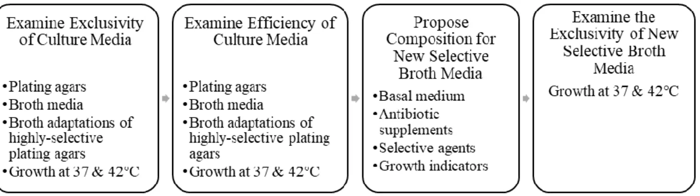
Experimental Design
The purpose of this study was to evaluate selective culture media for direct quantification of V. cholerae and to identify a culture medium appropriate for use in the CBT. An appropriate culture medium for an adapted Cholera CBT would be a broth medium that is selective against non-target microorganisms occurring in stored household drinking waters and natural surface water sources. The selectivity of the medium is imperative to limit false-positive results or inaccurate estimations of bacterial concentration of V. cholerae. An appropriate broth medium should enrich V. cholerae and be utilized for direct, efficient quantification of V. cholerae in drinking water samples. Selective growth of V. cholerae should be clearly visualized by chromogenic or fluorogenic change.
fecally-4
contaminated drinking water sample. In this study, we defined exclusivity as a feature of a selective agar medium; it describes the medium’s extent of inhibition of naturally-occurring non-target, non-vibrio organisms. The exclusivity of these plating agars was compared by incubation at both 37°C and 42°C.
The most selective and exclusive plating agars were adapted into broth media by centrifugation to eliminate the agar component. The exclusivity of these broth adaptations was compared to the exclusivity of previously-described selective broth enrichment media for Vibrio spp. and V. cholerae. Exclusivity was examined by quantifying the extent of growth of non-target organisms from a simulated fecally-contaminated drinking water sample. Exclusivity was compared for incubation temperatures of 37°C and 42°C.
Three selective media were compared based on their plating efficiency as both plating agars and broth media. Pure cultures of V. cholerae were plated onto modified taurocholate tellurite gelatin agar (TTGA), Thiosulfate-citrate-bile salts-sucrose agar (TCBS), CHROMagar Vibrio agar (CV), as well as non-selective Luria Broth (LB). The efficiency of these media was compared at incubation temperatures of 37°C and 42°C.
5
6
CHAPTER 2: REVIEW OF LITERATURE The Genus Vibrio
The genus Vibrio belongs to the family of Vibrionaceae, which consists of gram negative organisms that are oxidase-positive and have polar flagella (Drasar, B.S and Forrest, 1996). Historically, this family has also included the genera: Aeromonas, Photobacterium, and
Plesiomonas (Sneath, Mair, & Sharpe, 1986). Recently, molecular analyses have distinguished two new genera (Listonella and Shewanella), as well as a new family, Aeromonadaceae
(Koneman, Allen, & Janda, W.M. Schreckenberger, 1997).
Species belonging to the genus Vibrio are often classified as either V. cholerae or non-cholera Vibrios (Drasar, B.S and Forrest, 1996; Koneman et al., 1997). The primary habitat for most of these organisms is sea, brackish or freshwater (Forbes, Betty, Sahm, Daniel, Weissfeld, 2017; Thompson et al., 2006). A few vibrios can be isolated from freshwaters. These include V. cholerae, V. mimicus, V. fluvialis, V. navarrensis, and V. andguillarum (Thompson et al., 2006) Human infection by non-cholera Vibrios is typically associated with the consumption of
contaminated seafood or experiencing wounds that become infected by contaminated waters (Kaysner, Charles and DePaola, 2004).
7
dihydrolase-negative. Vibrio mimicus was once considered a subspecies of V. cholerae, but molecular analyses have distinguished the two species (Koneman et al., 1997). Vibrio mimicus may be differentiated from V. cholerae by the inability to ferment sucrose and a negative reaction on the Voges-Proskauer Test that is used to determine if an organism produces acetylmethyl carbinol from glucose fermentation (Kaysner, Charles and DePaola, 2004).
Vibrio cholerae
There are more than 140 strains of V. cholerae that are described and differentiated based on serogroup (Drasar, B.S and Forrest, 1996). Most of the strains associated with the cholera disease belong to the O1 serogroup (Drasar, B.S and Forrest, 1996). These strains produce the cholera enterotoxin, the main virulence factor associated with cholera (Thompson et al., 2006).
8
Figure 2. Classifications of V. cholerae by toxin-production, biovar, and serogroup. Adapted in part from Banerjee et al. 2014.
Strains of V. cholerae Non-O1 have been known to cause diarrheal illness similar to cholera (Drasar, B.S and Forrest, 1996). Some Non-O1 serotypes produce enterotoxins
indistinguishable from the cholera toxin (Dhiman Barua and William B. Greenough III, 1992). The O139 strain of V. cholerae emerged in 1993 during a large cholera outbreak across India and Bangladesh (Kaysner, Charles and DePaola, 2004). The O139 Bengal strain produces the same cholera toxin produced by those relevant strains of V. cholerae O1. Researchers suspect that the V. cholerae O139 strain may be responsible for an eighth cholera pandemic (Dhiman Barua and William B. Greenough III, 1992; Drasar, B.S and Forrest, 1996; Kaysner, Charles and DePaola, 2004). The relationship between these classifications of V. cholerae strains is described in Figure 2. Evidence suggests that mobile genetic elements can confer virulence to other Non-O1 and Non-O139 serogroups, and that these organisms may lead to potential disease outbreaks (Li, Shimada, Morris, Sulakvelidze, & Sozhamannan, 2002).
Vibrio cholerae
Serogroups that produce Cholera
Toxin
O1
Classical Inaba, Ogawa, Hikojima
El Tor Inaba, Ogawa, Hikojima
O139
Bengal
Calcutta
Serogroups that do not produce Cholera Toxin
9 Vibrio cholerae in the Aquatic Environment
Vibrio cholerae is a natural inhabitant of aquatic environments and may be isolated from fresh, brackish, and coastal waters (Dhiman Barua and William B. Greenough III, 1992; Huq, 2013; Thompson et al., 2006). V. cholerae can be isolated from environmental samples, even in regions where cholera is not endemic (Huq, 2013). The bacterium can be free-swimming, or found in association with sediment, zooplankton and shellfish. Sediment can harbor V. cholerae, and stressed bacteria can enter a dormant state to ensure long-term survival in natural waters (Dhiman Barua and William B. Greenough III, 1992; Huq, 2013). Vibrio species associate well with copepods and shellfish, as these zooplankton and shellfish facilitate increases in bacterial density (Huq, 2013; Thompson et al., 2006). The complex ecology of V. cholerae requires that sources of drinking water be monitored routinely for microbial quality and detection of the cholera bacterium, especially in areas where cholera occurs in humans and V. cholerae are present in environmental waters.
The Epidemiology of Cholera
10
severe dehydration and even death. Patients with acute cholera may excrete up to 20 liters of stool per day, containing viable organisms of V. cholerae (Dhiman Barua and William B. Greenough III, 1992). Without proper sanitation, these organisms can contaminate household and environmental waters.
Disease outbreaks are typically associated with poor or failing water and wastewater infrastructure (World Health Organization, 2018). Cholera can persist as an endemic disease, spreading through local populations in the same region over time. Today, cholera is endemic to Southern Asia, parts of Africa, the Middle East and Latin America (Thompson et al., 2006). Epidemic cholera emerges in new regions where local transmission is not often recognized or reported. The risk of disease transmission can increase during disaster events in the face of flooding, failing infrastructure, or displacement of large populations (Watson, Gayer, & Connolly, 2007). Humanitarian crises may similarly displace groups to overcrowded camps without provisions for clean water and sanitation (World Health Organization, 2018).
Disease control requires improvements in sanitation and provisions for clean drinking water in low-resource and disaster settings. Outbreaks from the seventh cholera pandemic have been associated with waters from rivers and other sources that are used without treatment
(Dhiman Barua and William B. Greenough III, 1992). Drinking water wells can be contaminated by fecal waste from cases of cholera within households and communities. Waters that are
11
Methods for Cultivation and Enumeration V. cholerae
Routine water quality monitoring can help reduce the risk of transmission of waterborne cholera. Microbial surveillance for V. cholerae can identify contamination in surface water sources, wells, or stored water supplies. Field-based
monitoring can evaluate the safety of drinking waters before use, examine the efficacy of point-of-use disinfection, or identify hotspots for V. cholerae
contamination. By identifying water contaminated with the organisms, remedial actions such as water treatment can be taken to eliminate the contamination.
Current methods for the detection and
quantification of V. cholerae include culture, molecular, immunochemical, or biochemical assays (Huq, 2013; Thompson et al. 2006). Many of these methods require specialized media and reagents, laboratory equipment and trained personnel that limit the applicability of these methods for routine field-based water quality monitoring. Recent advancements in field-based technologies may improve microbial surveillance in low-resource settings where prevention measures are often most needed.
To detect and isolate Vibrio cholerae from environmental samples by culture-based methods, the samples are often pre-enriched in non-selective and/or selective broth culture media and the cultures are then streaked onto selective agar plating media (Thompson et al., 2006).
Determine MPN based on tubes positive for V. cholerae based on biochemical or molecular analyses Confirm any growth on TCBS with
biochemical analyses or PCR specific for V. cholerae Streak 20µL from the pellicle of each
tube onto TCBS. Incubate at 37°C for 24 hours
Incubate tubes at 37°C for hours or 24 hours
Arrange 3-dilution MPN with 5 tubes of each: 5mL of 5X APW, 20mL undiluted sample 10mL of 2X APW, 10mL undiluted sample 10mL of 1X APW, 1mL undiluted sample
Figure 3. EPA Standard
12
Biochemical, immunological and/or molecular assays are often required to confirm that these presumptive isolates are V. cholerae and to determine their specific strain or type.
Vibrio cholerae can be quantified in environmental samples by traditional culture-based methods. Standard methods require multiple procedures, including pre-selective enrichment and biochemical or molecular confirmation of presumptive V. cholerae organisms. The methods described by the International Standards Organization (ISO) for the isolation of Vibrio spp. suggest that organisms from environmental media should be enriched in selective broth and then plated onto TCBS and an additional selective medium (Hartnell et al., 2018; ISO, 2017). Any presumptive colonies should then be confirmed using biochemical assays with an oxidase test, string test, or Voges-Proskaur Test. Molecular assays, such as the polymerase chain reaction (PCR) can confirm presumptive colonies as well.
The EPA Standard Analytical Protocol for V. cholerae O1 and O139 in Drinking Water and Surface Water describes a similar multi-step, culture-based method for quantification of V. cholerae. Using these methods, V. cholerae can be quantified by a multiple-tube assay
13
viable but nonculturable state (VBNC), during which they do not grow or form colonies on traditional culture media (Huq, 2013). Alternatively, V. cholerae can be enumerated by flow cytometry, colony hybridization, or culture-independent methods (Huq, 2013; Thompson et al., 2006).
Culture-independent methods may be used to enumerate V. cholerae from environmental samples. Fluorescence in situ hybridization (FISH) has been used to quantify Vibrios based on extracted nucleic acids (Huq, 2013; Thompson et al., 2006). Real-time PCR may be used to quantify V. cholerae (Thompson et al., 2006). The direct fluorescent antibody – direct viable count method can be used to distinguish viable from VBNC organisms of V. cholerae while estimating the count of organisms (Huq, 2013; Kahler et al., 2015).
These methods require laboratory equipment and trained personnel that limit the applicability of these methods for routine field-based water quality monitoring. Some of these methods may be adapted for field-use by implementing portable devices that employ the same culture-based or immunological assays. Recent advancements in such field-based technologies may improve microbial surveillance in low-resource settings where prevention measures are often most needed.
Field-Based Methods for Detection and Enumeration of V. cholerae
Field-based methods for detection of V. cholerae may improve microbial surveillance in low-resource settings. Emerging field-based methods implement both culture-based and
14
validated for surveillance of V. cholerae in household and stored drinking water (Rashid et al., 2017). The dipstick test utilizes antibodies that are specific to O1 and O139 serotypes to confirm the presence or absence of V. cholerae in a 1mL pre-enriched sample. Field-based
electrochemical immunosensors (biosensors) may be used for environmental surveillance of V. cholerae, again confirming the presence of the organism in a pre-enriched sample (Cecchini, Fajs, Cosnier, & Marks, 2016; Sharma, Goel, Singh, & Rao, 2006).
These alternative and novel methods may be useful tools for field-based monitoring of waters vulnerable to contamination. However, the methods above only analyze small volumes of sample (5 µL – 3mL) for the presence or absence of V. cholerae and they cannot be used readily to quantify the bacterial concentration in test waters unless modified to a multiple volume quantal assay format. A portable flow-cytometer may be used for rapid assays to enumerate V. cholerae from drinking water reservoirs, but the equipment may not be cost-efficient for low-resource settings (Righetto et al., 2015). Furthermore, many of these non-culture-based methods are based on detecting V. cholerae cells as physical objects or are based on detection of specific nucleic acids or antigens. They do not provide evidence that the detected cells or cell constituents are culturable or infectious unless applied after an initial culture procedure.
15 The Compartment Bag Test
The Aquagenx CBT is ideal for water quality monitoring in low-resource settings. The test kit is a low-cost, portable, and easy-to-use quantal method for quantification of culturable bacteria of E. coli or H2S -producing bacteria (Aquagenx, 2018). The CBT is a clear plastic bag designed for 100mL samples. Water samples are mixed with a selective culture medium and dispensed into the CBT. The bag is divided into multiple compartments of different volumes (1mL, 3mL, 10mL, 30mL, and 56mL). Samples are incubated overnight and these individual compartments are scored for growth based on chromogenic change of the culture medium. The results are reported as an most probable number, MPN/100mL, with corresponding confidence intervals as described in the user “look-up” tables described by Gronewold, Sobsey, & McMahan (2017). The CBT Kit instructions are shown in Figure 4.
Figure 4. CBT Kit Instructions. Accessed from Aquagenx LLC at https://www.aquagenx.com/how-to-use-the-cbt/.
16
Whalen, 2017). The Compartment Bag Test may be adapted for the direct quantification of V. cholerae if paired with an appropriate selective and differential culture medium.
Selective Culture Media for Vibrio and V. cholerae
Many broth media and plating agars have been described for the selective enrichment and culture of Vibrio spp. (Dhiman Barua and William B. Greenough III, 1992; Donovan & van Netten, 1995; Koneman et al., 1997; Thompson et al., 2006). Several additional commercially-available media may be utilized for selective isolation of Vibrio spp. These media are described in Table 1. Almost none of these media are capable of selecting for a single species of Vibrio (Thompson et al., 2006).
The most commonly used medium for V. cholerae is TCBS. The major disadvantage of this medium is its limited selectivity and specificity against non-vibrios (Donovan & van Netten, 1995). Species of Acinetobacter, Aeromonas, Alcaligenes, Enterobacter, Escherichia coli, Pasteurella, Pseudomonas, Salmonella, and Proteus are all able to grow on TCBS (Donovan & van Netten, 1995). Another limitation of TCBS is that this medium cannot distinguish
specifically between V. cholerae and other sucrose fermenting Vibrios such as V. alginolyticus, V. fluvialis, V. furnissii, and V. metschnikovii, (Thompson et al., 2006).
non-17
target non-Vibrio organisms, and distinguish between V. cholerae and V. mimicus. Such a medium would employ nutrients, specific substrates, selective agents, and growth indicators for the selective broth culture of V. cholerae.
Basal Media
Of the Vibrio media previously described in literature, basal media are composed of peptone alone or in combination with beef extract, yeast extract, or cellobiose. Columbia Blood Agar Base or Brain Heart Infusion Broth may be appropriate for culturing V. cholerae as well (Beazley, 1992; Dhiman Barua and William B. Greenough III, 1992). Gelatin may be
incorporated into differential media, as most vibrios hydrolyze gelatin (Kaysner, Charles and DePaola, 2004). However, other non-target organisms like Aeromonas spp. may also hydrolyze gelatin (Dhiman Barua and William B. Greenough III, 1992).
Vibrio species grow best under alkaline conditions (Kaysner, Charles and DePaola, 2004). While many Vibrios are halophilic, V. cholerae and V. mimicus do not require addition of NaCl in culture media; these organisms can utilize sodium ions from other constituents that make up most media (Kaysner C. and DePaola A. 2004). Multiple strains of V. cholerae demonstrate optimal growth at NaCl levels between 0.5% and 5% NaCl (Griffitt & Grimes, 2013).
Selective Agents
Selective media for Vibrio spp. incorporate selective agents and differential components that inhibit gram-positive organisms and distinguish Vibrios from other gram-negative
18
Bile salts. Bile salts may be incorporated into culture media for the selective growth of gram-negative, enteric organisms. Originating from the gastrointestinal tract, these animal-origin agents inhibit most gram-positive organisms and some gram-negative organisms. Among the most common bile salts used for selective culture of V. cholerae are: oxgall/oxbile, sodium cholate, sodium deoxycholate, and sodium taurocholate. Ox bile is a mixture of conjugated bile salts. Sodium deoxycholate is a conjugated bile salt and the most potent of the animal-origin inhibitors (EMD Millipore Corporation, 2008). While gram-negative bacteria are relatively resistant to bile salts, sodium deoxycholate may exhibit an initial killing effect on gram-negative bacteria (Paul D ’Mello, 1980).
Tellurite. Tellurium (Te) compounds have been used as antimicrobial agents (Taylor, 1999). Microbes that are resistant to tellurite may convert the agent to a less toxic form, tellurium. These microbes produce black colonies on selective media. Potassium tellurite has been described as a selective agent for the isolation of Corynebacterium diphtheriae,
Staphylococcus aureus, and Shigella spp. Tellurite resistance has been occasionally reported for Pseudomonas spp. Potassium tellurite has no inhibitory effect on V. cholerae at a concentration of 1:200,000 (Monsur, 1961).
Media prepared with potassium tellurite must be monitored for quality assurance, as tellurite converts to tellurite over time and diminishes the potency of the medium (Bolinches, Romalde, & Toranzo, 1988; Thompson et al., 2006). It is best practice to use media with
potassium tellurite within 24 hours of preparation (Thompson et al., 2006). This reagent is filter sterilized (Atlas, 1997).
19
may be used in place of bile salts (Jameson, J.E., 1956). Cationic detergents are more effective against gram-positive bacteria, but also inhibit gram-negative organisms (Baker, Harrison, & Miller, 1941; Salton, 1960). A killing effect of gram-negative organisms has been observed for anionic detergents at concentrations exceeding 1:30,000 (Baker et al., 1941).
Selective Dyes. Selective dyes may be incorporated into culture media for the inhibition of non-target microorganisms. Crystal violet is a basic dye that inhibits growth of gram-positive organisms, while still allowing growth of gram-negative organisms (Fung & Miller, 1973). Bromothymol blue is an acid dye that inhibits gram-positive organisms (Fung and Miller 1973). This dye may be implemented as a pH indicator in Vibrio media.
Sodium Citrate. Sodium citrate has antimicrobial properties for the inhibition of gram-positive bacteria, but shows little activity against gram-negative bacteria (Lee, Cesario, Owens, Shanbrom, & Thrupp, 2002).
Antibiotics. Additional antimicrobials may improve the selectivity of a culture medium, depending on the antibiogram of the target organism and competing non-target organisms. Polymixin B and colistin may be implemented in selective culture media for V. cholerae, but some serogroups of V. cholerae (like the Classical biovars) are susceptible to colistin (Dhiman Barua and William B. Greenough III, 1992; Thompson et al., 2006). V. cholerae has
20
known to cause false-positives in media for V. cholerae (Donovan & van Netten, 1995; Monsur, 1961; Popovic, Steinort, Pillai, & Joukhadar, 2010).
Selective Culture Conditions
High pH. Vibrio species grow best under alkaline conditions (Centre for Disease Control and Prevention (CDC);, 2004; Dhiman Barua and William B. Greenough III, 1992; Thompson et al., 2006). Optimal growth is observed at pH between 7.6 and 8.6, but may be supported at a pH range between 5.6 and 9.6 (Dhiman Barua and William B. Greenough III, 1992). High pH is thought to improve the selectivity of culture media for Vibrio spp. but this assertion has also been contested (Donovan & van Netten, 1995; Thompson et al., 2006).
Incubation Temperature. Vibrio cholerae grows optimally at incubation temperatures between 20°C and 45°C (Martinez, Megli, & Taylor, 2010). Increased incubation temperature may select against non-target environmental or enteric organisms. V. cholerae does not persist at 4°C for extended periods; refrigeration or cold storage of samples may result in V. cholerae becoming VBNC (Martinez et al., 2010; USEPA, 2010).
Growth Indicators and Specific Substrates
Chromogenic and fluorogenic indicators are chemical agents and metabolizable
21
(Kaysner, Charles and DePaola, 2004). Phenol red turns yellow in acidic conditions. However, sucrose fermentation is not specific to V. cholerae or Vibrio spp. The vibrios V. alginolyticus, V. fluvialis, V. furnissii, and V. metschnikovii are all sucrose-positive, as are the Enterobacteriaceae Klebsiella pneumonia and Proteus vulgaris (Dhiman Barua and William B. Greenough III, 1992; Kaysner, Charles and DePaola, 2004; Koneman et al., 1997). V. mimicus is a non-fermenting vibrio, and may be distinguished from V. cholerae based on an indicator for sucrose fermentation (Dhiman Barua and William B. Greenough III, 1992; Kaysner, Charles and DePaola, 2004).
Colwell et al. described that 4-methylumbelliferyl-β-D-galactoside (4-MU-Gal) may be incorporated into selective and differential media for the culture of V. cholerae (1985). The substrate fluoresces when cleaved by enzymatically-active β-D-galactosidase in viable cells of V. cholerae. The β-galactosidase enzyme is not specific to V. cholerae, however, and may
contribute to high numbers of false-positive results when examining contaminated water samples (Tryland & Fiksdal, 1998). The vibrios V. fluvialis, V. furnissii and V. mimicus are all capable of β-gal activity (Kaysner, Charles and DePaola, 2004). The β-galactosidase enzyme has been identified in several families and genera of gram-negative organisms including the
Enterobacteriaceae, Pseudomonadaceae, and Neisseriaceae (Tryland & Fiksdal, 1998).
22
HardyCHROM Vibrio, CHROMagar Vibrio, chromID, and HiChrome Vibrio utilize such proprietary chromogenic substrates (Table 1).
Criteria for Cholera CBT Medium
An appropriate medium for quantification of V. cholerae with the CBT should enrich the growth of V. cholerae to the exclusion non-target microorganisms. An appropriate medium may differentiate between V. cholerae and organisms of non-concern. This medium must perform as a broth medium that can be applied to an MPN format to enrich the growth of V. cholerae and accurately estimate the concentration of organisms in a drinking water sample. Such a broth medium should incorporate selective agents such as bile salts, detergents, selective dyes, or antibiotics to inhibit gram-positive and other gram-negative organisms. The medium may
maintain a pH at or above 8.6 to improve selectivity. A chromogenic or fluorogenic substrate and indicator should be incorporated for clear visualization of V. cholerae in the CBT.
Culture media were included in this study based on the above criteria, and the ease of access of media components. Media selected for examination are described in Table 2. Samples of the commercial media HardyCHROM Vibrio Agar, HiChrome Vibrio Agar, and CHROMagar Vibrio Agar were donated by their manufacturers. These media were examined for their
23
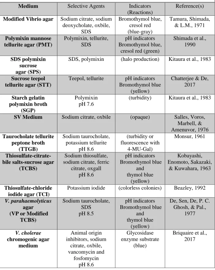
Table 1. Composition of selective culture media described for V. cholerae. Adapted in part from Donovan & van Netten, 1995 and Thompson et al., 2006.
Medium Selective Agents Indicators (Reactions)
Reference(s) Alkaline peptone
water
pH (turbidity) Furniss, Lee, & Donovan, 1978 Cellobiose,
polymyxin, colistin agar (CPC)
Colistin, polymixin B pH indicators Bromothymol blue
and Cresol red (green)
Massad & Oliver, 1987
CHROMagar Vibrio Salts mixture pH 8.6
Chromogenic substrates
(blue)
CHROMagar
chromID Vibrio Not disclosed Chromogenic substrate (blue-green) bioMerieux Fe-EDTA enrichment broth Fe-EDTA pH 9.0
(turbidity) Kida, Suzuki, & Taguchi, 1995 Gelatin phosphate
saline broth (GPS)
- (turbidity) Madden,
McCardell, & Morris, 1989 Glucose salt teepol
broth (GSTB)
Methyl violet, teepol, 3% NaCl
pH 9.4
(turbidity) Akiyama et al., 1963
Glucose-salt-tellurite-crystal violet medium
(GSTC)
crystal violet, potassium tellurite, 3%
NaCl pH 8.4 pH indicator Bromothymol blue (yellow) Bolinches, Romalde, & Toranzo, 1988 HardyCHROM Vibrio agar
Sea salt mixture, oxbile, sodium citrate,
sodium pyruvate Chromogenic mixture (purple) Hardy Diagnostics HiChrome Vibrio agar Sodium citrate, sodium cholate pH 8.5 Chromogenic mixture (purple) HiMedia Labs Modified taurocholate tellurite gelatin agar
(TTGA) Sodium taurocholate, potassium tellurite pH 8.6 4-MU-Gal (blue fluorescence) Monsur, 1961; O’Brien & Colwell,
24
Table 1 (Continued). Composition of selective culture media described for V. cholerae. Adapted in part from Donovan & van Netten, 1995 and Thompson et al., 2006.
Medium Selective Agents Indicators (Reactions)
Reference(s) Modified Vibrio agar Sodium citrate, sodium
deoxycholate, oxbile, SDS Bromothymol blue, cresol red (blue-gray) Tamura, Shimada, & L.M., 1971 Polymixin mannose
tellurite agar (PMT)
Polymixin, tellurite, SDS
pH indicators Bromothymol blue,
cresol red (green)
Shimada et al., 1990 SDS polymixin
sucrose agar (SPS)
SDS, polymixin (halo production) Kitaura et al., 1983
Sucrose teepol tellurite agar (STT)
Teepol, tellurite pH indicators Bromothymol blue
(yellow)
Chatterjee & De, 2017 Starch gelatin polymixin broth (SGP) Polymixin pH 7.6
(turbidity) Kitaura et al., 1983
SV Medium Sodium citrate, oxbile (opaque) Salles, Voros, Marbell, & Amenuvor, 1976 Taurocholate tellurite peptone broth (TTGB) Sodium taurocholate, potassium tellurite pH 8.6 (turbidity or fluorescence with 4-MU-Gal) Monsur, 1961 Thiosulfate-citrate-bile salts-sucrose agar
(TCBS)
Sodium thiosulfate, sodium citrate, ferric
citrate, oxgall pH 8.6 pH indicators Bromothymol blue and thymol blue (yellow) Kobayashi, Enomoto, Sakazaki,
& Kuwahara, 1963
Thiosulfate-chloride iodide agar (TCI)
Potassium iodide (colorless colonies) Beazley, 1992 V. parahaemolyticus
agar (VP or Modified
TCBS) Sodium taurocholate, SDS pH 8.5 pH indicators Bromothymol blue and thymol blue (yellow)
De, Sen, De, P. C. Ghosh, & Pal.,
1977 V. cholerae chromogenic agar medium Animal origin inhibitors, sodium citrate, oxbile, vancomycin and fosfomycin pH 8.6 Glycosidase enzyme substrate (blue)
25
CHAPTER 3: METHODS Sample Collection and Preparation
1% Primary Effluent-Surface Water Sample
Primary sewage effluent collected from the Mason Farm Wastewater Treatment Plant (Orange County, NC) was combined at a volume ratio of 1:100 (1% final concentration) with natural surface water from Morgan Creek (Chapel Hill, NC). This combined water matrix was prepared to achieve a diversity of microorganisms in a small-volume inoculum and to simulate samples from impaired watersheds and fecally-contaminated drinking water sources. Cholera is not endemic in the United States and neither primary effluent nor surface water samples were expected to contain V. cholerae. The effluent and surface water may contain other non-target organisms that interfere with culture-based methods to detect V. cholerae. Therefore, any organisms cultured from the primary effluent are presumptive non-vibrios and non-V. cholerae.
Primary sewage samples were collected in sterile polypropylene bottles by the staff at the wastewater treatment facility. Samples were stored at 4°C overnight. A 1mL sample of primary effluent was spiked into 99mL of natural surface water before each experiment.
Natural Surface Waters
26
Trail, a public green space with paved and wooden walking trails. Samples were collected in sterile polypropylene bottles and stored at 4°C overnight. Morgan Creek is an inland stream that is not expected to have a population of Vibrio spp. Therefore, any organisms cultured from Morgan Creek are presumptive non-vibrios and non-Vibrio cholerae.
Media Preparation
Enrichment broths and plating agars examined for this study are described in Table 2. Media were prepared according to The Handbook of Microbiological Media (1997). Media that were commercially-sourced were prepared according to manufacturer’s instructions. Broth and agar adaptations of Taurocholate-Tellurite-Gelatin Agar (TTGA) were prepared according to modifications of Monsur’s agar described by Colwell et al. (1986), which incorporates 4-methylumbelliferyl-β-D-galactopyranoside (4-MU-Gal) for detection of β-Gal activity. A homemade Thiosulphate-Citrate-Bile Salts-Sucrose (TCBS) broth was prepared according to R. Atlas (1997), without the agar component. Thiosulphate-Chloride-Iodide (TCI), was prepared according to C. Pfeffer and J.D. Oliver (2003).
In preparing Glucose Salt Teepol Broth (GSTB), gelatin was substituted for glucose as a carbon source. Teepol and sodium dodecyl sulfate were substituted with sodium lauryl sulfate. This altered composition will be referred to as Gelatin Salt Lauryl Sulfate (GSLS) broth. The concentration of gelatin in TTGA was reduced from 30g to 20g in both agar and broth
compositions.
Broth adaptations of TCI, TCBS, and CV were prepared by removing the agar
27
centrifugation, the insoluble agar component sediments out of the suspension and the remaining liquid medium can be recovered. The liquid medium was removed and DI water was added in equal parts. The solution was centrifuged again, and once more DI was added in equal parts to the recovered liquid medium for a final 1x concentration (approximate). A 2x broth medium was prepared in the same manner, but without the final DI water dilution.
The pH of each medium was adjusted prior to sterilization through boiling or autoclaving. Media containing sucrose or other heat-sensitive components were brought to a boil not
exceeding 100°C. All other media were autoclaved at 115 psi and 121°C for 15 minutes. All antibiotics and enzyme substrates were added aseptically after media cooled. Potassium tellurite was filter-sterilized and added aseptically after media cooled. All media were prepared and stored at 4°C. Selective media were stored no more than 24 hours before use.
Table 2. Vibrio-specific media examined.
Non-Selective Media Luria-Bertani Broth (Difco)
Luria-Bertani Agar (Broth with Bacto Agar) Vibrio-Specific
Agar Media
Cellobiose, Polymyxin, Colistin (CPC) Agar (HiMedia) CHROMagar Vibrio Agar (CHROMagar)
HardyCHROM Vibrio Agar (Hardy Diagnostics) HiCrome Vibrio (HiMedia)
Sucrose Teepol Tellurite (STT) Agar
Taurocholate Tellurite Gelatin Agar (TTGA) Thiosulphate Chloride Iodide (TCI) Agar
Thiosulphate-Citrate-Bile Salts-Sucrose (TCBS) Agar (Difco) Vibrio parahaemolyticus (VP) Agar
Vibrio-Specific Enrichment Broths
Gelatin Phosphate Saline (GPS) Gelatin Salt Lauryl Sulfate (GSLS) Taurocholate Tellurite Peptone (TTGB) Broth Adaptations of
Vibrio-Specific Agar Media
CHROMagar Vibrio (CHROMagar)
28
Examining the Exclusivity of Media for Culturing V. cholerae
Enumeration of Non-Target Organisms from 1% Primary Effluent-Surface Water Matrix on Solid Agar Media
An appropriate medium for quantification of V. cholerae with the CBT should enrich the growth of V. cholerae as evidence of sensitivity by exclude non-target microorganisms as evidence of specificity. An appropriate medium should differentiate between V. cholerae and organisms of non-concern, with both high sensitivity (detects all of the target organisms) and specificity (does not detect any of the non-target organisms).
29
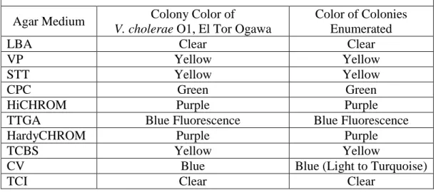
Table 3. Criteria for enumerating colonies appearing the same as V. cholerae from the 1% primary effluent matrix on solid agar media.
Agar Medium Colony Color of
V. cholerae O1, El Tor Ogawa
Color of Colonies Enumerated
LBA Clear Clear
VP Yellow Yellow
STT Yellow Yellow
CPC Green Green
HiCHROM Purple Purple
TTGA Blue Fluorescence Blue Fluorescence
HardyCHROM Purple Purple
TCBS Yellow Yellow
CV Blue Blue (Light to Turquoise)
TCI Clear Clear
Quantification of Non-Target Organisms from 1% Primary Effluent-Surface Water Matrix in Broth Media
Selective broths were examined for their inhibition or differentiation of non-target
organisms from a 1% primary effluent-natural water matrix as evidence of specificity. Culturable organisms from 1% primary effluent in natural water were quantified by an adapted MPN
method, using 24-well plates to perform a 3-well, 3-dilution MPN assay. The 1% primary effluent-natural water sample was serially diluted to 10-4 in Standard Methods phosphate buffer. Wells were prepared with 0.9mL of the appropriate selective broth medium and inoculated with 100μL of the diluted sample. Well plates were incubated for 24 and 48 hours at 37°C and 42°C. Wells were scored for growth; wells were considered positive if presented any visible
30
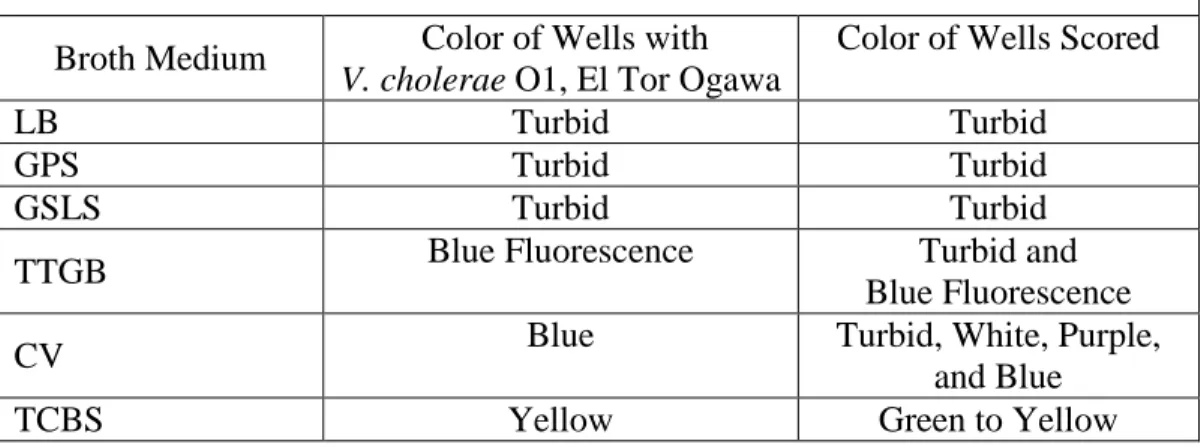
Table 4. Criteria for scoring wells for growth of all non-target organisms from the 1% primary effluent-surface water matrix.
Broth Medium Color of Wells with V. cholerae O1, El Tor Ogawa
Color of Wells Scored
LB Turbid Turbid
GPS Turbid Turbid
GSLS Turbid Turbid
TTGB Blue Fluorescence Turbid and
Blue Fluorescence
CV Blue Turbid, White, Purple,
and Blue
TCBS Yellow Green to Yellow
Quantification of Non-Target Organisms from Surface Waters
31
Table 5. Criteria for scoring tubes for growth of all non-target organisms from the natural surface waters.
Broth Medium Color of Tube with V. cholerae O1, El Tor Ogawa
Color of Tubes Scored
LB Turbid Turbid
TTGB Blue Fluorescence Turbid and
Blue Fluorescence
CV Blue Turbid, White, Purple,
Blue
TCBS Yellow Green to Yellow
Identification of Non-Target Organisms in Taurocholate Tellurite Peptone (TTGB) To further examine the lack of exclusivity of the TTGB medium, non-target organisms cultured from natural surface waters were isolated and purified as individual colonies and then identified by MALDI-TOF Mass Spectrometry. Natural surface waters were collected and 0.5mL of the sample was spiked into 15 sterile tubes each containing 8mL of TTGB. Tubes were
32
Examining the Efficiency of Media for Culturing V. cholerae
Quantification and Enumeration of V. cholerae (Multiple Tube Format)
An appropriate medium for the Cholera CBT must perform as a broth medium that can be applied to an MPN format to enrich the growth of V. cholerae and accurately estimate the
concentration of organisms in a drinking water sample. To examine the efficiency of selective broth media, pure cultures of V. cholerae were quantified by multiple-tube test (MTT) and also enumerated by direct spread plate. Initial experiments quantified pure cultures of V. cholerae by MTT using the TTGB medium. The MPN results were compared to Colony-Forming Units (CFU) from direct spread plate on non-selective LBA, selective TCBS, and selective TTGA agar media.
Overnight cultures of V. cholerae O139 (ATCC 51395), V. cholerae O1, El Tor Ogawa (ATCC BAA-2163) and V. cholerae Non-O1 (ATCC 35971) were prepared separately in alkaline peptone water (APW) and incubated at 37°C for 24 hours. Each overnight culture was serially-diluted in phosphate buffer. A 5-tube, 3-dilution MTT assay was prepared by inoculating 100μL sample into sterile tubes with 10mL of TTGB. Multiple Tube Tests were prepared in triplicate. A 100μL inoculum was plated in duplicate on LBA, TTGA, and TCBS by spread plate. Tubes and plates were incubated at 37°C for 18-24 hours. Tubes were incubated on a shaker tray.
Quantification and Enumeration of V. cholerae (Multiple Well Format)
33
were compared to the enumeration of colonies by agar versions of the original media. These candidate media were compared to the enumeration and quantification of V. cholerae by non-selective LBA and LB. An adapted MPN assay was prepared in 48-well plates for a 3-well, 4-dilution MPN assay. Overnight cultures of V. cholerae O139, V. cholerae O1, El Tor Ogawa and V. cholerae Non-O1 were incubated in APW at 37°C for 18-24 hours. Each overnight culture was serially-diluted in phosphate buffer. A 100μL sample was inoculated into 0.9mL of selective broth media in the corresponding well. The multiple-well assays were prepared in duplicate. A 100μL sample was also plated in duplicate on solid agar media by spread plate. A set of duplicate well plates and solid agar plates were incubated at both 37°C and 42°C for 18-24 hours.
Examining the Efficiency of Basal Media for Culturing V. cholerae
Screening Candidate Basal Media for Growth of V. cholerae
An appropriate broth medium for the CBT should consist of a basal medium that will enrich the growth of V. cholerae to provide
reliable results when quantifying organisms from drinking water samples. Brain Heart Infusion Broth (BHI Broth), peptone, and a combination of peptone, yeast and beef extract were evaluated as basal media for quantification of V. cholerae by MPN assay. These media were evaluated at different concentrations of NaCl (Table 6).
Table 6. Constituents of basal media. Basal Medium (g/L) Added NaCl
(% weight/volume) Brain Heart
Infusion Broth (37g/L)
+0% NaCl +1% NaCl +3%NaCl +5% NaCl Peptone (10g/L) +0% NaCl +1% NaCl +3% NaCl +5% NaCl Peptone (10g/L)
+ Yeast Extract (5g/L) + Beef Extract (5g/L)
34
Each basal medium was screened for growth of pure cultures of V. cholerae. Overnight cultures of V. cholerae O139, V. cholerae O1, El Tor Ogawa and V. cholerae Non-O1 were incubated in APW at 37°C for 18-24 hours. The overnight cultures were touched with a loop and transferred to a well containing 0.9mL of the basal medium. Well plates were incubated at 42°C for 18-24 hours and visualized for changes in turbidity. Turbid wells indicated positive growth.
Efficiency of Basal Media for Quantification of V. cholerae
35
Antibiotic Susceptibility Testing of V. cholerae and Non-V. cholerae Bacteria
Antibiotics may be incorporated into culture media to improve selectivity and specificity by preventing the growth of non-target bacteria. Four antibiotics – fosfomycin, streptomycin, ampicillin, and vancomycin – were evaluated for their selective properties in broth culture media. The minimum inhibitory concentration (MIC) was determined for three strains of V. cholerae, one strain of V. mimicus,
and seven non-Vibrio bacteria suspected to interfere in Vibrio-specific media (Table 7). The MIC values were obtained following the EUCST methods for determination of minimum inhibitory
concentrations (MICs) of
antibacterial agents by broth dilution (EUCAST Discussion Document E. Dis 5.1, March 2003).
Preparation of Antibiotics
Solid salts of each antibiotic were dissolved in distilled water at a final concentration of 5120mg/L and stored at -20°C. Antibiotic stock solutions were diluted in Muller-Hinton Broth (MHB) to final concentrations of: 1mg/L, 2mg/L, 4mg/L, 8mg/L, 16mg/L, 32mg/L, 64mg/L, 128mg/L, 256mg/L, 512mg/L. Of these solutions, 175µL was transferred into 96-well plates. The antibiotics were plated in order of increasing concentration.
Table 7. Organisms included in antibiotic susceptibility testing for the MIC determination of fosfomycin, streptomycin, ampicillin, and vancomycin. Vibrio spp.
Vibrio cholerae 0139 (ATCC 51395)
Vibrio cholerae O1, El Tor Ogawa (ATCC BAA-2163) Vibrio cholerae Non-O1 (ATCC 35971)
Vibrio mimicus (ATCC 33653) Gram-Negative Organisms
Aeromonas hydrophila (ATCC 7966) Proteus mirabilis (ATCC 9921)
Pseudomonas aeruginosa (ATCC 12175) Escherichia coli (ATCC 25922)
Gram-Positive Organisms
Bascillus pumilus (Environmental)
36
When mixed with an equal volume of inoculum in the wells, the final concentrations of each antibiotic were: 0.5mg/L, 1mg/L, 2mg/L, 4mg/L, 8mg/L, 16mg/L, 32mg/L, 64mg/L,
128mg/L, 256mg/L. The solution with 256mg/L of antibiotic was plated as the antibiotic control, mixed with an equal volume of sterile MHB. Stock solutions were discarded after use.
Preparation of Inoculum
Pure cultures of each test organism were streaked onto LBA plates and incubated at 37°C for 18-24 hours. Pure cultures of Aeromonas hydrophila were streaked onto LBA and incubated at 28°C for 18-24 hours. From each plate, four or five colonies were suspended into 8mL of MHB in sterile glass culture tubes. Cultures were then incubated at 37° for 2-4 hours until the turbidity was equal to that of a 0.5 McFarland standard, or the absorbance at 625nm was in the range of 0.08-0.10 as measured by a UV-VIS Spectrophotometer. Within 30 minutes of this standardization, 0.1mL of the culture was transferred into 9.9mL of MHB. Of this dilution, 175µL was transferred into each well of the 96-well plate, excluding the antibiotic control wells. Positive control wells contained 175µL of the bacterial strain as well as 175µL of the MHB. The final inoculum concentration was approximately 3 – 7 X 10⁵ CFU/mL. For quality control, a 10µL inoculum of the positive control well was transferred into 10mL of MHB, and 100µL of this solution was spread onto LBA plates and incubated at 37°C to ensure that the inoculum concentration was within this range.
MIC Determination
37
37°C for 18-24 hours. Wells were observed for turbidity compared to the positive control. The MIC100 was recorded as the lowest concentration of the antibiotic that completely inhibited growth.
Examining the Exclusivity of Newly-Proposed Culture Media for V. cholerae
Six new compositions of culture media were proposed for the selective culture of V. cholerae (Table 8). These compositions incorporate new selective agents to existing culture media (CV Plus), modify previously described media (modified TTGB), or incorporate different selective agents than previously described media. Detailed compositions of these media are described in Appendix A.
To examine the exclusivity of these media, three strains of V. cholerae, nine
gram-negative organisms, six gram-positive organisms, and V. mimicus were tested for growth in these broth media (Table 9). In addition, growth was tested in LB, TCBS, TTGB, and CV.
38
Table 8. Composition of novel culture media for the selective culture of V. cholerae.
Medium Selective Agents Indicators
CV Plus Salts mixture,
streptomycin, fosfomycin pH 8.6
Chromogenic Substrate
Cholera CBT Medium A Sodium taurocholate, sodium citrate, sodium lauryl sulfate,
crystal violet pH 8.6
pH indicator (Bromothymol Blue)
Cholera CBT Medium A-Plus Sodium taurocholate, sodium citrate, sodium lauryl sulfate,
crystal violet streptomycin, fosfomycin
pH 8.6
pH indicator (Bromothymol Blue)
Cholera CBT Medium B Oxgall, sodium taurocholate, sodium citrate, sodium
desoxycholate Streptomycin, ampicillin
pH 8.6
4-MU-Gal
Cholera CBT Medium C Sodium citrate, oxgall, sodium taurocholate, potassium tellurite
pH 8.6
pH indicator (Bromothymol Blue)
Cholera CBT Medium D Sodium taurocholate, sodium carbonate, potassium tellurite
Streptomycin, ampicillin pH 8.6
39
Table 9. Test organisms to examine the exclusivity of culture media for Vibrio spp. and V. cholerae.
Vibrio spp.
Vibrio cholerae 0139 (ATCC 51395)
Vibrio cholerae O1, El Tor Ogawa (ATCC BAA-2163) Vibrio cholerae Non-O1 (ATCC 35971)
Vibrio mimicus (ATCC 33653) Gram-Negative Organisms
Aeromonas hydrophila (ATCC 7966) Proteus mirabilis (ATCC 9921)
Pseudomonas aeruginosa (ATCC 12175) Escherichia coli (ATCC 25922)
Shigella flexneri (ATCC 12661) Shigella spp. (ATCC 23354)
Klebsiella pneumoniae (ATCC 23357) Salmonella typhimurium LT2
Raoultella terrigena Gram-Positive Organisms B. cereus (ATCC 1778) S. aureas (ATCC 29213) E. faecalis (ATCC 29212)
Bascillus pumilus (Environmental)
40
Table 10. Criteria for scoring wells for growth of known, relevant test organisms.
Broth Medium Color of Well with V. cholerae O1, El Tor Ogawa
Color of Wells Scored
LB Turbid Turbid
TTGB Blue Fluorescence Blue Fluorescence
CV Blue Blue
CV Plus Blue Blue
TCBS Yellow Yellow
Cholera CBT Medium A Yellow Yellow
Cholera CBT Medium A Plus
Yellow Yellow
Cholera CBT Medium B Blue Fluorescence Blue Fluorescence
Cholera CBT Medium C Yellow Yellow
Cholera CBT Medium D Blue Fluorescence Blue Fluorescence
Data Analysis
41
CHAPTER 4: RESULTS
Examining the Exclusivity of Media for Culturing V. cholerae
Enumeration of Non-Target Organisms from 1% Primary Effluent-Surface Water Matrix on Solid Agar Media
42
Figure 5. Enumeration of naturally-occurring, non-target organisms with Vibrio appearance cultured from 1% primary effluent in surface water on selective agar media for Vibrio spp. as determined by direct spread plate (n = 3). Values marked with (*) were estimated from plates with no exhibited growth from an undiluted inoculum. Examination of TTGB and LB were
conducetd seperately from those other media; these experiments are not paired.
Of the nine Vibrio-specific media examined, TCBS (Difco), CV (CHROMagar), and TCI agars completely inhibited the growth of any non-target organisms from the plated inoculum of surface water with 1% primary effluent. Other media allowed the growth of non-target microbes appearing the same as V. cholerae, with STT and VP allowing the highest levels of growth. Incubation at 42°C gave less growth of non-target organisms and thereby improved inhibition of non-target organisms.
To further examine the exclusivity of these agar media, we describe the frequency of observing non-target growth on each selective agar medium that appears the same as colonies of V. cholerae on the medium. This frequency is described across the three replicate trials. These frequencies are described in Table 11. The most appropriate selective medium would give no growth across any of the three trials. Based on these results, TCBS, CV and TCI are effective
43
solid agar media for the inhibition of non-target organisms from the 1% primary effluent matrix at incubation of 37°C or 42°C. HardyCHROM agar is additionally effective at 42°C incubation.
Table 11. Frequency of observing non-target growth appearing the same as V. cholerae on selective agar media for across three replicate trials. Frequency is reported below as # Trials (out of three total). Values represent the number of trials for which growth appearing the same as V. cholerae was observed on selective agar media, when inoculated with a 1% primary effluent in surface water matrix.
Medium 37°C 42°C
LBA 3 3
VP 3 3
STT 3 3
CPC 3 3
HiCHROM 3 3
TTGA 3 3
HardyCHROM 3 0
TCBS 0 0
CHROMagar 0 0
TCI 0 0
Quantification of Non-Target Organisms from 1% Primary Effluent-Surface Water Matrix in Broth Media
The exclusivity of Vibrio-specific broth media was examined by quantifying the non-target organisms from a 1% primary effluent in surface water matrix plated in a multiple-well MPN assay. Growth was indicated by turbidity, chromogenic change, or flourogenic change. Wells were scored if any growth was observed in the broth medium. The quantification of these organisms is reported in Figure 6.
44
acquired from Difco. Although GSLS was among the most selective media, bacterial growth was difficult to visualize, especially at 42°C incubation. This medium does not incorporate any chromogenic or fluorogenic indicators to aid in visualization of bacteria growth. Selective agents from the most effective of these media may be considered for new adaptations of selective Vibrio media.
CHROMagar Vibrio ranks among the more selective media examined, limiting non-target growth. Across all media, incubation at 42°C improved exclusivity, reducing the growth of non-target organisms.
Figure 6. Quantification of all non-target organisms from 1% primary effluent in surface water in selective broth media as determined by multiple well MPN assay (n = 3). Log MPN values
marked with (*) were estimated from plates with no exhibited growth from an undiluted inoculum. Examination of TTGB and LB were conducted separately from the other media; so,
these experiments are not paired.
To further examine the exclusivity of these broth media, we describe the frequency of observing any non-target growth in each selective broth medium. Growth was observed and reported based on any changes in turbidity, or chromogenic or fluorogenic changes in the
45
medium. This frequency is described across the three replicate trials. These frequencies are described in Table 12. The most appropriate selective medium would give no growth of any bacteria across any of the three trials. Only one medium, GSLS was consistently effective at inhibiting non-target organisms from the 1% primary effluent-surface water matrix across three replicate trials, at an incubation of 42°C for 24 hours. The TTGB medium was moderately effective at inhibition of non-target organisms at 42°C for 24 or 48 hours; non-target organisms were detected in only one of the three replicate trials.
Table 12. Frequency of observing any non-target growth in selective broth media for Vibrio spp. across three replicate trials. Values represent the number of trials for which growth was observed on selective broth media, when inoculated with a 1% primary effluent in surface water matrix. Frequency is reported below as number of trials (out of three total trials).
Medium 37°C - 24 Hr 37°C - 48 Hr 42°C - 24 Hr 42°C - 48 Hr
LB 3 3 3 3
GSP 3 3 3 3
TCI 3 3 3 3
GSLS 3 3 0 3
CV 3 3 3 3
TCBS 3 3 3 3
TCBS Homemade 3 3 3 3
TTGB 2 3 1 1
Quantification of Non-Target Organisms from Surface Waters
46
an adapted CBT while attempting to quantify V. cholerae from water samples. An ideal
candidate medium would exclude or inhibit the growth of all non-target organisms in a 100-mL sample volume.
Of the three selective broth media examined, CV was most selective in inhibiting the growth of non-target microorganisms from a 100-mL-volume surface water sample. The average MPN and 95% confidence interval of interfering from undiluted 100 mL water samples was 4.6 (0.74, 16.2) MPN/100mL (n = 3). The mean concentration includes an estimated MPN from a trial