In vitro Adherence
of Microorganisms
to Denture
Base Resin with
Different
Surface
Texture
Mutsuo YAMAUCHI*, Khoji YAMAMOTO**, Manabu WAKABAYASHI** and Joji
KAWANO*
*First Department of Prosthodontics, Asahi University, School of Dentistry
**Operative
Dentistry, Asahi University, School of Dentistry
1851-1
Hozumi-cho, Motosu-gun,
Gifu 501-02,
Japan
Received
on July 31, 1989
Accepted on April 13, 1990
We examined the effects of various denture base resin surface textures on the adherence of microorgan-isms.
S. sanguis and B. gingivalis adhered in greater amounts to the denture base resin than the other microorganisms tested. As to bacterial adherence according to polishing state, S. oralis, B. gingivalis C-101, and B. intermedius C-001 more adhered to the No.400 paper-polished surface than to the buff-polished and smoothening-treated surfaces. S. sanguis less adhered to the smoothening-treated surface. S. mitis and C. albicans, on the other hand, more adhered to the smoothening-treated surface. For the other microorganisms tested, no relationship was observed between surface texture and bacterial adherence. The fall-off test revealed no remarkable differences in the fall-off of S. sanguis and B. gingivalis C-101 by the types of surface treatment. However, the fall-off of C. albicans was poorest from the No.400 paper-polished surface. These results indicate that smoothening the denture base surface is important for denture plaque control. Key words: Denture base resin, Surface texture, Bacterial adherence
INTRODUCTION
It has been reported that microorganisms inhabiting denture plaque, in particular the Candida species, play a role in the development of denture stomatitis1-4). Thus adherence of Candida to denture base resin has been studied by a number of researchers5-11). Since caries and periodontitis of abutment teeth are likely to develop after inserting a partial denture, examining the adherence of pathogenic bacteria is essential10).
After inserting a denture, its surface changes, and these changes promote the adherence of denture plaque12). There are few reports, however, on the relationship between the surface texture of the denture base resin and the adherence of denture plaque13).
In this study we examined the effects of various base resin surface textures on the adherent ability of microorganisms which cause caries, periodontitis and denture stomatitis.
MATERIALS AND METHODS
Specimens
Both sides of 10mm•~10mm•~1mm square heat-cured denture base resin# plates were
treated in one of the following three ways: polishing with emery paper No.400##; polishing
with emery paper No.2000## and subsequent buff polishing; or polishing with emery paper
No.2000 followed by surface smoothening treatment###. The mean roughness (Ra) of the No.
400 paper-polished resin, buff-polished resin, and surface-smoothening-treated resin were 1.12
m, 0.09ƒÊm, and 0.22ƒÊm respectively. The contact angles of distilled water on No.400
paper-polished resin, buff-polished resin, and surface-smoothening-treated resin were 68.9, 67. 6, and 60.9 degrees respectively. All specimens were examined after immersion in 37•Ž water for 7 days.
Microorganism culture and labeling
Streptococcus mutans ATCC 25175, Streptococcus sanguis ATCC 10556, Streptococcus
oralis ATCC 35037, Streptococcus mitis ATCC 33399, Streptococcus salivarius ATCC 25975,
Bacteroides gingivalis ATCC 33277, Bacteroides gingivalis C-101 (Clinical), Bacteroides
inter-medius ATCC 15032, Bacteroides intermedius C-001 (Clinical), and Candida albicans IFO1060
were used in these experiments.
A 0.1ml aliquot of a C. albicans suspension was inoculated to 20ml of Sabouraud glucose
broth•• containing 5ƒÊCi/ml •k6-3H•l -glucose$$. The broth was then incubated at 37•Ž for
24 hours under aerobic conditions. Other bacteria were added to 20ml of trypticase soy
broth$$$ with 0.5% yeast extract! containing 2ƒÊCi/ml [6-3H] -thymidine!!, and incubated in an anaerobic jar!!! at 37•Ž for 24 hours. The suspensions were centrifuged at 8,000G for 20min and radiotagged cells were washed gently two successive portions of pH 7.0 phosphate buffer solution.
Adherence of bacteria
According to the ƒ³ rstavik et al.14) method, specimens were coated with saliva in a water bath incubator at 37•Ž for 2 hours. Subsequently each specimen was immersed in 1ml of the radiotagged cell suspension (108cells/ml) and then gently shaken at 37•Ž for 2 hours.
Speci-mens were then washed gently with two successive portions of pH 7.0 phosphate buffer
solution and then incinerated in an automatic sample combustion system\. The amount of
adherent bacteria was determined by measuring [6-3H] -radioactivity (DPM) with a liquid
scintilation counter\\.
Data were analyzed by one-way analysis of variance for each microorganisms. Fall-off test
While ultrasonically treating a piece of denture base resin with adhered S. sanguis, B. gingivalis C-101, and C. albicans for 1min, the rate of bacteria fall-off was examined.
##Nippon Coated Abrasive , Aichi, Japan
###Perma cure system , GC Dental Industrial Corp., Tokyo, Japan $Nissui , Tokyo, Japan
$$TRK-85, Amersham International plc , Buckinghamshire, England. $$$BBL , London, England
! Difco, Laboratories, Detroit, Michigan, U.S.A
!!TRK-61 , Amersham International plc, Buckinghamshire, England. !!!Gaspack
, BBL, London, England \ ASC-113, Aloka, Tokyo, Japan \\ LSC-903SP, Aloka, Tokyo, Japan
RESULTS
Adherence of microorganisms
Table 1 shows the adherence of microorganisms to the surfaces of resins treated in each
of the three ways. S. sanguis, B. gingivalis ATCC, and B. gingivalis C-101 adhered in greater
amounts to each of the polishing surfaces than the other microorganism. As to bacterial
adherence among the surface texture, S. oralis, B. gingivalis C-101, and B. intermedius C-001
more adhered to the No.400 paper-polished surface. S sanguis less adhered to the
smoothen-ing-treated surface. S. mitis and C. albicans, on the other hand, more adhered to the
smoothening-treated surface than to the other polished surfaces. No relationship was found
between surface treatment and adherence of S. mutans ATCC, S. salivarius, B. gingivalis
ATCC, and B. intermedius ATCC.
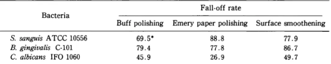
Fall-off test
For S. sanguis and B. gingivalis C-101, we found no remarkable difference between the
polishing state and the fall-off rate. However, the fall-off of C. albicans was poorest from the
No.400 paper-polished surface (Table 2).
Table 1 Bacterial adherence on denture base resin under each polishing condition
*:Mean value of three specimens
Table 2 Fall-off rate of bacteria from denture base resin under each polishing condition by ultrasonic treatment
*:(1- DPM after ultrasonic treatment/
DISCUSSION
In this study, S. oralis, B. gingivalis C-101, and B. intermedius C-001 adhered to the No.
400 paper-polished surface, which had the largest surface roughness value. These results
suggest that care should be taken in polishing and plaque control for resin adjacent to the
abutment teeth of a partial denture, otherwise caries and periodontitis near abutment teeth
may be accelerated. Badawi et al.13) reported that plaque adherence is significantly greater
on unpolished fitting surfaces of upper complete dentures than on polished surfaces.
Therefore, increased surface roughness can lead to problems.
Satou et al.15) found that the contact angles of restoratives and the adherent cells showed
high positive correlation for S. sanguis. Minagi et al.9) showed that adherence of Candidas
increases when the surface free energy of the resin calculated by the contact angle is
equivalent to that of Candidas. In the present study there was no relationship between
contact angle and bacterial adherence except for S. sanguis, S. oralis, S. mitis, B. gingivalis
C-101, B. intermedius C-001 and C. albicans; this may be explained by the lack of any
remarkable difference in contact angles among the various polished surfaces and difference
of hydrophobicity of each microorganism.
C. albicans, pathogenic microorganisms of denture stomatitis, lowest adhered to the resin
surface than the other microorganisms; however, C. albicans more adhered to the
surface-smoothening-treated resin than to the other polished resins. Minagi et al.9) reported that C.
albicans is strong hydrophobic bacteria, and best adhered to the hydrophobic materials.
Because surface-smoothening-treated resin has smaller contact angle than the other polished
resins, namely, it is more hydrophobic than the other polished resins, C. albicans more
adhered to the surface-smoothening-treated resin than the other polished resins.
Budtz-Jorgensen et al.16) inserted the complete dentures in which half of fitting surface
is treated with surface-smoothening and remaining is not treated, and monitored plaque
accumulation on fitting surfaces, the number of bacterial colonies, and the proliferation of
yeast after 1 week and 1 month. At 1 week all treated surface values were significantly
smaller than the non-treated surface values, whereas after 1 month no differences were
observed. There are sevral probable explanations for disagreement with our results that C.
albicans well adhered to the surface-smoothening-treated resin than buff-polished resin.
Firstly, they studied only fitting surface which had different environments for plaque
accumu-lation and anaerobically. The period of exposure to microorganisms was far shorter in our
experiments since we wanted to study early adherence events. Lastly, denture base resins
were coated with pellicle in their experiment.
In the fall-off test, S. sanguis and B. gingivalis C-101 were used because they adhered in
greater amounts to the resin surface than the other microorganisms, and these
microorgan-isms induce caries or periodontitis. Also, C. albicans was used as an example of a pathogenic
bacteria of denture stomatitis. The polishing state of the denture base resin did not influence
the fall-off rate of S. sanguis and B. gingivalis C-101. The fall-off rate of C. albicans was
equivalent on the surface-smoothening-treated and the buff-polished resins. Based on these
observations, surface smoothening-treatment appears to be the effective for removal of
adhered to smoothening-treated surfaces is easily removed. We confirmed that plaque is readily removed from the surface of smoothening-treated dentures17).
These results indicate the importance of polishing the denture base surface to minimize adherence of microoragnisms.
CONCLUSIONS
The effects of the denture surface texture on the adherence of oral microorganisms were studied using Streptococci, Bacteroides, and Candida albicans as test microorganisms. Their adherence to and release from resin surfaces treated with either No.400 emery paper-polishing, buff-polishing, or surface-smoothening was examined.
S. sanguis, B. gingivalis ATCC, and B. gingivalis C-101 adhered in greater amounts to the denture base resin than the other microorganisms tested. As to bacterial adherence among the polishing conditions, S. oralis, B. gingivalis C-101, and B. intermedius C-001 more adhered to the No.400 paper-polished surface. S. sanguis less adhered to the smoothening-treated surface. On the other hand, S. mitis and C. albicans more adhered to the smoothening-treated surface than to the other polished surfaces. In S. mutans, S. salivarius, B. gingivalis ATCC,
and B. intermedius ATCC, no correlation was observed between surface texture and bacterial adherence.
The fall-off rate of S. sanguis, B. gingivalis C-101 and C. albicans from variously treated resin surfaces by ultrasonication was examined. In S. sanguis and B. gingivalis C101, no
remarkable differences were noted among the surface treatments. However, the fall-off rate of C. albicans was poorest on the No.400 paper-polished surface.
REFERENCES
1) Davenport, J.C.: The oral distribution of Candida in denture stomatitis, Br Dent J, 129 (4): 151-156, 1970.
2) Budtz-Jorgensen, E.: The siginificans of Candida albicans in denture stomatitis, Scand J Dent Res, 82 (2): 151-190, 1974.
3) Olsen, I.: Denture stomatitis, Occurrence and distribution of fungi, Acta Odontol Scand, 32 (5): 329-333, 1974.
4) Renner, R.P., Andors, L. and McNamara, T.F.: The role of C. albicans in denture stomatitis, Oral Surg, 47 (4): 323-328, 1979.
5) Samaranayake, L.P. and MacFarlane, T.W.: An in vitro study of the adherence of Candida albicans to acrylic surfaces, Arch Oral Biol., 25 (8/9): 603-609, 1980.
6) Samaranayake, L.P., McCourtie, J. and MacFarlane, T.W.: Factors affecting the in vitro adherence of Candida albicans to acrylic surfaces, Arch Oral Biol, 25 (8/9): 611-615, 1980.
7) McCourtie, J. and Douglas, L.J.: Relationship between cell sutface composition Candida albicans and adherence to acrylic after growth on different carbon sources, Infect Immun, 32 (3): 1234-1241, 1984. 8) McCourtie, J., MacFarlane, T.W. and Samaranayake, L.P.: Effect of chlorhexidine gluconate on the
adherence of Candida species to denture acrylic, J Med Microbiol 20 (1): 97-104, 1985.
9) Minagi, S., Miyake, Y., Inagaki, K., Tsuru, H. and Suginaka, H.: Hydrophobic interaction in Candida albicans and Candida tropicalis adherence to various denture base resin materials, Infect Immun, 47 (1): 11-14, 1985.
10) Yoshimura, K.: Investigation on the adherence of oral microorganisms to prosthetic materials and removal of in vitro plaque, J Osaka Odontol Soc, 50 (3): 287-333, 1987. (in Japanese)
11) Verran, J. and Motteram, K.L.: The effect of adherent oral streptococci on the subsequent adherence of Candida albicans to acrylic in vitro, J Dent, 15 (2): 73-76, 1987.
12) Hamada, T.: Denture plaque control, 1st. ed., Nagasue pub., Kyoto, 1983, pp. 24-25. (in Japanese) 13) Badawi, M.S., Nada, M. and Kadry, S.: The effect of polishing the fitting surface of maxillary denture
on denture plaque accumulation, Egypt Dent J 32 (4): 303-315, 1986.
14) ƒ³ rstvik, D., Kraus, F.W. and Henshaw, L.C.: In vitro attachment of Streptcocci to the tooth surface, Infect Immun, 9 (5): 794-800, 1974.
15) Satou, J., Fukunaga, A., Satou, N., Shintani, H. and Okuda, K.: Streptococcal adherence on various restorative materials, J Dent Res, 67 (3): 588-591, 1988.
16) Budtz-Jorgensen, E. and Habber, S.: Clinical effects of using an ultraviolet curing method,Scand J Dent Res, 94 (6): 569-574, 1986.
17) Yamauchi, M., Takigawa, H., Uematsu, K., Kawano, J., Sakai, M., Nakazato, G. and Andoh, M.: Clinical observation of acrylic resin dentures treated with surface smoothening and hardening agents, J Gifu Dent Soc, 13 (2): 340-350, 1986. (in Japanese)
In vitroに お け る 異 な る 表 面 性 状 の 床 用 レ ジ ン へ の 細 菌 付 着 に つ い て 山 内 六 男,山 本 宏 治 *,若 林 学 *,川 野 襄 二 朝 日大学歯学部歯科補綴学第1講 座 *朝 日大学歯学部歯科保存学第1講 座 エ メリペ ーパー400番 研磨,バ フ研磨お よび表面 滑沢 硬 化 処 理 を 行 っ た 床 用 レ ジ ン へ のStreptococci,
Black-pigmented Bacteroidesお よ びCandida albicansの 付 着 に つ い て 検 討 し た 。
S. sanguisお よ びB. gingivalisは,他 の 供 試 菌 に 比 べ 床 用 レ ジ ン へ よ く付 着 し て い た 。 床 用 レ ジ ン の 表 面 性 状 別 に み た 場 合,S. oralis, B. gingivalis C-101お よ びB. intermedius C-001は,表 面 粗 さ の 最 も 大 き い400番 研 磨 面 へ の 付 着 が 表 面 粗 さ の 小 さ い バ フ研 磨 面 お よ び 表 面 滑 沢 硬 化 処 理 面 よ り も 多 く,S. sanguisで は 表 面 滑 沢 硬 化 処 理 面 へ の 付 着 が 少 な か っ た 。S. mitisお よ びC. al-bicansで は 表 面 滑 沢 硬 化 処 理 面 へ の 付 着 が 多 か っ た。 そ の 他 の 供 試 菌 で は,表 面 粗 さ と細 菌 付 着 と の 間 に 有 意 な 関 連 は み ら れ な か っ た 。 一 方 ,S. sanguis, B. gingivalis C-101お よ びC. albicansの 各 研 磨 面 か ら の 脱 離 性 に つ い て み た 場 合,C. albicansは エ メ リペ ー パ ー400番 研 磨 面 か ら の 脱 離 率 が 悪 か っ た 。 これ ら の 結 果 か ら,デ ン チ ャ ー ・プ ラ ー ク に と っ て は,義 歯 床 表 面 を 滑 沢 す る こ と が 必 要 で あ る こ と が 示 唆 さ れ た 。