Research Article
Effect of perinatal asphyxia on level of thyroid hormones
in term neonates
Nishant Prabhakar
1*, Avyact Agrawal
1, Neha Jain
2, Ashok Kumar Ahirwar
3INTRODUCTION
Perinatal asphyxia is the one of major public health problem in the world. It is the major cause of morbidity and mortality. It accompanies one of the three most common causes of death along with prematurity and bacterial infections.1 It causes decreased perfusion to the tissues causes diving seal reflex which causes shunting of blood away from lung, kidney, GIT, skin to brain, adrenals and heart.2 Cellular dysfunction occurs as a result of diminished oxidative phosphorylation and ATP production. Impaired oxidative phosphorylation can occur during the primary hypoxic ischemic insults as well as during a secondary energy failure that usually occurs approximately 6 to 24 hours after the initiating insult.
Cell death can be either immediate or delayed, and either necrotic or apoptotic.
In hypoxic adults and older children effect of hypoxia on thyroid metabolism has been studied.3-8,24 Effect of hypoxia on thyroid indices in term new-born have also been evaluated with conflicting results.8-10 For the development of central nervous system thyroid hormone plays a pivotal role. In non-thyroidal illnesses low level of thyroid hormones are associated with poor prognosis. The present study was conducted to evaluate the effect of perinatal asphyxia on level of thyroid hormones by comparing thyroid profile in cord blood and venous blood at 18-24 hour after birth in new-borns with and without asphyxia.
ABSTRACT
Background: To evaluate how perinatal asphyxia alters the thyroid function in term newborn by comparing cord blood and 18-24 hours venous blood in between newborns with and without asphyxia.
Methods: A prospective case-control study was carried out at Tertiary care pediatric center in central India. 60 Full term newborn with perinatal asphyxia requiring bag and mask ventilation for ≥1 minute or APGAR at 1 and 5 minute ≤7 or required intubation for resuscitation as cases and control -60 normal term newborn with APGAR ≥7 at 1 and 5 minute were selected. Cord blood and 18 to 24 hour after birth venous blood were collected in both groups and compared for level of thyroid hormones (T3, T4, TSH) via radioimmunoassay.
Results: There was no significant difference observed in cord blood thyroid hormones (P>0.05). But a significant lower level of thyroid hormones observed in asphyxiated group as compared to control group in venous sample 18-24 hours after birth [mean± standard deviation; T3 case=68.87 ± 21.08, T3 control=79.93 ± 20.52, P=0.004; T4 case 6.48 ± 2.41, T4 control=8.89 ± 1.46, P<0.001; TSH case=4.33 ± 4.36; TSH control=8.45 ± 1.70, P<0.001].
Conclusions: Perinatal asphyxia depresses the TSH level which further leads to decrease in T3 and T4 suggestive of central hypothyroidism.
Keywords: Perinatal asphyxia, Term newborns, Thyroid function, Non-thyroidal illness syndrome
1Department ofPediatrics, NSCB Medical College and Hospital, Jabalpur, MP, India 2
Department ofObstetrics and Gynaecology, JN Medical College and Hospital, AMU, Aligarh, UP, India
3Department ofBiochemistry, AIIMS, New Delhi, India
Received: 28 March 2016
Accepted: 09 May 2016
*Correspondence:
Dr. Nishant Prabhakar,
E-mail: [email protected]
Copyright: © the author(s), publisher and licensee Medip Academy. This is an open-access article distributed under the terms of the Creative Commons Attribution Non-Commercial License, which permits unrestricted non-commercial use, distribution, and reproduction in any medium, provided the original work is properly cited.
METHODS
It was a prospective case control study conducted on randomly selected 60 asphyxiated neonates (cases) and 60 full term healthy non-asphyxiated neonates (controls) born in NSCB MCH, Jabalpur, Madhya Pradesh, India from November 2014 to October 2015 after getting approval from the Institutional ethical committee. Sample size was calculated considering a significance of 0.05 and a statistical power of 90% based on data presented by Pareira et al.19 Continuous variables were described through means, median, and standard deviations and the categorical variables were described through the proportion of data obtained during the study from sample.
Cases selected were full term asphyxiated new-born who required bag and mask ventilation for at least 1 minute immediately after birth or required intubation for resuscitation or 1 and 5 minute APGAR score <7 and controls were every 5th full term normal new-born who cried immediately after birth and 1 and 5 minute APGAR score ≥7. Cases and controls were matched in terms of gestational age, type of delivery, ethnic group, birth weight and sex. New-borns with any congenital malformation or diseases or born to a mother with abnormal thyroid profile or if mother taking any drugs like antihypertensive, diuretics, corticosteroids and antithyroid drugs or parents not giving consent were excluded from the study.
Gestational age was determined according to obstetrical formula and confirmed by physical examination (new Ballard scoring system). When the difference between obstetric gestational age and clinical parameters were higher than 2 weeks, clinical evaluation was considered.
Immediately after birth, umbilical cord was clamped at two different sites and blood was collected for analysis and determination of T3, T4 and TSH. After 18 to 24
hours of birth, venous blood sample was collected for levels of thyroid hormones of each new-born in both groups (cases and controls). The levels of hormones in both the groups were compared in cord blood and 18-24 hour sample respectively. Severity of hypoxic ischemic encephalopathy assessed by Sarnat and Sarnat Staging and asphyxiated baby were graded as HIE I, HIE II and HIE III. T4, T3, and TSH were measured by radioimmunoassay method. The values of T3 were expressed in ng/dl, TSH in mIU/dl, and those of total T4 in µg/dl.
All the new-borns with birth asphyxia were admitted in neonatal intensive care unit under strict observation for vitals, urine output and kept nil by mouth. Hydration was maintained and given intravenous fluid. All healthy term new-born were given to mother and received feeding on demand. Both the groups were kept under observation till discharge or death.
Descriptive statistical analysis was done and continuous variables were described as mean and standard deviation and categorical variables in number (%). Student's t test has been used to assess continuous variables for pair-matched samples with a confidence limit of 95%. Significance is assessed at a level of 5%. For categorical variables chi square test was used.
RESULTS
There was no significant difference between asphyxiated and non-asphyxiated babies in terms of mother age, mother's thyroid status, mode of delivery, parity of mother, sex of baby, and birth weight of babies (Table 1). All the babies (in both the groups) were term babies with no significant difference in gestational age.
Table 1: Profile of cases and control in our study.
Case (mean + SD)
Control (mean ± SD)
p value
Mother age (years)
Case (mean + SD)
Control (mean ± SD)
p value
Parity No. % No. %
0.788
1 36 60.0 38 63.3%
2 18 30.0 14 23.3%
3 3 5.0 5 8.3%
4 3 5.0 3 5.0%
Mode of
delivery No. % No. %
0.346
Vaginal 35 58.3% 40 66.7%
LSCS 25 41.7% 20 33.3%
Sex of
baby No. % No. %
0.358
Female 24 40.0% 29 48.3%
Male 36 60.0% 31 51.7%
Thyroid hormone levels
(mean ± SD) (mean ± SD)
T3 129.15 ± 21.12 131.97 ± 20.49 0.46
T4 8.10 ± 1.47 7.70 ± 0.82 0.07
TSH 4.08 ± 0.89 4.23 ± 0.99 0.39
Table 2: Mean level of thyroid hormones in cord blood.
All the asphyxiated babies had APGAR score <7 at 1 and 5 minute after birth. Most of the new-borns had APGAR <3 (81.7%, 49/60) at 1 minute after birth out of them 3/60
Case (mean± sd)
Control
(mean±sd) T p value
T3
80.86 ± 18.82
80.42 ±
14.10 0.14 0.88
T4
8.70 ±
1.47 8.71 ± 1.25 0.04 0.97
TSH 8.92 ±
persisted to have APGAR <3 at 5 minutes after birth. All asphyxiated babies admitted in neonatal intensive care unit and supportive care were given. According to Sarnat and Sarnat criteria for HIE staging 5% (3/60) were in HIE stage I, 50% (30/60) in HIE stage II, and 45% (27/60) in HIE stage III.
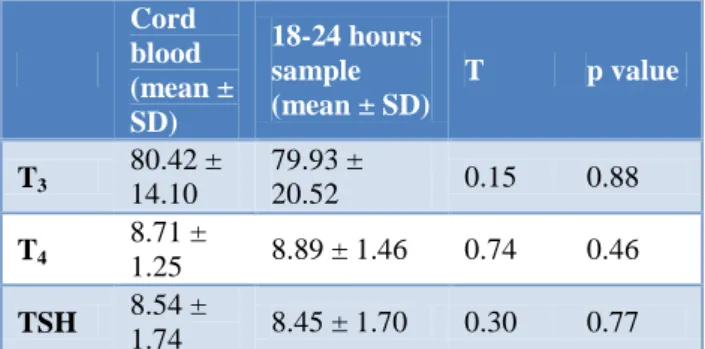
Table 3: Mean level of thyroid hormones at 18-24 hours after birth.
Table 4: Comparison of thyroid hormones in cord blood and 18-24 hrs sample in cases.
Table 5: Comparison of thyroid hormones in cord blood and 18-24 hrs sample in controls.
In cord blood of asphyxiated and non-asphyxiated babies mean level of T3, T4 and TSH were 80.86, 8.7, 8.92 and
80.42, 8.71, 8.54 with p value 0.88, 0.97, 0.49 respectively, which was statistically not significant (Table 2). Again at 18-24 hours after birth venous blood collected and in this sample it was observed that mean level of T3, T4 and TSH in asphyxiated and
non-asphyxiated neonates were 68.87, 6.48, 4.33 and 79.93, 8.89, 8.45 with p value of 0.004, <0.0001, <0.0001 respectively which showed statistically significant
difference (Table 3). Among asphyxiated new-borns when we compared mean level of T3, T4 and TSH in cord
blood and 18-24 hour sample after birth it was 80.86, 8.70, 8.92 and 68.87, 6.48, 4.33 with p value 0.0001, <0.0001, <0.0001 respectively (Table 4) and in non-asphyxiated neonates mean level of T3, T4 and TSH in
cord blood and 18-24 hour sample after birth were 80.42, 8.71, 8.54 and 79.93, 8.89, 8.45 with p value 0.88, 0.46, 0.77 respectively (Table 5).
DISCUSSION
Several studies evaluated the effect of hypoxia in alteration of thyroid function with conflicting results. It may be due to methodological differences among the studies. Several factors studied to influence the thyroid function. Various perinatal factors such as gestational age, weight, sex, mode of delivery, Eclampsia, APH, Birth Asphyxia, PROM, HIV status, maternal age and thyroid status etc. have been studied for influence on cord blood T3, T4 and TSH.11-15
Moshang et al compared alteration of thyroid function in acute versus chronic hypoxia in children around 2-16 years and found elevated serum rT3 and decreased serum
T3 concentrations (indicating extra thyroid metabolism).7
Warner S et al, they observed that hypoxia leads to activation of deiodinase type 3 in turn inactivates the peripheral conversion of T4 to T3.16
Borges et al reported that in spite of the maximal TSH surge, in asphyxiated new-borns serum fT3 and fT4 levels
failed to increase and concluded that the alterations in the thyroid function observed in asphyxiated new-borns may be caused by the low consumption of oxygen with low metabolic rate, suggesting that asphyxia plays an important role in thyroid metabolism.17
Procianoy RS et al suggested that decreased levels of T4
and T3 18-24 hours after birth in asphyxiated new-borns
is due to a diminished TSH level.18 Pareira DN and Procianoy RS found that serum concentration of TSH, T4,
T3, and fT4 are lower in asphyxiated new-borns than in
the normal new-borns between 18 and 24 hour of life.19 Frank et al observed that prenatal treatment with thyroxine and thyroid releasing hormone (TRH) accelerate surfactant system maturation and has opposite effect on the antioxidant enzyme in the lung.21 Gupta et al observed that neonates with low APGAR at 1 minute resulted in significantly raised cord blood TSH (p<0.01).11
Tahivoric HF et al studied the serum concentration of thyroid hormone levels (T4, fT4 , rT3 , T3, TSH and TBG)
in cord blood at birth and serum on 5th day of life he concluded that neither hypoxia nor the method of delivery had any influence on the peripheral metabolism of thyroid hormones.21
Case (mean± sd)
Control
(mean±sd) T p value
T3
68.87 ±
21.08 79.93±20.52 2.91 0.004
T4
6.48 ±
2.41 8.89 ± 1.46 6.60 <0.001
TSH 4.33 ±
4.36 8.45 ± 1.70 6.82 <0.001
Cord blood (mean± SD)
18-24 hours sample (mean ± SD)
T p value
T3
80.86 ± 18.82
68.87 ±
21.08 4.21 0.0001
T4
8.70 ±
2.38 6.48 ± 2.41 4.91 <0.0001
TSH 8.92 ±
3.94 4.33 ± 4.36 6.99 <0.0001
Cord blood (mean ± SD)
18-24 hours sample (mean ± SD)
T p value
T3
80.42 ± 14.10
79.93 ±
20.52 0.15 0.88
T4
8.71 ±
1.25 8.89 ± 1.46 0.74 0.46
TSH 8.54 ±
In this study we matched both the groups with respect to gestational age, mother's age and thyroid status, baby's sex, birth weight, type of delivery, ethnic group and parity of mother so that the maximum difference was in exposure to birth asphyxia and probability of confounding bias could be minimized. There was also difference in receiving enteral nutrition, although this variable has no significant effect on hormonal levels unless malnutrition is present.
In the resource limited setting we had to rely on APGAR score. Those with APGAR <7 at 1 and 5 minute of life or required intubation or required bag and mask ventilation for >1 minute after birth, according to Sarnat and Sarnat criteria HIE staging was done. Such asphyxiated new-borns compared with the normal new-new-borns for mean level of thyroid hormone in the cord blood at birth and venous blood 18-24 hours after birth. There was no significant difference observed for the level of thyroid hormones between the groups in the cord blood while at 18-24 hours after birth there was significant decrease in all T3, T4 and TSH was observed in asphyxiated
new-borns. Which suggest low level of T3 and T4 are
secondary to low TSH concentration. It may be due to central hypothyroidism which is most commonly observed in first 24 hours. Further evaluation of these alterations could not be done due to some practical issues which required multiple blood samples at different durations, both in asphyxiated and non-asphyxiated new-borns.
Non thyroid illness syndrome (NTIS) is a form of combined central and peripheral hypothyroidism often associated with other crucially important hormone deficiencies. In this T3 and T4 are deficient without an
elevation of TSH.22 The level of involvement of thyroid function correlates with the severity of the disease. This sickness may also be called as euthyroid sick syndrome. It can also occur in protein energy malnutrition, postoperative period of large surgeries, sepsis, meconium aspiration, and asphyxia. This syndrome is also associated with use of certain drugs, such as corticosteroids, dopamine, and ionized contrasts.23
Joshi et al also studied influence of perinatal factors on thyroid profile.25 They collected umbilical cord blood sample for assessment of TSH and T4 in 830 new-borns.
They found that birth asphyxia had significant influence on cord blood TSH as compared to normal new-born.
Durga et al measured cord blood TSH in 100 live new-born infants.26 They observed that requirement of resuscitation in initial steps and low APGAR scores at 1 minute result in significantly raised cord blood TSH (P<0.05).
To substantiate our findings, studies with larger sample size and multiple numbers of blood samples at different duration after birth are needed to establish whether these effects are transient or permanent. Further studies to
supplement thyroid hormone may be considered since it’s a curable cause via thyroid replacement.
Funding: No funding sources Conflict of interest: None declared
Ethical approval: The study was approved by the Institutional Ethics Committee
REFERENCES
1. Black RE, Cousens S, Johnson HL, Lawn JE, Rudan I, Bassani DG, et al. Child Health Epidemiology Reference Group of WHO and UNICEF. Global, regional and national causes of child mortality in 2008: a systematic analysis. Lancet. 2010;375(9730):1969-87.
2. Phibbs RH. Delivery room management. In: Avery GB, Fletcher MA, MacDonald MG. Neonatology, Pathophysiology and Management of the Newborn. 5th ed. Philadelphia: Lippincott Williams and Wilkins; 1999. p. 279-99.
3. Varela V, Houssay AB, Lopardo MI. Modification of the pituitary-thyroid axis induced by hypobaric hypoxia. Acta Physiol Lat Am. 1982;32(1):53-8. 4. Curbelo HJM, Karliner EC, Houssay AB. Effect of
acute hypoxia on blood TSH levels. Horm Metab Res. 1979;11:155-7.
5. d'A Semple P, Beastall GH, Watson WS, Hume R. Hypothalamicpituitary dysfunction in respiratory hypoxia. Thorax. 1981;36:605-9.
6. Mordes J, Blume M, Boyer S, Braverman LE. High-altitude pituitary thyroid dysfunction on Mount Everest. N Engl J Med. 1983;308:1135-8.
7. Moshang T, Chance KH, Kaplan MN, Utiger RD. Effects of hypoxia on thyroid function tests. J Pediatr. 1980;97:602-4.
8. Borges M, Lanes R, Moret LA, Balochi D, Gonzalez S. Effect of asphyxia on free thyroid hormone levels in full term newborns. Pediatr Res 1985;19:1305-7.
9. Pereira DN, Procianoy RS. Transient elevation of aldosterone levels in perinatal asphyxia. Acta Paediatr. 1997;86:851-3.
10. Rashmi, Seth A, Sekhri T, Agarwal A. Effect of perinatal factors on cord blood thyroid stimulating hormone levels. J Pediatr Endocrinol Metab. 2007;20:59-64.
11. Gupta A, Srivastava S, Bhatnagar A. Cord Blood Thyroid Stimulating Hormone Level – Interpretation in Light of Perinatal Factors. Indian Pediatr. 2014;51:32-6 .
12. Kim EY, Park SK, Song CH, Lim SC. Perinatal factors affecting thyroid stimulating hormone (TSH) and thyroid hormone levels in cord blood. Korean J Pediatr. 2005;48:143-7.
14. Chan I, Chiu P, Lau T. Antepartum conditions af-fecting cord blood TSH level. Acta ob-stet. 2001;33:344-5.
15. Armanian A, Hashimepour M, Esnaashari A. Influence of perinatal factors on TSH hormone level in cord blood. Advance Biomedical Research. 2013;2(2):1-4.
16. Warner S, Simonides, Michelle A, Mulcahey, Everaldo M, Redout, et al. Huang Hypoxia-inducible factor induces local thyroid hormone inactivation during hypoxic-ischemic disease in disease in rats. I Clin Invest. 2008;118(3):973-83. 17. Borges M, Lanes R, Moret LA, Balochi D,
Gonzalez S; Effect Of Asphyxia On Free Thyroid Hormone Levels In Full Term Newborn: pediatr research. 1985;1305-7.
18. Procianoy RS, Pareira DN. Effect of Birth Asphyxiaon Thyroid Hormone in Full Term Infants. American Pediatr Society and Society for the Pediatric Research. 1998;43:190.
19. Procianoy RS, Pareira DN. Effect of Perinatal Asphyxia on Thyroid Stimulating Hormone and Thyroid Hormone levels. Acta Pediatr. 2003;92(3):339-45.
20. Frank L 1998. Development of the antioxidant in fetal life. Semin Neonatol. 3:173-182.
21. Tahirovic HF. Transient hypothyroxemia in neonates with birth asphhyxia on free thyroid hormone levels in full term newborns. Pediatr Res. 1985;19:1305-7.
22. DeGroot LJ. Non-thyroidal illness syndrome is functional central hypothyroidism, and if severe, hormone replacement is appropriate in light of present knowledge. J Endocrinol Invest. 2003;26:1163-70.
23. Procianoy RS, Pareira DN. Effect of perinatal asphyxia on thyroid hormones. J Pediatr Rio J. 2001;77(3):175-8.
24. Galton VA. Some effects of altitude on thyroid function. Endocrinology. 1972;91:1393-7.
25. Joshi G, Menon R. Profile of umbilical cord blood TSH, T4 and influence of perinatal factors on thyroid functions in newborns. J Clin Biomed Sci. 2014;4(2):282-5.
26. Durga D, Rudrappa S, Kumar R, Manjunath SN. Prenatal factors influencing the interpretation of cord blood TSH levels. International Journal of Scientific Study. 2015;2(12):104-9.