R E S E A R C H
Open Access
Genome-wide analysis of the mouse lung
transcriptome reveals novel molecular gene
interaction networks and cell-specific
expression signatures
Rudi Alberts
1, Lu Lu
2,3, Robert W Williams
2and Klaus Schughart
1*Abstract
Background:The lung is critical in surveillance and initial defense against pathogens. In humans, as in mice, individual genetic differences strongly modulate pulmonary responses to infectious agents, severity of lung disease, and potential allergic reactions. In a first step towards understanding genetic predisposition and pulmonary molecular networks that underlie individual differences in disease vulnerability, we performed a global analysis of normative lung gene expression levels in inbred mouse strains and a large family of BXD strains that are widely used for systems genetics. Our goal is to provide a key community resource on the genetics of the normative lung transcriptome that can serve as a foundation for experimental analysis and allow predicting genetic predisposition and response to pathogens, allergens, and xenobiotics.
Methods:Steady-state polyA+ mRNA levels were assayed across a diverse and fully genotyped panel of 57 isogenic strains using the Affymetrix M430 2.0 array. Correlations of expression levels between genes were
determined. Global expression QTL (eQTL) analysis and network covariance analysis was performed using tools and resources in GeneNetwork http://www.genenetwork.org.
Results:Expression values were highly variable across strains and in many cases exhibited a high heri-tability factor. Several genes which showed a restricted expression to lung tissue were identified. Using correlations between gene expression values across all strains, we defined and extended memberships of several important molecular networks in the lung. Furthermore, we were able to extract signatures of immune cell subpopulations and characterize co-variation and shared genetic modulation. Known QTL regions for respiratory infection susceptibility were investigated and severalcis-eQTL genes were identified. Numerous cis- andtrans-regulated
transcripts and chromosomal intervals with strong regulatory activity were mapped. TheCyp1a1 P450transcript had a strongtrans-acting eQTL (LOD 11.8) on Chr 12 at 36 ± 1 Mb. This interval contains thetranscription factor
Ahrthat has a critical mis-sense allele in theDBA/2J haplotype and evidently modulates transcriptional activation by AhR.
Conclusions:Large-scale gene expression analyses in genetic reference populations revealed lung-specific and immune-cell gene expression profiles and suggested specific gene regulatory interactions.
* Correspondence: [email protected]
1Department of Infection Genetics, Helmholtz Centre for Infection Research & University of Veterinary Medicine Hannover, Inhoffenstr. 7, D-38124 Braunschweig, Germany
Full list of author information is available at the end of the article
Background
The lung is the first line of defense against many patho-gens and inhaled xenobiotics and is therefore a key part of the immune system. Host defense is strongly influ-enced by genetic differences and several studies have shown that the genetic background and sequence differ-ence among humans and other host species modulate susceptibility and resistance to infectious diseases, aller-gens, and xenobiotics. Systems genetics is a modern extension of complex trait analysis that jointly analyzes and integrates large sets of genotypes and phenotypes to explain and predict variation in outcome measures and disease severity (for review see [1,2]). A typical systems genetics study relies on extensive single nucleotide poly-morphism (SNP) data sets, matched data on RNA expression in key cells, tissues, or organs and a core set of key dependent measures such as disease susceptibility [3]. These data are collected across a panel or popula-tion of genetically diverse individuals or strains. This group of individuals represents a natural genetic pertur-bation, with well defined genotype and haplotype differ-ences comprising the “treatment.” The independent measurements in this case can consist either of the gen-otype or of crucial intervening variables such as the expression of genes and proteins.
In this study, we exploited a very well characterized panel of inbred strains of mice (a mouse genetic refer-ence panel) that consists of two parts–a small set of standard inbred strains and a larger family of BXD type recombinant inbred strains. The genome of each BXD strain represents a mixture of the C57BL/6J and DBA/2J parental background and is homozygous at almost every genomic location. The genomic make-up of each BXD line has been determined by extensive mapping with molecular markers. After performing microarray expres-sion analysis for each of the BXD mice, the expresexpres-sion level of each gene can be used as a quantitative trait (e. g.[4-6]). By comparing these expression values for all BXD mice with their molecular markers data along the genome, genomic expression quantitative trait loci (eQTL) can be identified that are likely to regulate the expression of one or several genes [2,5,7-12]. When an eQTL is located at the same genomic position as the gene itself (within a 10Mb interval of the gene) it is con-sidered as acis-eQTL. In this case, variations in the pro-moter sequence or in elements that determine the stability of the mRNA of the gene are the most likely causes for the observed differences in expression levels. If the eQTL is at a distant location from the regulated gene, the eQTL is referred to as atrans-eQTL.
Here, we performed a global gene expression analysis from the lungs of 47 BXD and eight widely used inbred strains. The aim of our study was to reveal genes and gene networks in mouse lung in steady state condition.
We found that many genes had high variation in expres-sion and that often this variation was highly heritable. This allowed us to identify many cis-andtrans-eQTLs. In addition, we used the correlation structure in the data to obtain expression signatures for specific cell types within the lung.
Methods
Mouse strains and sample preparation
C57BL/6J, BALB/cByJ, FVB/NJ, and WSB/EiJ, as well as B6D2F1 and D2B6F1 lines were obtained from the Uni-versity of Tennessee Health Science Center (UTHSC). DBA/2J, 129X1/SvJ, LP/J and SJL/J were obtained from The Jackson Laboratory. Mice from 38 BXD recombi-nant inbred strains were obtained from UTHSC and mice from nine BXD strains were obtained from The Jackson Laboratory. All animals were housed at UTHSC before sacrifice. Mice were killed by cervical dislocation and whole lungs including blood were removed and placed in RNAlater. Total RNA was extracted from the lungs using RNA STAT-60 (Tel-Test Inc.). RNA from two to five animals per strain were pooled and used for gene expression analysis. Animals used in this study were between 49 and 93 days of age. All inbred strains were profiled for both sexes, and for a given BXD strain either males or females were used. Mice were main-tained under specific pathogen free conditions. All pro-tocols involving mice were approved by the UTHSC Animal Care and Use Committee.
Microarray analysis
Gene expression profiling was performed using Affyme-trix GeneChip Mouse Genome 430 2.0 Arrays at UTHSC. Samples were amplified according to the recommended protocols by the manufacturer (Affyme-trix, Santa Clara, CA, USA). In all cases, 4-5μg of each biotinylated cRNA preparation was fragmented and included in a hybridization cocktail containing four bio-tinylated hybridization controls (BioB, BioC, BioD, and Cre), as recommended by the manufacturer. Samples were hybridized for 16 hours. After hybridization, Gene-Chips were washed, stained with SAPE, and read using an Affymetrix GeneChip fluidic station and scanner.
Data preprocessing and analysis
Data analysis was performed using the GeneNetwork web service [13], a large resource with phenotypes and mRNA expression data for several genetic reference populations and multiple organisms. The expression data were preprocessed like all other datasets in Gene-Network: adding an offset of 1 unit to each signal inten-sity value to ensure that the logarithm of all values were positive, computing the log2 value, performing a
arrays using the same initial steps used by the RMA transform [14], computing the Z scores for each cell value, multiplying all Z scores by 2 and adding 8 to the value of all Z scores. The advantage of this variant of a Z transformation is that all values are positive and that 1 unit represents approximately a 2-fold difference in expression as determined using the spike-in control probe sets (see [8] for details). For correlation analyses we used Pearson’s correlation unless otherwise stated. Heritability was determined using ANOVA with one factor mouse strain, and by dividing the mean mouse-strain variance by the sum of the mean between-mouse strain variance plus the mean within-strain variance.
QTL Mapping and expression analyses
All probe sets were mapped using standard interval mapping methods at 1 cM intervals (~2 Mb) along all autosomes and the X chromosome. This procedure gen-erates estimates of linkage between variation in tran-script expression levels and chromosomal location. The entire set of values can be used to construct a set of QTL maps for all chromosomes (except Chr Y and the mitochondrial genome) in which position is plotted on the x-axis and the strength of linkage–the likelihood ratio statistic (LRS) or log of the odds ratio (LOD)-is plotted on the y-axis. An LRS of 18 or higher is signifi-cant at a genome-wide p value of < 0.5. To compute LRS values we exploited the computationally efficient Haley-Knott regression equations [15] and a set of 3796 SNPs and microsatellite markers that we and others have genotyped over the past decade [16,17]. In order to rapidly map all 45,101 probe sets we used our custo-mized QTL Reaper code http://qtlreaper.sourceforge. net/. QTL Reaper performs up to a million permuta-tions of an expression trait to calculate the genome-wide empirical p value and the LRS scores associated with each interval or marker. The peak linkage value and position was databased in GeneNetwork and users can rapidly retrieve and view these mapping results for any probe set. Any of the QTL maps can also be rapidly regenerated using the same Haley-Knott methods, again using functions imbedded in GeneNetwork. GeneNet-work also enable a search for epistatic interactions (pair scanning function) and composite interval mapping with control for a single marker.
Data quality control
We used two simple but effective methods to confirm correct sample identification of all data entered into GeneNetwork. Expression of theXist transcript (probe set 1427262_at) was used to validate the sex of the sam-ple. Xist is involved in the inactivation of one X chro-mosome in females [18] and is only expressed at high
levels in females. Other genes that show strong sex-spe-cific expression areEif2s3y,Jarid1d andDdx3y. In addi-tion, we investigated several genes that exhibit a strongly bimodal Mendelian expression pattern, meaning that one parental allele exhibits a high expression level whereas the other allele exhibits a low expression and only the F1 hybrids are intermediate. The expression level of such transcripts is directly correlated with the genotype at this locus and they can collectively be used to confirm sample genotype and hence strain. For exam-ple, expression of the Rpgrip1 transcript (probe set 1421144_at) has a distinctly bimodal distribution, inter-mediate values for F1 animals, and is associated with a LOD score peak of 50 that corresponds precisely to the location of the cognate gene on Chr 14 at 52.5 Mb.
Results
Variation in gene expression
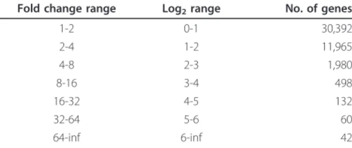
The Affymetrix M430 2.0 array that we used includes 45,101 probe sets and provides consensus estimates of expression for the vast majority of all protein coding genes. Table 1 gives an overview of the range of varia-tion across strains in each of the probe sets used. Strik-ingly, more than 2,000 genes showed a range of expression that was larger than four-fold different between the strain with the lowest and the highest expression. Among the genes with the most extreme range in expression levels were Krt4(keratin 4),Krt13
(keratin 13) and Krtdap (keratinocyte differentiation associated protein). Another gene with highly variable expression wasCftr(cystic fibrosis transmembrane con-ductance regulator homolog). This important lung dis-ease-causing gene showed a four-fold variation in expression levels between strains. Several other genes with high variation were sex-specifically expressed genes, like Xist (inactive X specific transcripts), Ddx3y
(DEAD (Asp-Glu-Ala-Asp) box polypeptide 3, Y-linked) andSerpina1b(serine (or cysteine) preptidase inhibitor, clade A, member 1B).
Table 1 Variation in gene expression for 45,101 probe sets.
Fold change range Log2range No. of genes
1-2 0-1 30,392
2-4 1-2 11,965
4-8 2-3 1,980
8-16 3-4 498
16-32 4-5 132
32-64 5-6 60
64-inf 6-inf 42
Heritability of variation in gene expression
To investigate to which extent the variation in expres-sion was due to genetic effects, we calculated the herit-ability for each of the genes, which is the fraction of variation in expression caused by genetics. The heritabil-ity values ranged from as high as 0.96 (most of the var-iance was associated with between-strain differences) until as low as 0.01. Genes with the largest heritability were Cdk17/Pctk2 (cyclin-dependent kinase 17, probe set 1446130_at), Gm1337 (predicted gene 1337, 1443287_at) andPdxdc1/KIAA0251 (pyridoxal-depen-dent decarboxylase domain containing 1, 1452705_at), all having a value above 0.99. High heritability values indicate that it is likely to successfully map QTLs that influence gene expression values.
Lung-specific genes
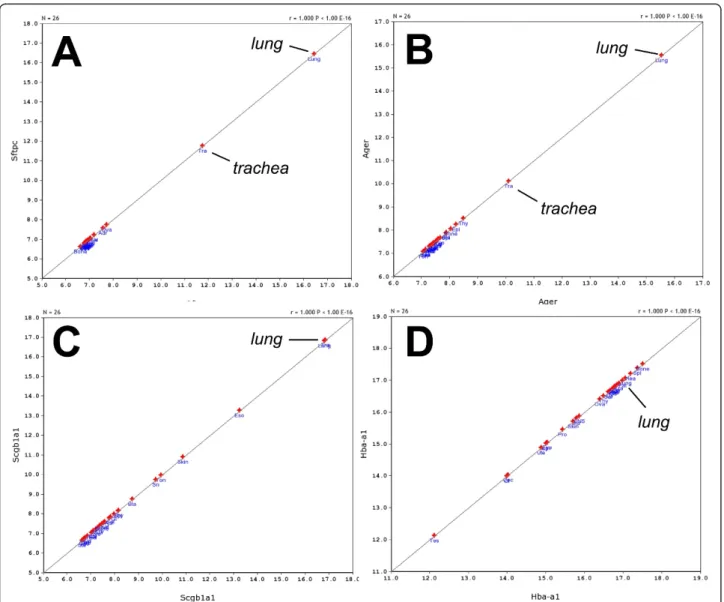
The large dataset in GeneNetwork and its built-in fea-tures allowed us to compare the gene ex-pression pat-terns in the lung with data from 25 other tissues. First, we identified the most highly expressed genes in lung (Table 2 lists the 15 highest expression signals). Two of these genes were highly restricted to the lung and tra-chea: Sftpc (surfactant associated protein C) andAger
(advanced glycosylation end product-specific receptor) (Figure 1A, B) whereas Scgb1a1 (secretoglobin, family 1A, member 1 (uteroglobin)) was highly expressed in lung but also showed expression in some other tissues (Figure 1C). On the other hand, Hba-a1 (hemoglobin alpha, adult chain 1) was expressed at high levels in most tissues (Figure 1D). We then usedStfpc in a tissue
correlation analysis to identify other genes that may not be as highly expressed but still be restricted to lung tis-sue. The first 70 probe sets found were then analyzed as above for lung-specific expression, and 15 genes were identified (Table 3). A comparison to the expression patterns described in the BioGPS database [19] con-firmed that the majority was only expressed in lung, most of them at high level. Two genes were not restricted to the lung according to BioGPS, and five genes were also found at lower levels in one other tissue (Table 3).
Identification of gene networks using correlations
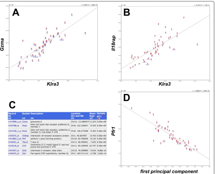
The large data set for expression values for ~39,000 transcripts in 57 mouse strains allowed us to calculate correlations between any pair of genes. A Spearman rank correlation analysis identified 12,985 pairs of genes with a correlation value above 0.8, and 604 pairs showed a correlation value of 0.9 or higher. For example, the expression of Klra3 (killer cell lectin-like receptor
subfamilily A, member 3) was strongly correlated with the expression of Gzma (granzyme A) (Figure 2A).
Klra3 also appeared to be strongly correlated with
Il18rap (interleukin 18 receptor accessory protein, Figure 2B). We then calculated the first principal com-ponent of theKlra3,Gzma, Il18rapandKlrg1(killer cell lectin-like receptor subfamily G, member 1) genes and used it to determine the correlations with all other genes in the lung data set. In this way, we could identify a network of nine genes exhibiting a correlation of >= 0.8 with this principal component (Figure 2C). One of the newly identified genes wasPrf1(perforin 1) which was correlated with a p-value of < 10-16with the princi-pal component (Figure 2D). If genes exhibit a strong correlation of their expression values, one may hypothe-size that they are involved in the same biological process or pathway, or they may be expressed in the same cell type.
In a similar way, we identified another gene network of 20 genes that exhibited very high correlations of their
Table 2 List of 15 probe sets with highest expression signals in the lung
Probe set Symbol Description Location
(Chr, Mb)
Mean Expr
Tissue-specific expression
1428361_x_at Hba-a1 hemoglobin alpha, adult chain 1 Chr11:
32.184441
15,10 MT
1418639_at Sftpc surfactant associated protein C Chr14:
70.920826
14,92 LS
1452543_a_at Scgb1a1 secretoglobin, family 1A, member 1 (uteroglobin) Chr19: 9.158206
14,78 LHOT
1417184_s_at Hbb-b2 hemoglobin, beta adult minor chain Chr7: 110.976103
14,74 MT
1441958_s_at Ager advanced glycosylation end product-specific receptor Chr17: 34.737745
14,69 LS
AFFX-b-ActinMur/ M12481 _3_at
Actb actin beta, cytoplasmic Chr5:
143.665528
14,67 MT
1452757_s_at Hba-a1 hemoglobin alpha, adult chain 1 Chr11:
32.196742
14,66 MT
1416642_a_at Tpt1 tumor protein, translationally-controlled 1 Chr14: 76.246098
14,62 MT
1418509_at Cbr2 carbonyl reductase 2 Chr11:
120.628111
14,62 LHOT
1436996_x_at Lzp-s P lysozyme structural and lysozyme Chr10: 116.724902
14,62 ND
1416624_a_at Uba52 ubiquitin A-52 residue ribosomal protein fusion product 1 Chr8: 73.032191
14,58 MT
1427021_s_at Fth1 ferritin heavy chain 1 Chr19:
10.057382
14,57 ND
AFFX-MURINE_B2_at
B2 AFFX-MURINE_B2_at short interspersed nuclear element (SINE) class of repeat (probes target Chr 1 and Chr 2 most heavily)
N/A 14,52 ND
1415906_at Tmsb4x thymosin, beta 4, X chromosome ChrX:
163.645132
14,51 MT
1449436_s_at Ubb ubiquitin B Chr11:
62.366564
14,50 MT
expression levels across all mouse strains. All possible pairs of genes in this network showed a correlation above 0.95 (Figure 3). The network contained two kera-tin genes, Krt4 (keratin 4) andKrt13 (keratin 13) and genes involved in cytoskeleton functions, again pointing to a possible interaction of these genes in the same pathway or biological process. Further gene networks found by correlation studies were related to B and T cells (see below).
Correlation analysis identified gene expression signatures for T and B cells
The hemoglobin genesHba-a1(hemoglobin alpha, adult chain 1) and Hbb-b2 (hemoglobin beta, adult minor chain) were among the top 10 genes with highest expression values in our lung data set. The high levels of hemoglobin transcripts suggested that circulating blood cells, including immune cells, may also be ana-lyzed in our data set. Therefore, we investigated the gene expression networks of known immune cell
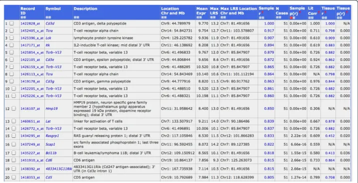
markers, e.g.Cd3genes as specific markers for T cells. We calculated the correlations of Cd3d(Cd3 antigen, delta polypeptide) expression levels over all BXD lines with all other genes. This analysis revealed 20 genes with a very highly correlated expression value (p-value below 10-14, Figure 4). Most of these genes were known T cell markers or involved in T cell regulation. Eight out of the 12 genes with the strongest correlations were also exclusively expressed in T cells according to the BioGPS database (Wu et al., 2009): Cd3d, Itk, Tcrb-13V Cd3e, Cd3g, Scap1, Cd6 and Cd5(see Figure 4 for full gene names). Similarly, we searched for B cell-specific signatures starting with the B cell marker gene Cd19
(CD19 antigen). The probe set“1450570_a_at” detected
Cd19 mRNA levels and showed a mean expression level of 9.3. We found 14 probe sets with a correlation above 0.80 (p-value < 10-14, Figure 5). A comparison with the BioGPS database revealed that eight of them, Cd19, Cd79b, Faim3, Cd79a, Blk, B3gnt5, Cd22 and Blr1 (see Figure 5 for full gene names) were also exclusively
Table 3 List of genes with lung-restricted expression found by tissue correlation analysis withSftpc
Probe set Symbol Description Location
(Chr, Mb)
Mean Expr
BioGPS expression
1418639_at Sftpc surfactant associated protein C Chr14: 70.920826
14,92 high in lung, low in nucleus accumbens
1437028_at Sftpb surfactant associated protein B (nonciliated bronchiolar and alveolar type 2 cell signature)
Chr6: 72.260763
13,68 high in lung only
1422334_a_at Sftpa1 surfactant associated protein A1 Chr14: 41.946994
14,24 high in lung only
1422346_at Nkx2-1 (Titf1)
thyroidtranscription factor 1 Chr12: 57.634187
8,07 lung only
1417057_a_at Lamp3 lysosomal-associated membrane protein 3 Chr16: 19.653875
12,09 high in lung, low in ES cells and some cell lines
1421404_at Cxcl15 chemokine (C-X-C motif) ligand 15 Chr5: 91.230349
13,87 high in lung only
1441958_s_at Ager advanced glycosylation end product-specific receptor Chr17: 34.737745
14,69 high in lung only
1436787_x_at Sec14l3 SEC14-like protein 3 Chr11:
3.978573
13,21 only data for human available -not lung specific
1425218_a_at Scgb3a2 secretoglobin, family 3A, member 2 Chr18: 43.924081
14,17 high in lung only
1449428_at Cldn18 claudin 18 Chr9:
99.591247
12,70 highest in lung, lower in stomach
1449525_at Fmo3 flavin containing monooxygenase 3 Chr1: 164.884088
10,90 high in lung, maybe weak in some other tissues 1425814_a_at Calcrl calcitonin receptor-like Chr2:
84.170818
12,91 high in lung, weak in macrophages 1421373_at Cox4i2 cytochrome c oxidase subunit IV isoform 2 Chr2:
152.582819
9,24 not specific for lung
1419699_at Scgb3a1 secretoglobin, family 3A, member 1 Chr11: 49.477871
13,68 high in lung only
1451604_a_at Acvrl1 activin A receptor, type II-like 1 Chr15: 100.968668
11,86 high in lung only
1420347_at Plunc palate, lung, and nasal epithelium carcinoma associated Chr2: 153.973359
13,42 high in lung, low in heart
expressed in B cells. Therefore, these genes can be con-sidered as T and B cell signature genes which may be used to follow the presence and infiltration of T and B cells in the lung under normal and pathological conditions.
Identification of candidate genes regulating phenotypic traits in the lung
Once a QTL for a phenotypic trait has been found, it will be important to identify the underlying quantitative gene (QTG) which is causing the variation. Searching
cis-eQTLs in the QTL interval represents one suitable approach [8]. As a prototype for this approach in our lung data set, we examined two traits for which lung phenotypes were studied in the BXD population and
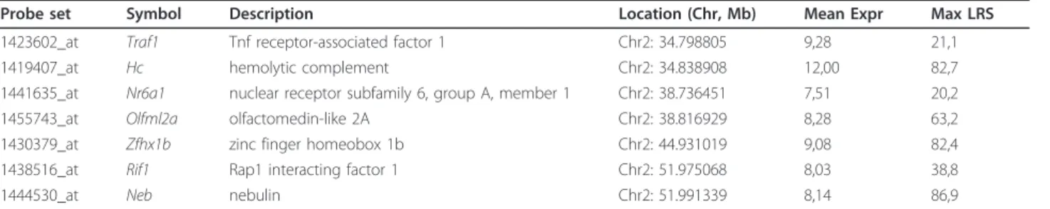
which were available in GeneNetwork. Boon et al. [20] described several QTLs for the susceptibility of BXD mice to influenza A infections. We analyzed one signifi-cant QTL peak on chromosome 2 and two suggestive peaks on chromosomes 7 and 17. Seven cis-eQTL regu-lated genes were found in the chromosome 2 QTL interval (Table 4), including the Hc(hemolytic comple-ment) gene which was shown to contribute to influenza susceptibility [20]. The analysis of the QTL region on chromosome 7 revealed 12 cis-regulated genes in the lung, includingTrim12(tripartite motif protein 12) and
Trim34 (tripartite motif protein 34) which were also described as potential candidate QTGs by [20]. In the chromosome 17 QTL region, we found 17 cis-eQTL genes, of which Prkcn (protein kinase C, nu), Qpct
(glutaminyl-peptide cyclotransferase (glutaminyl cyclase) and Mta3 (metastasis associated 3) were suggested as potential QTGs by [20]. Another lung-specific pheno-type in the GeneNetwork database is “Mycoplasmosis susceptibility, alveolar exudate” (GeneNetwork ID
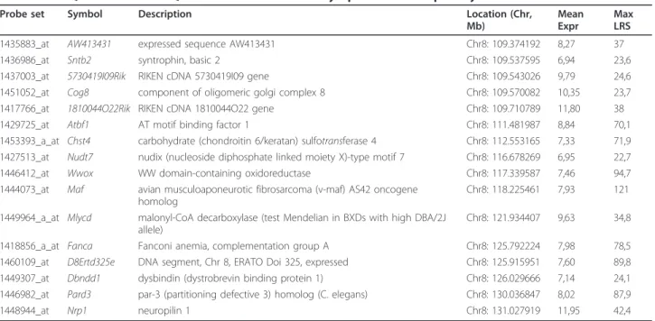
10692, [21] and Cartner et al. unpublished). This trait showed a significant QTL on chromosome 10, between 105 and 130 Mb. The analysis of our lung expression data revealed 16 cis-eQTLs in the genomic interval (Table 5). Three of the cis-QTL genes have been
Figure 3The cytokeratin network. Pearson correlations (listed below the diagonal) showed very high correlations between all pairs of the 20 selected genes. Spearman Rank correlations are shown above the diagonal.
associated previously with immune functions and thus represent suitable candidates to regulate this trait:Chst
(carbohydrate (keratan sulfate Gal-6) sulfotransferase 1) was found to exhibit a critical role in lymphocyte traf-ficking during chronic inflammation [22]. The transcrip-tion factorMaf(avian musculoaponeurotic fibrosarcoma (v-maf) AS42 oncogene homolog) was shown to play a role in the transcriptional regulation of cytokine expres-sion and immune cell markers, e.g. [23-29]. Nrp1 (neu-ropilin 1) has been primarily described as neuronal receptor but appears also to play a role in the primary immune response and formation of the immunological synapse [30,31].
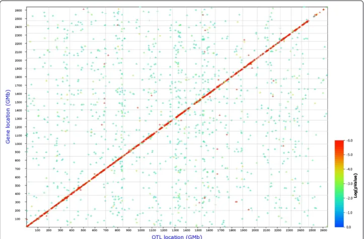
Cis- and trans-eQTLs
We then performed a search for eQTLs on a global level, for all probe sets. In this analysis 5,214 cis- and 15,485 trans-regulated genes were identified at an LRS threshold of 12 (Table 6 and Figure 6). When the LRS threshold was increased to 50, 1,332cis-regulated genes were found, whereas the number of trans-regulated genes was reduced to 15. This observation indicates that
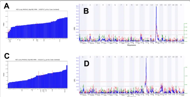
many of thetrans-eQTL showed a much lower signifi-cance value than thecis-eQTL. Next, we present exam-ples for one cis- and onetrans-eQTL. A strong eQTL was detected on chromosome 14, at 52 megabases (Mb; Figure 7B) regulating the expression levels of Ang
(angiogenin, ribonuclease, RNase A family, 5) (Figure 7A). Since Ang is located at the same position as the eQTL (51.7 Mb on chromosome 14) it represents acis -eQTL. Furthermore, a strong eQTL was found on chro-mosome 12 regulating the expression levels of the
Cyp1a1gene (cytochrome P450, family 1, subfamily a, polypeptide 1) (Figure 7C,D). Cyp1a1 is located on chromosome 9 and the corresponding eQTL was found on chromosome 12 (trans-eQTL). The eQTL signifi-cance interval contained nine genes, four of which were expressed in lung at a level above 10. Ahr (aryl-hydro-carbon receptor) was one of the four genes and was at the top of the QTL peak (Figure 8). It is the most likely candidate forCyp1a1regulation. In conclusion, our data set contained a large number of genes whose expression levels are likely to be influenced by allelic variations in the genomes of C57BL/6J and DBA/2J. Therefore, the
Figure 5Gene signatures for B-cells. List of the strongest correlates forCd19(probe set 1450570_at), all correlated at p < 10-12.
Table 4Cis-eQTLs identified in QTL inteval on chromosome 2 for influenza susceptibility
Probe set Symbol Description Location (Chr, Mb) Mean Expr Max LRS
1423602_at Traf1 Tnf receptor-associated factor 1 Chr2: 34.798805 9,28 21,1
1419407_at Hc hemolytic complement Chr2: 34.838908 12,00 82,7
1441635_at Nr6a1 nuclear receptor subfamily 6, group A, member 1 Chr2: 38.736451 7,51 20,2
1455743_at Olfml2a olfactomedin-like 2A Chr2: 38.816929 8,28 63,2
1430379_at Zfhx1b zinc finger homeobox 1b Chr2: 44.931019 9,08 82,4
1438516_at Rif1 Rap1 interacting factor 1 Chr2: 51.975068 8,03 38,8
1444530_at Neb nebulin Chr2: 51.991339 8,14 86,9
presence of pairs of regulated genes and their corre-sponding eQTLs predicts possible regulatory interac-tions and will allow searching for yet unknown regulatory networks.
Discussion
Here, we performed global gene expression profiling in eight inbred mouse strains and a cohort of BXD recom-binant inbred strains from whole lung tissues. Our stu-dies identified several lung-specific genes, large variations in gene expression levels, and a strong herit-ability in many gene expression traits. Correlation analy-sis of gene expression and genotypes identified potential gene interaction networks, pairs of trans- and cis -eQTLs, and genes with cis-eQTLs that may represent candidate genes involved in susceptibility to respiratory infections. In addition, one specific gene interaction pathway was identified in which Ahr regulates the
Cyp1a1gene.
Using tissue correlations of gene expression patterns across the BXD strains, we identified 16 genes with a
highly restricted expression in the lung of which 14 could be validated by comparison to the BioGPS data-base [19]. The second most strongly expressed gene in the lung tissues wasSftpcwhich has been shown to play a role in lung development and the prevention of pneu-monitis and emphysema [32,33]. Also, Sftpc deficiency increases the severity of respiratory syncytial virus-induced pulmonary inflammation [34]. Furthermore,
Scgb1a1andAger were amongst the five most strongly expressed genes.Scgb1a1is expressed in lung clara cells and its deficiency results in enhanced susceptibility to environmental agents [35]. Scgb3a1 (secretoglobin, family 3A, member 1) and Scgb3a2 (secretoglobin, family 3A, member 2) were shown by others to be highly expressed in the lung and lower levels in other organs [36]. Scgb3a2is down-regulated in inflamed air-ways [37] and plays an important role in lung develop-ment [38]. Sftpb(surfactant associated protein B (non-ciliated bronchiolar and alveolar type 2 cell signature) is a hydrophobic peptide which enhances the surface prop-erties of pulmonary surfactant and is expressed in non-ciliated bronchiolar and aleveolar type 2 cells [39]. Maintenance ofSftpb expression is critical for survival during acute lung injury [40] and reduction of alveolar expression causes surfactant dysfunction and respiratory failure [41]. Plunc (palate, lung, and nasal epithelium carcinoma associated) is expressed in the oral, lingual, pharyngeal and respiratory epithelia [42] and members of thePluncgene family are thought to pay a role in the innate immune response [43]. The presence of Plunc protein in the lung decreases the levels ofMycoplasma Table 5Cis-eQTLs identified in QTL on chromosome 8 for Mycoplasmosis susceptibility trait
Probe set Symbol Description Location (Chr,
Mb)
Mean Expr
Max LRS
1435883_at AW413431 expressed sequence AW413431 Chr8: 109.374192 8,27 37
1436986_at Sntb2 syntrophin, basic 2 Chr8: 109.537595 6,94 23,6
1437003_at 5730419I09Rik RIKEN cDNA 5730419I09 gene Chr8: 109.543026 9,79 24,6 1451052_at Cog8 component of oligomeric golgi complex 8 Chr8: 109.570082 10,35 23,7 1417766_at 1810044O22Rik RIKEN cDNA 1810044O22 gene Chr8: 109.710789 11,80 38
1429725_at Atbf1 AT motif binding factor 1 Chr8: 111.481987 8,84 70,1
1453393_a_at Chst4 carbohydrate (chondroitin 6/keratan) sulfotransferase 4 Chr8: 112.553165 7,33 71,9 1427513_at Nudt7 nudix (nucleoside diphosphate linked moiety X)-type motif 7 Chr8: 116.678269 6,95 22,7 1446412_at Wwox WW domain-containing oxidoreductase Chr8: 117.339587 7,46 94,7 1444073_at Maf avian musculoaponeurotic fibrosarcoma (v-maf) AS42 oncogene
homolog
Chr8: 118.225461 7,93 121
1449964_a_at Mlycd malonyl-CoA decarboxylase (test Mendelian in BXDs with high DBA/2J allele)
Chr8: 121.934407 9,63 34,8
1418856_a_at Fanca Fanconi anemia, complementation group A Chr8: 125.792224 7,98 78,5 1460109_at D8Ertd325e DNA segment, Chr 8, ERATO Doi 325, expressed Chr8: 125.915951 7,60 89,8 1449307_at Dbndd1 dysbindin (dystrobrevin binding protein 1) Chr8: 126.029666 7,14 24,1 1446982_at Pard3 par-3 (partitioning defective 3) homolog (C. elegans) Chr8: 130.036847 8,02 87,9
1448944_at Nrp1 neuropilin 1 Chr8: 131.027919 11,95 42,4
For each gene, only the highest LRS is shown. Mean Expr: mean expression in lung for BXD strains.
Table 6 Amount ofcis- andtrans-regulatedtranscripts for different significance thresholds
Threshold (LRS) No. ofciseQTLs No. oftranseQTLs
12 5,214 15,485
16 4,391 3,149
20 3,666 536
30 2,500 48
pneumoniaeand its levels are reduced in allergic inflam-matory conditions [44]. Thus, the lung data set allowed us to find important genes that are expressed primarily in the lung and are important for lung homeostasis and prevention of disease.
It should be noted that our analysis of genes with
“restricted expression to the lung” is not ex-clusive; it only refers to the tissues that are represented in Gene-Network and BioGPS. Also, the analysis performed here should not be considered to be comprehensive. More sophisticated approaches may be employed to identify additional genes which also fulfill the criterion of“ lung-restricted” expression.
Furthermore, genes may not be apparent in the lung transcriptome because they are expressed only in a small fraction of cells within the lung. This issue of dilu-tion of expression signals is an important one and we have studied it in several tissues with considerable care (eye, retina, and numerous brain regions) using the same genetic methods and the same array platform. We were consistently able to detect expression of genes that are only expressed in very small cell subpopulations
(<0.1%) such as rare amacrine cell subclasses in the retina [8] or very rare oxytocin-expressing neurons (<2000) in whole brain samples. The reason for the increased sensitivity is that with such large sample sizes (~70 lung arrays) the signal-to-noise ratios are much better than standard studies using Affymetrix arrays. These stuides typically use far fewer arrays and do not use genetic methods to“validate”the source of signal.
The strong signal for hemoglobin and lymphocyte-specific genes clearly showed that gene ex-pression pat-terns of circulating blood cells are readily detectable in the lung transcriptome. This raises the question if an organ should be studied with or without containing blood. The correct answer to this question depends of course on the particular circumstances. However, we feel strongly that a global systems and genetic approach requires the analysis of the entire organ. The expression of genes is not cell-autonomous and depends on cellular micro envi-ronment, physical factors (gas pressure and gradients, etc), pathogen exposure, and many types of interactions. These factors also influence the expression of genes in blood cells. Therefore, we think that it is
Figure 7Examples for variations of expression levels in different BXD and inbred mouse strains and ge-nome-wide analysis of cis-andtrans-eQTLs. (A) Log2expression levels forAng1and (B) correspondingcis-eQTL signal. The numbers at the top are chromosomes. The blue line represents the significance level of the QTL expressed as LRS score (likelihood ratio statistic). A positive additive coefficient (green line) indicates that DBA/2J alleles increased trait values. A negative additive coefficient (red line) indicates that C57BL/6J alleles increased trait values. The two horizontal lines mark the genome-wide significance levels at p < 0.05 (red line) and p < 0.63 (gray line).Ang1is located on Chr 14 (triangle) and the QTL peak is at the same location as the gene. (C) Log2expression levels forCyp1a1and (D) correspondingtranseQTL peak. Cyp1a1is on chromosome 9 (triangle) and the QTL was found on chromosome 12.
imperative to look simultaneously at all cells in a func-tion unit: in this case the whole lung plus its containing blood.
In conclusion, the combined analysis of expression levels and correlations in a variety of tissues tissue allowed us to determine genes with restricted or prefer-ential expression in the lung. For several of these genes, an important function in the lung has been described and the same may be assumed for the others. This information will also contribute to a better understand-ing of the biological function of these genes.
Many phenotypic traits have been studied for the BXD mouse populations and several QTLs were identified which influence diseases or vulnerability in the lung. The detection of cis eQTLs in the very same tissue is one method to identify potential candidate genes under the QTL which may causally influence the trait. Here, we investigated two traits in more detail, susceptibility to influenza virus and susceptibility to mycoplasmosis. Several cis-eQTLs were found in the corresponding QTL regions and in each case, genes could be identified with a presumed role in the host immune defense (dis-cussed already in the results section). Thus, the study of
cis-eQTLs in our data set may provide valuable candi-dates for other quantitative trait genes that influence important lung phenotypes. Furthermore, we found 13 BXD lines with low expression signals for Krt4, Krt13
and Krtdap. Krt4 and Krt13 have been shown to be responsible for White sponge nevus (WSN), also known as Cannon’s disease, which is an autosomal dominant skin condition in humans [45-47]. We propose that the 13 mouse strains have genetic alterations which result in low transcript levels of these genes and they may represent a good model for Cannon’s disease. It should be noted, however, that nocis-eQTLs found were found for any of theKrtgenes.
We also identified a set of genes for which the expres-sion levels correlated highly with members of the Klr
gene family. Klra3 and Klrg1 are killer cell lectin-like receptors that are exclusively expressed on natural killer cells (NK cells). NK cells form a major component of the innate immune system and kill cells by releasing small cytoplasmic granules of proteins called perforins and granzymes [48]. Both Gzmaand Prf1 were in the gene network that we identified. In addition, correla-tions can also be used to expand already known gene networks in specific cell populations. When starting with theCd3T cell marker and calculating correlations with all other transcripts measured, we identified a strongly correlated network of genes, in which most of the genes were known as T cell markers or to be involved in T cell activation or homeostasis. In a similar way, when starting with the Cd19 B cell marker, we could identify a strongly correlated network of B cell
signature genes. The analysis of these T cell and B cell expression signatures in the Bi-oGPS data base with expression profiles in mouse tissues revealed that indeed >90% of the T and B cell markers were specifically expressed in either T or B cells. Furthermore, most of the T and B cell signature genes represented genes with known function in B and T cell differentiation, activa-tion and homeostasis. For example, the T cell signature included genes encoding subunits of the T cell receptor:
Cd3d (CD3 antigen, delta po-lypeptide), Cd3g (CD3 antigen, gamma polypeptide), Tcra (T-cell receptor alpha chain) andTcrb-V13(T-cell receptor beta, variable 13) and Lat(linker for activation of T cells) which are involved in T cell activation. The B cell signature con-tained components of the B cell antigen receptor com-plex, Cd19(CD19 antigen) andCd79a(CD79A antigen (immunoglobulin-associated alpha)), as well as Blk (B lymphoid kinase) tyrosine kinase which is associated with the receptors. Also, the correlations for both signa-tures in the spleen expression data set in GeneNetwork could indeed confirm that the signatures were strongly correlated (data not shown). In summary, these studies demonstrate that correlation analyses are able to identify genes which very likely interact in a common network or biological process. The approach used here may thus have a great potential to identify new networks and bio-logical processes in the lung. In addition, starting with a known bona-fide cell-specific gene and then analyzing gene expression values across strains, it is possible to identify a set of highly correlated genes. These gene sets genes can now be used as cell-specific signature genes in complex transcriptome studies,e.g.to detect infiltrat-ing immune cells in the lungs after infection.
The genetic mapping of lung expression profiles revealed many cis- and trans-eQTLs, indicating that many gene expression patterns in lung have a strong genetic component.Trans-eQTLs allow the identifica-tion of gene-gene regulatory networks. As an example, we found that the transcription factor Ahrwas present in a trans-eQTL region detected for the Cyp1a1gene.
Ahris a transcription factor known to induce Cyp1a1
transcription levels after ligand binding [49-51]. Six binding sites for the Ahr receptor ligand have been revealed in the 700-basepair DNA domain upstream of
Cyp1a1[52]. However, a critical leucine-to-proline sub-stitution in Ahrresults in a 15 to 20-fold reduction in the binding affinity of the proline variant found in DBA/ 2J compared to the leucine variant found in C57BL/6J [53]. Indeed, in our data set, expression values for
interaction between Cyp1a1 and Ahr as such is not a new finding, it is quite remarkable that the interaction becomes apparent in lungs which were not exposed to an inducing xenobiotic. Furthermore, we do not see this relationship in several other tissues, such as liver. There-fore, our observation suggests that in the lung, which is potentially exposed to many xenobiotics, the Ahr recep-tor may always be activated at a low level. Alternatively,
Ahr expression may be stimulated by yet unknown ligands that are also present under normal environmen-tal conditions.
Conclusions
Here, we showed that whole genome expression analysis of the lungs from a large set of strains of the BXD mouse population can be exploited to identify important gene regulatory networks. We found a large number of expression correlations and QTLs which can be further investigated to better understand molecular interaction networks in the lung. The search forcis-eQTLs in geno-mic intervals that were identified previously as QTLs for infectious diseases revealed several quantitative trait candidate genes. In addition, we demonstrated that the analysis of gene expression correlations, starting with a few cell-specific genes, could identify a larger set of genes which allows detecting the presence of B and T cells within the transcriptome of the whole lung. Such expression signatures will be very important to follow normal and abnormal host responses during infections and other diseases of the lung.
Acknowledgements
This work was supported by intra-mural grants from the Helmholtz-Association (Program“Infection and Immunity”) and a research grant “FluResearchNet”(No. 01KI07137) from the German Ministry of Education and Research to KS. RWW acknowledges the support of the UTHSC Center for Integrative and Translational Genomics and NIH grant P20DA21131 and U01AA13499, and LL was supported by the NIH grant U01AA014425. We thank Dr. Yan Jiao and Weikuan Gu at UTHSC and the VA Medical Center, Memphis, for running the Affymetrix arrays in their core facility. We also thank the GeneNetwork development team, including Arthur Centeno, Xiaodong Zhou, Ning Liu, Zachary Sloan, and Lei Yan, for their help in integrating and error-checking the lung transcriptome data. We also thank Samuel C. Cartner for making the data on the Mycoplasmosis susceptibility phenotype available to us prior to publication.
Author details
1Department of Infection Genetics, Helmholtz Centre for Infection Research & University of Veterinary Medicine Hannover, Inhoffenstr. 7, D-38124 Braunschweig, Germany.2Department of Anatomy and Neurobiology, University of Tennessee Health Science Center, Memphis, Tennessee, USA. 3Jiangsu Key Laboratory of Neuroregeneration, Nantong University, Nantong, China.
Authors’contributions
RA performed the bioinformatics analysis and wrote the manuscript. KS designed the experiments, performed the bioinformatics analysis and wrote the manuscript. LL and RWW prepared the study material and supervised the expression array studies. RW contributed to writing of the manuscript. All authors have read and approved the final manuscript.
Competing interests
The authors declare that they have no competing interests.
Received: 14 January 2011 Accepted: 2 May 2011 Published: 2 May 2011
References
1. Schughart K:SYSGENET: a meeting report from a new European network for systems genetics.Mamm Genome2010,21(7-8):331-336.
2. Morahan G, Peeva V, Mehta M, Williams R:Systems genetics can provide new insights in to immune regulation and autoimmunity.J Autoimmun 2008,31(3):233-236.
3. Threadgill DW, Hunter KW, Williams RW:Genetic dissection of complex and quantitative traits: from fantasy to reality via a community effort. Mamm Genome2002,13(4):175-178.
4. Alberts R, Fu J, Swertz MA, Lubbers LA, Albers CJ, Jansen RC:Combining microarrays and genetic analysis.Brief Bioinform2005,6(2):135-145. 5. Chesler EJ, Lu L, Shou S, Qu Y, Gu J, Wang J, Hsu HC, Mountz JD,
Baldwin NE, Langston MA,et al:Complex trait analysis of gene expression uncovers polygenic and pleiotropic networks that modulate nervous system function.Nat Genet2005,37(3):233-242.
6. Jansen RC, Nap JP:Genetical genomics: the added value from segregation.Trends Genet2001,17(7):388-391.
7. Rosen GD, Pung CJ, Owens CB, Caplow J, Kim H, Mozhui K, Lu L, Williams RW:Genetic modulation of striatal volume by loci on Chrs 6 and 17 in BXD recombinant inbred mice.Genes Brain Behav2009, 8(3):296-308.
8. Geisert EE, Lu L, Freeman-Anderson NE, Templeton JP, Nassr M, Wang X, Gu W, Jiao Y, Williams RW:Gene expression in the mouse eye: an online resource for genetics using 103 strains of mice.Mol Vis2009, 15:1730-1763.
9. Gaglani SM, Lu L, Williams RW, Rosen GD:The genetic control of neocortex volume and covariation with neocortical gene expression in mice.BMC Neurosci2009,10:44.
10. Gatti D, Maki A, Chesler EJ, Kirova R, Kosyk O, Lu L, Manly KF, Williams RW, Perkins A, Langston MA,et al:Genome-level analysis of genetic regulation of liver gene expression networks.Hepatology2007,46(2):548-557. 11. Bystrykh L, Weersing E, Dontje B, Sutton S, Pletcher MT, Wiltshire T, Su AI,
Vellenga E, Wang J, Manly KF,et al:Uncovering regulatory pathways that affect hematopoietic stem cell function using‘genetical genomics’.Nat Genet2005,37(3):225-232.
12. Bao L, Wei L, Peirce JL, Homayouni R, Li H, Zhou M, Chen H, Lu L, Williams RW, Pfeffer LM,et al:Combining gene expression QTL mapping and phenotypic spectrum analysis to uncover gene regulatory relationships.Mamm Genome2006,17(6):575-583.
13. Chesler EJ, Lu L, Wang J, Williams RW, Manly KF:WebQTL: rapid exploratory analysis of gene expression and genetic networks for brain and behavior.Nat Neurosci2004,7(5):485-486.
14. Irizarry RA, Hobbs B, Collin F, Beazer-Barclay YD, Antonellis KJ, Scherf U, Speed TP:Exploration, normalization, and summaries of high density oligonucleotide array probe level data.Biostatistics2003,4(2):249-264. 15. Haley CS, Knott SA:A simple regression method for mapping
quantitative trait loci in line crosses using flanking markers.Heredity 1992,69(4):315-324.
16. Shifman S, Bell JT, Copley RR, Taylor MS, Williams RW, Mott R, Flint J:A high-resolution single nucleotide polymorphism genetic map of the mouse genome.PLoS Biol2006,4(12):e395.
17. Williams RW, Gu J, Qi S, Lu L:The genetic structure of recombinant inbred mice: high-resolution consensus maps for complex trait analysis. Genome Biol2001,2(11):RESEARCH0046.
18. Brockdorff N, Ashworth A, Kay GF, Cooper P, Smith S, McCabe VM, Norris DP, Penny GD, Patel D, Rastan S:Conservation of position and exclusive expression of mouse Xist from the inactive X chromosome. Nature1991,351(6324):329-331.
19. Wu C, Orozco C, Boyer J, Leglise M, Goodale J, Batalov S, Hodge CL, Haase J, Janes J, Huss JW,et al:BioGPS: an extensible and customizable portal for querying and organizing gene annotation resources.Genome Biol2009,10(11):R130.
21. Cartner SC, Simecka JW, Briles DE, Cassell GH, Lindsey JR:Resistance to mycoplasmal lung disease in mice is a complex genetic trait.Infect Immun1996,64(12):5326-5331.
22. Hiraoka N, Kawashima H, Petryniak B, Nakayama J, Mitoma J, Marth JD, Lowe JB, Fukuda M:Core 2 branching
beta1,6-N-acetylglucosaminyltransferase and high endothelial venule-restricted sul-fotransferase collaboratively control lymphocyte homing.J Biol Chem 2004,279(4):3058-3067.
23. Cao S, Liu J, Chesi M, Bergsagel PL, Ho IC, Donnelly RP, Ma X:Differential regulation of IL-12 and IL-10 gene expression in macrophages by the basic leucine zipper transcription factor c-Maf fibrosarcoma.J Immunol 2002,169(10):5715-5725.
24. Cao S, Liu J, Song L, Ma X:The protooncogene c-Maf is an essential transcription factor for IL-10 gene expression in macrophages.J Immunol 2005,174(6):3484-3492.
25. Hiramatsu Y, Suto A, Kashiwakuma D, Kanari H, Kagami S, Ikeda K, Hirose K, Watanabe N, Grusby MJ, Iwamoto I,et al:c-Maf activates the promoter and enhancer of the 21 gene, and TGF-beta inhibits c-Maf-induced IL-21 production in CD4+ T cells.J Leukoc Biol2010,87(4):703-712. 26. Nakamura M, Hamada M, Hasegawa K, Kusakabe M, Suzuki H, Greaves DR,
Moriguchi T, Kudo T, Takahashi S:c-Maf is essential for the F4/80 expression in macrophages in vivo.Gene2009,445(1-2):66-72. 27. Pot C, Jin H, Awasthi A, Liu SM, Lai CY, Madan R, Sharpe AH, Karp CL,
Miaw SC, Ho IC,et al:Cutting edge: IL-27 induces the transcription factor c-Maf, cytokine IL-21, and the costimulatory recep-tor ICOS that coordinately act together to promote differentiation of IL-10-producing Tr1 cells.J Immunol2009,183(2):797-801.
28. Voice J, Donnelly S, Dorsam G, Dolganov G, Paul S, Goetzl EJ:c-Maf and JunB mediation of Th2 differentiation induced by the type 2 G protein-coupled receptor (VPAC2) for vasoactive intestinal peptide.J Immunol 2004,172(12):7289-7296.
29. Xu J, Yang Y, Qiu G, Lal G, Wu Z, Levy DE, Ochando JC, Bromberg JS, Ding Y:c-Maf regulates IL-10 expression during Th17 polarization. J Immunol2009,182(10):6226-6236.
30. Tordjman R, Lepelletier Y, Lemarchandel V, Cambot M, Gaulard P, Hermine O, Romeo PH:A neuronal receptor, neuropilin-1, is essential for the initiation of the primary immune response.Nat Immunol2002, 3(5):477-482.
31. Wulfing C, Rupp F:Neuropilin-1: another neuronal molecule in the “immunological synapse”.Nat Immunol2002,3(5):418-419. 32. Bridges JP, Wert SE, Nogee LM, Weaver TE:Expression of a human
surfactant protein C mutation associated with interstitial lung disease disrupts lung development in transgenic mice.J Biol Chem2003, 278(52):52739-52746.
33. Glasser SW, Detmer EA, Ikegami M, Na CL, Stahlman MT, Whitsett JA: Pneumonitis and emphysema in sp-C gene targeted mice.J Biol Chem 2003,278(16):14291-14298.
34. Glasser SW, Witt TL, Senft AP, Baatz JE, Folger D, Maxfield MD, Akinbi HT, Newton DA, Prows DR, Korfhagen TR:Surfactant protein C-deficient mice are susceptible to respiratory syncytial virus infection.Am J Physiol Lung Cell Mol Physiol2009,297(1):L64-72.
35. Stripp BR, Reynolds SD, Boe IM, Lund J, Power JH, Coppens JT, Wong V, Reynolds PR, Plopper CG:Clara cell secretory protein deficiency alters clara cell secretory apparatus and the protein composition of airway lining fluid.Am J Respir Cell Mol Biol2002,27(2):170-178.
36. Porter D, Lahti-Domenici J, Torres-Arzayus M, Chin L, Polyak K:Expression of high in normal-1 (HIN-1) and uteroglobin related protein-1 (UGRP-1) in adult and developing tissues.Mech Dev2002,114(1-2):201-204. 37. Chiba Y, Kusakabe T, Kimura S:Decreased expression of
uteroglobin-related protein 1 in inflamed mouse airways is mediated by IL-9.Am J Physiol Lung Cell Mol Physiol2004,287(6):L1193-1198.
38. Kurotani R, Tomita T, Yang Q, Carlson BA, Chen C, Kimura S:Role of secretoglobin 3A2 in lung development.Am J Respir Crit Care Med2008, 178(4):389-398.
39. Bohinski RJ, Huffman JA, Whitsett JA, Lattier DL:Cis-active elements controlling lung cell-specific expression of human pulmonary surfactant protein B gene.J Biol Chem1993,268(15):11160-11166.
40. Bein K, Wesselkamper SC, Liu X, Dietsch M, Majumder N, Concel VJ, Medvedovic M, Sartor MA, Henning LN, Venditto C,et al: Surfactant-associated protein B is critical to survival in nickel-induced injury in mice.Am J Respir Cell Mol Biol2009,41(2):226-236.
41. Melton KR, Nesslein LL, Ikegami M, Tichelaar JW, Clark JC, Whitsett JA, Weaver TE:SP-B deficiency causes respiratory failure in adult mice.Am J Physiol Lung Cell Mol Physiol2003,285(3):L543-549.
42. LeClair EE, Nomellini V, Bahena M, Singleton V, Bingle L, Craven CJ, Bingle CD:Cloning and expression of a mouse member of the PLUNC protein family exclusively expressed in tongue epithe-lium.Genomics 2004,83(4):658-666.
43. Bingle CD, Craven CJ:PLUNC: a novel family of candidate host defence proteins expressed in the upper airways and nasopharynx.Hum Mol Genet2002,11(8):937-943.
44. Chu HW, Thaikoottathil J, Rino JG, Zhang G, Wu Q, Moss T, Refaeli Y, Bowler R, Wenzel SE, Chen Z,et al:Function and regulation of SPLUNC1 protein in Mycoplasma infection and allergic inflam-mation.J Immunol 2007,179(6):3995-4002.
45. Canon AB:White sponge nevus of the mucosa (nevus spongiosus albus mucosa).Arch Dermatol Syphilol1935,31:365-370.
46. Terrinoni A, Rugg EL, Lane EB, Melino G, Felix DH, Munro CS, McLean WH: A novel mutation in the keratin 13 gene causing oral white sponge nevus.J Dent Res2001,80(3):919-923.
47. Rugg EL, McLean WH, Allison WE, Lunny DP, Macleod RI, Felix DH, Lane EB, Munro CS:A mutation in the mucosal keratin K4 is associated with oral white sponge nevus.Nat Genet1995,11(4):450-452.
48. Trapani JA, Smyth MJ:Functional significance of the perforin/granzyme cell death pathway.Nat Rev Immunol2002,2(10):735-747.
49. Nebert DW, Dalton TP, Okey AB, Gonzalez FJ:Role of aryl hydrocarbon receptor-mediated induction of the CYP1 enzymes in environmental toxicity and cancer.J Biol Chem2004,279(23):23847-23850.
50. Okey AB, Riddick DS, Harper PA:The Ah receptor: mediator of the toxicity of 2,3,7,8-tetrachlorodibenzo-p-dioxin (TCDD) and related compounds. Toxicol Lett1994,70(1):1-22.
51. Sutter CH, Yin H, Li Y, Mammen JS, Bodreddigari S, Stevens G, Cole JA, Sutter TR:EGF receptor signaling blocks aryl hydrocarbon receptor-mediated transcription and cell differentiation in human epidermal keratinocytes.Proc Natl Acad Sci USA2009,106(11):4266-4271. 52. Denison MS, Fisher JM, Whitlock JP Jr:Protein-DNA interactions at
recognition sites for the dioxin-Ah receptor complex.J Biol Chem1989, 264(28):16478-16482.
53. Chang C, Smith DR, Prasad VS, Sidman CL, Nebert DW, Puga A:Ten nucleotide differences, five of which cause amino acid changes, are associated with the Ah receptor locus polymorphism of C57BL/6 and DBA/2 mice.Pharmacogenetics1993,3(6):312-321.
doi:10.1186/1465-9921-12-61
Cite this article as:Albertset al.:Genome-wide analysis of the mouse lung transcriptome reveals novel molecular gene interaction networks and cell-specific expression signatures.Respiratory Research201112:61.
Submit your next manuscript to BioMed Central and take full advantage of:
• Convenient online submission
• Thorough peer review
• No space constraints or color figure charges
• Immediate publication on acceptance
• Inclusion in PubMed, CAS, Scopus and Google Scholar
• Research which is freely available for redistribution