ABSTRACT
PURPOSE: To evaluate changes induced by stan-dard laser in situ keratomileusis (LASIK) for hyper-opia on total and corneal optical quality.
METHODS: Total and corneal aberrations were measured before and after standard hyperopic LASIK in 13 eyes (preoperative spherical equiva-lent refractive error +3.17 ± 1.10 D). The Chiron Technolas 217C laser with PlanoScan was used. Total aberrations (measured using laser ray trac-ing) and corneal aberrations (estimated from a videokeratoscope) were described using Zernike terms. Root-mean-square wavefront error for both total and corneal aberrations, and through-focus Strehl ratio for the point spread function of the whole eye were used to assess optical changes induced by surgery.
RESULTS: Third and higher order aberrations increased significantly after hyperopic LASIK (by a factor of 2.20 for total and 1.78 for corneal aberra-tions, for a 6.5-mm pupil). Spherical aberration changed to negative values (corneal average decreased by -0.85 ± 0.48 µm and total average by -0.70 ± 0.30 µm). Best Strehl ratio for the whole eye decreased by a factor of 1.84. Hyperopic LASIK induced larger changes than myopic LASIK, com-pared to an equivalent group of myopic eyes from a previous study. Induced corneal spherical aberra-tion was six times larger after hyperopic LASIK, for a similar range of correction, and of opposite sign.
As with myopic LASIK, changes in internal spheri-cal aberration are of opposite sign to those induced on the corneal anterior surface.
CONCLUSIONS: Hyperopic LASIK induced sig-nificant amounts of aberrations. The largest increase occurred in spherical aberration, which showed a shift (toward negative values) of opposite sign; increase was greater than for myopic LASIK. [J Refract Surg2004;20:203-216]
A
lthough studies have been published on opti-cal changes induced by standard laser in situ keratomileusis (LASIK) for myopia, reports on objective evaluation of change in optical aberra-tions and optical quality with standard LASIK for hyperopia are scarce. There are few reports on hyperopic eyes prior to treatment, either in terms of geometric structure (biometry1, corneal shape2-6) oroptical aberrations5, and some reports are
contra-dictory.
As in correction of myopia, LASIK is a popular surgical option for the correction of hyperopia. A hinged flap is created by means of a microkeratome and folded back to expose the stroma. An excimer laser is then used to ablate the stroma, increasing corneal refractive power in the case of a hyperopic correction. To achieve an effective steepening of the cornea, the laser removes a ring of tissue in the mid-peripheral zone of the corneal stroma.7-10The
abla-tion profile for hyperopes requires a smooth transi-tion zone to prevent an abrupt step at the peripher-al edge.11The result after LASIK for hyperopia is a
cone-like corneal profile.
Published studies on change of aberrations with refractive surgery for myopia report an increase of total12-14 and corneal14-17 third and higher order
aberrations (ie, all aberrations excluding tilt, defo-cus, and astigmatism). This increase is mainly due to an increase of spherical aberration toward more positive values, although a significant increase in coma attributed to decentration in the ablation pat-tern18 was also found. The increase of corneal
Total and Corneal Optical Aberrations Induced by
Laser in situ Keratomileusis for Hyperopia
Lourdes Llorente, OD; Sergio Barbero, PhD; Jesus Merayo, MD, PhD; Susana Marcos, PhD
From the Instituto de Optica "Daza de Valdés", Consejo Superior de Investigaciones Científicas, Madrid, Spain (Llorente, Barbero, Marcos) and Instituto Universitario de Oftalmobiología Aplicada, Universidad de Valladolid, Valladolid, Spain (Merayo).
This research was supported by grants BFM2002-02638 and CAM08.7/004.1/2003 to S. Marcos. S. Barbero thanks the Ministerio de Educación, Cultura y Deportes, Spain for a predoctoral fellowship. The authors acknowledge the Unidad Asociada Instituto de Optica, CSIC-IOBA, Universidad de Valladolid.
The authors have no proprietary interest in the materials presented herein.
Correspondence: Lourdes Llorente, OD, C/. Serrano, 121, 28006 Madrid, Spain. Tel: 34.91.5616800; Fax: 34.91.5645557; E-mail: [email protected]
Received: July 21, 2003 Accepted: January 9, 2004
spherical aberration accounted for most of the changes observed in the total aberration pattern.14
However, changes in the posterior surface of the cornea and the aberrations of the crystalline lens played a significant role and can explain individual outcomes for certain subjects.14
Earlier studies of hyperopic correction with excimer laser also suggest an increase of optical aberrations with the procedure.19,20 Oliver and
col-leagues19 studied anterior corneal aberrations
induced by photorefractive keratectomy (PRK) for hyperopia in nine eyes. They reported a change in corneal spherical aberration, which was positive in all eyes prior to surgery, toward negative values for 5.5-mm and 7-mm pupil diameters. A significant increase in coma root-mean-square (RMS) was also reported. Comparing the results of this study with those obtained in a previous study on myopic PRK17
they found that the change of anterior corneal aber-rations following PRK for hyperopia was greater than those after myopic PRK. Chen and colleagues21
studied corneal asphericity for 33 eyes before and after hyperopic LASIK. They found a significant change in corneal asphericity toward more negative values. This change in asphericity toward negative values results in a shift of spherical aberration toward negative values. Ma and colleagues22
com-pared wave aberrations in control eyes with eyes after LASIK and lensectomy corrections (with intraocular lens implantation) for hyperopia. The LASIK group had the highest RMS aberration, and the most negative corneal and total spherical aber-ration. In addition, they found significant
differ-ences in the internal spherical aberration in the LASIK group.
We present corneal and total optical quality in the same group of eyes measured before and after standard LASIK for hyperopia. Estimation of inter-nal aberrations before and after LASIK in the same eyes allows us to account for changes on the posteri-or cposteri-orneal surface induced by the surgical procedure.
PATIENTS AND METHODS Patients
Thirteen eyes from seven patients (mean age ± standard deviation: 37 ± 11 years; range 24 to 54 yr) were measured before (15 ± 17 days) and after (68 ± 43 days; range 35 to 150 days) LASIK for hyperopia. Preoperative spherical equivalent refraction ranged from +1.38 to +4.50 diopters (D) (mean +3.17 ± 1.10 D) and preoperative astigmatism was less than 2.50 D in all patients. Left and right eyes were ana-lyzed independently for each patient. All patients were properly informed and signed a written con-sent, which met the tenets of the Declaration of Helsinki; informed consent was obtained before enrollment in the study. This consent form was approved by an institutional review board. All data for both eyes were generally collected during the same experimental session at Instituto de Óptica (C.S.I.C.), Madrid, Spain.
Surgery
Standard LASIK procedures and clinical follow-up were performed at Instituto de Oftalmobiología
Table
Clinical Data for 13 Eyes After LASIK for Hyperopia
Eye No. Patient Axial Optical Zone Treatment Attempted Attempted Keratometry Age (yr) Length Diameter (mm) Zone Diameter Spherical Spherical (mm)
(mm) (mm) Equivalent (D) Correction (D) 1 (OS) 48 23.08 5 8.5 1.375 0.25 7.76 x 7.52 2 (OD) 48 23.18 5 8.5 1.5 0.5 7.64 x 7.50 3 (OD) 43 21.79 5 8.5 2.125 1.5 7.50 x 7.34 4 (OS) 43 21.70 5 8.5 2.375 2 7.48 x 7.41 5 (OS) 32 22.51 6 12.8*9.4 2.375 2.75 7.80 x 7.66 6 (OS) 24 22.47 6.5 10 3.5 3 8.26 x 7.94 7 (OD) 36 22.98 6 9.7 3.5 3.5 8.07 x 7.98 8 (OS) 36 23.14 6 9.7 3.5 3.5 8.04 x 7.98 9 (OS) 54 21.66 5 8.6 4 4 7.66 x 7.46 10 (OD) 24 22.33 6.5 10 4 3.75 8.16 x 8.00 11 (OD) 54 21.70 5 8.7 4.25 4.25 7.75 x 7. 50 12 (OS) 25 23.34 5 8.5 4.25 3.5 7.89 x 7.66 13 (OD) 25 23.42 5 8.5 4.5 4 7.90 x 7.83
Aplicada (IOBA), Universidad de Valladolid, Spain, for four patients (eyes #1, 2, 3, 4, 9, 11, 12 and 13) and at Centro Oftalmológico de Madrid (COM), Madrid, Spain for three patients (eyes #5, 6, 7, 8 and 10).
All procedures were performed by the same sur-geon, using the same laser system (a narrow beam, flying spot excimer laser, Chiron Technolas 217-C, equipped with the PlanoScan software; Bausch & Lomb Surgical, Munich, Germany). The laser had an emission wavelength of 193 nm, a fixed pulse repetition rate of 50 Hz, and a radiant exposure of 400 mJ. The flap, which was created using a Hansatome microkeratome (Bausch & Lomb) was 9.5 mm in diameter with an intended depth of 160 µm for all eyes except three (#5, 12 and 13), in which the intended depth was 180 µm. The hinge was always superior. The Table shows clinical data including age, axial length, optical and transition zone diameters, attempted spherical correction, attempted spherical equivalent refraction, and ker-atometric power.
Total Aberrations
Total aberrations were measured using a laser ray tracing technique developed at the Instituto de Óptica in Madrid (Spain). This technique has been described23-25as well as its application as an
evalu-ation tool in myopic LASIK.13,14,26In this technique,
parallel narrow laser beams are delivered sequen-tially through different positions of the pupil, and simultaneously a high resolution coupled charge device (CCD) camera records the retinal spot image corresponding to each entry pupil location. The cen-troid for each image was computed. The deviation of each centroid from that of the principal ray (entry pupil position corresponding to the center of the pupil) is proportional to the slope of the wave aber-ration. Each run consists of 37 rays sampling a 6.51-mm effective pupil in 1-mm steps, arranged in a hexagonal pattern. A single run lasts about 4 sec-onds, and each measurement is repeated five times. The illumination source used in the measurement was a diode laser coupled to an optical fiber (Schäfter + Kirchhoff, Hamburg, Germany) with a wavelength of 786 nm and a nominal output power of 15 mw. The use of infrared wavelength has some advantages over visible light and the results are equivalent to using visible light (except for defocus) within the accuracy of the technique.27 The laser
was attenuated by means of neutral-density filters so that light exposure was at least one order of mag-nitude below safety limits.28
Pupils were dilated with one drop of tropicamide 1% prior to measurement. Subjects' stabilization was achieved by means of a dental impression and a forehead rest, and the pupil was monitored with a CCD camera to ensure its center was aligned with the optical axis of the system. A spherical trial lens (+4 D) was used to compensate for spherical error in eye #12. The raw data (derivatives of the wave aber-ration) were fit to a seventh-order Zernike polyno-mial expansion, using a least-mean-square proce-dure, to obtain the wave aberration. The recommen-dations of the Committee for Standardization of the Optical Society of America were followed regarding ordering and notation for Zernike coefficients.29We
analyzed individual Zernike terms, such as fourth order spherical aberration (Z0
4), and computed the
RMS wavefront error, such as third and higher order RMS, ie, excluding piston (Z0
0), tilts (Z 1 1 and Z-1 1) and defocus (Z 0 2), and astigmatism (Z 2 2and Z -2 2).
We also used computed through-focus Strehl ratios for preoperative and postoperative data.
Corneal Aberrations
The procedure to estimate corneal aberrations has been described in detail.14,30,31 Height data of
the anterior surface of the cornea were obtained from a videokeratoscope (Atlas Mastervue; Humphrey Instruments-Zeiss, San Leandro, CA). These numerical data were processed using custom software (Matlab; Matworks, Natick, MA) and exported to an optical design program (Zemax V.9; Focus software, Tucson, AZ), which performs a vir-tual ray tracing and computes the optical aberra-tions due to the anterior surface of the cornea. Indices of refraction were taken as those of the air and the aqueous humor (1.3391) for a wavelength set to 786 nm, as in total aberrations computation. The corneal wave aberration was described using a seventh-order Zernike polynomial expansion. Custom routines in Matlab were used to shift the reference axis of the corneal wave aberration to the line of sight, to ensure common centration of the total and corneal wave aberration patterns, as has been described in detail.14,30,31Corneal wave
aberra-tions were also computed for a 6.51-mm pupil. Only one corneal map per eye was obtained. In a control experiment with one normal eye (RMS 0.59 µm, for third and higher order terms) a mean Zernike coef-ficient standard deviation (mean across terms) of 0.016 µm was found.
As for total aberrations, individual Zernike terms and RMS were used as optical quality metrics. For convenience we use the term “corneal aberrations”
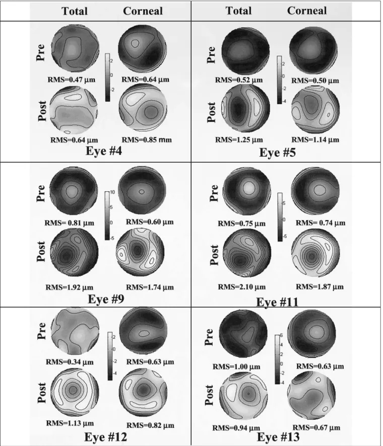
Figure 1. Wave aberration maps for third and higher order aberrations, before and after hyperopic LASIK. For each eye, the maps on the upper
row show the wave aberrations before surgery and the maps on the lower row show the aberrations after hyperopic LASIK. The maps on the right correspond to corneal (anterior surface) aberrations and on the left to total (whole eye) aberrations. All four maps corresponding to the same patient are plotted in the same scale. Contour lines are plotted every 0.2 mm. Pupil size is 6.5 mm.
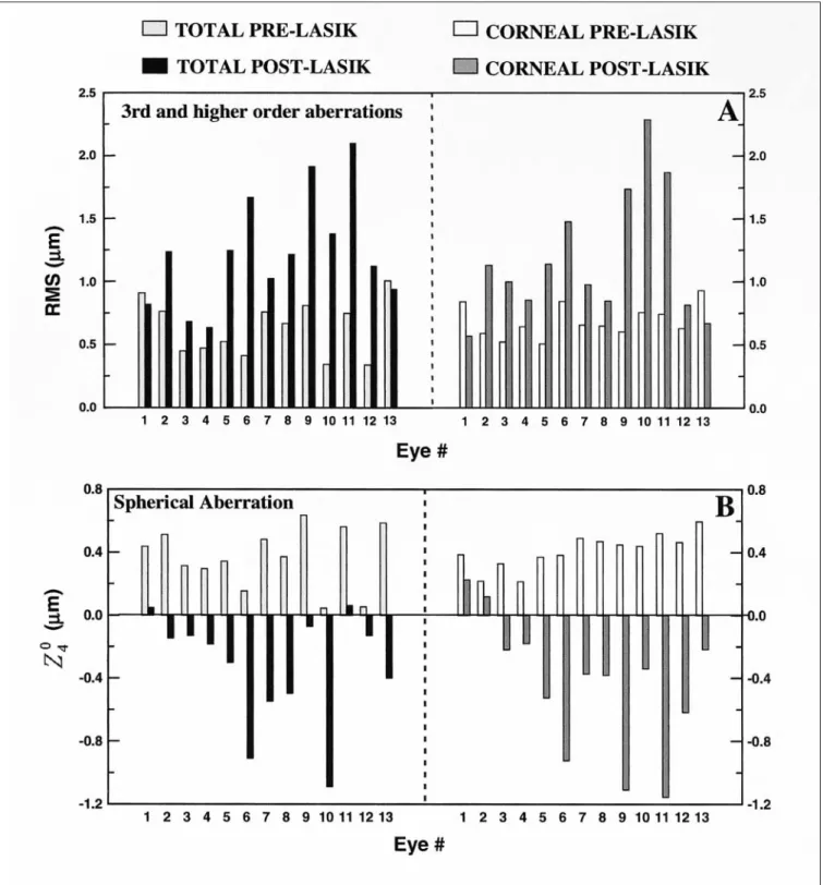
Figure 2. Total (left) and corneal (right) third and higher order: A) RMS wavefront error, and B) spherical aberration, before (light bars) and after
(dark bars) hyperopic LASIK for the 13 eyes in the study. Eyes are sorted by increasing preoperative spherical equivalent refractive error. Pupil size is 6.5 mm.
when we refer to the aberrations of the anterior sur-face of the cornea.
Internal aberrations were computed as the subtraction—term by term—of corneal aberrations from total aberrations.
Additional Measurements
Asphericity and corneal radius were obtained by fitting videokeratoscope height data to a conicoid using custom software written in Matlab.
RESULTS
Figure 1 shows the total (left) and corneal (right) wave aberration patterns before (upper row) and after (lower row) LASIK for hyperopia of six repre-sentative eyes from the study. Only third and high-er ordhigh-er abhigh-errations are represented. Pupil diame-ter is 6.51 mm and contour lines are plotted at at every 0.2 µm. The same scale was used for the four diagrams corresponding to each eye. The num-ber below each map indicates the RMS for third and higher order aberrations.
In some of preoperative eyes, there was an inter-esting similarity between total and corneal wave aberration patterns, which are dominated by posi-tive spherical aberration. This similarity indicates that corneal spherical aberration is the major con-tributor to optical degradation in these eyes, and that the crystalline lens does not play a significant role in counteracting the aberrations of the cornea. This behavior (absolute value of internal spherical aberration <0.1 µm) occurred in 8 of the 13 eyes (62%) in the study. The mean preoperative third and higher order RMS (ie, excluding tilts, defocus, and astigmatism) across all eyes in the study was 0.63 ± 0.22 µm for total aberrations and 0.68 ± 0.13 µm for corneal aberrations.
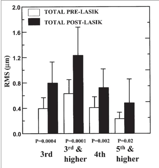
Total and corneal postoperative wave aberration patterns were similar, showing the dominance of corneal aberrations after the procedure. There was a significant increase of aberrations after surgery, indicated by the increase in the number of contour lines of the diagram, and by the corresponding RMS wavefront error. Figure 2A shows total (left) and Figure 3. Preoperative (white bars) and
post-operative (black bars) RMS wavefront error, averaged across all patients, for third and higher order aberrations, third order aberra-tions, fourth order aberraaberra-tions, and fifth and higher order aberrations, for a 6.5-mm pupil.
corneal (right) RMS before (light bars) and after (dark bars) LASIK for third and higher order aber-rations in the 13 eyes, ie, for the best correction of defocus and astigmatism. Total and corneal third and higher order aberrations increased significantly after surgery. The average increase factor was 2.2 for total aberrations and 1.8 for corneal aberrations.
Figure 2B shows total (left) and corneal (right) spherical aberration (Z0
4) before (light bars) and
after (dark bars) LASIK for hyperopia. Total and corneal spherical aberration, which were positive in all preoperative eyes (0.37 ± 0.19 µm and 0.41 ± 0.11 µm, respectively), changed significantly (P<.00001 for both total and corneal) toward more negative values (-0.33 ± 0.35 µm and -0.44 ± 0.43 µm, respectively) after surgery, turning into negative values in 11 of 13 eyes. Corneal spherical aberration decreased on average by -0.85 ± 0.48 µm and total spherical aberration decreased on average by -0.70 ± 0.30 µm.
Figure 3 shows mean preoperative and postoper-ative total RMS for several individual and combined orders. RMS for third and higher order aberrations increased on average by a factor of 2.2 ± 1.1; third order RMS increased by a factor of 2.2 ± 0.9; fourth-order RMS increased by a factor of 2.5 ± 2.4; and fifth and higher order RMS by 2.2 ± 1.5 on average. All these values were for a 6.5-mm diameter pupil; differences were statistically significant for all aber-rations. We did not find that aberrations in eyes with smaller optical zones (5 mm as opposed to 6 or 6.5 mm) increased more than in those with the largest optical zone. We recalculated the aberrations of all patients for a 5-mm pupil and obtained similar increase factors: 2.1 for third and higher order aber-rations, and 2.2 for third order aberrations alone. In addition, we did not find that spherical aberration in eyes with smaller optical zones (5 mm) was greater than in eyes with larger optical zones (6 or 6.5 mm), for either the cornea (P=.99) or the total eye (P=.67). Time after surgery ranged from approx-imately 1 to 3 months. Within this sample of eyes, we did not find any correlation between postopera-tive spherical aberration (P=.54 for the cornea, P=.58 for the total eye) and time after surgery.
DISCUSSION
Comparison of Change in Aberrations With Myopic and Hyperopic LASIK
We compared the outcomes of standard hyperop-ic LASIK with the outcomes of standard myophyperop-ic LASIK from a previous study conducted in our
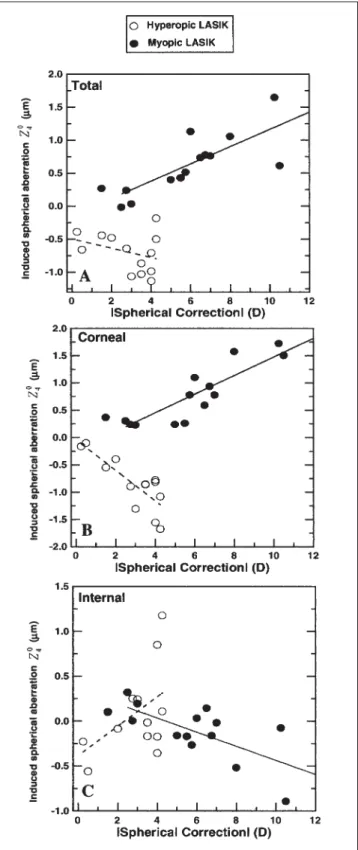
Figure 4. A) Total, B) corneal, and C) internal spherical aberration
induced by hyperopic LASIK (open circles, from this study) in com-parison with the spherical aberration induced by myopic LASIK (black circles, from a previous study14) as a function of absolute
spherical correction, for a 6.5-mm pupil. Shaded areas indicate eyes included in the averages reported in the text.
laboratory using similar techniques.14 Figure 4
shows the total (4A), corneal (4B), and internal (4C) spherical aberration induced by myopic (black cir-cles) and hyperopic (white circir-cles) LASIK. Myopic preoperative spherical errors were as high as -12.00 D; the hyperopic procedure was limited to lower amounts of hyperopia (+4.50 D). In both groups cylinder was less than 2.50 D. Also, the myopic patients were younger than the hyperopic patients (mean age 26 ± 1.45 yr for myopic patients and 37 ± 11 yr for hyperopic patients).
Total and corneal induced spherical aberrations were always positive in myopes and negative in hyperopes, as shown in Figure 4. The induced corneal spherical aberration was well correlated with attempted spherical correction for both myopic (r=-0.87, P<.0001) and hyperopic eyes (r=-0.81, P=.0003). The rate of total spherical aberration increment per diopter of attempted spherical correc-tion tended to be higher for the myopic procedure (+0.13 mm/D of myopic error and -0.07 mm/D of hyperopic error). However, the average induced total spherical aberration in a subgroup of hyperop-ic (n=4) and myophyperop-ic (n=4) eyes of similar absolute attempted correction (1.5 to 3.00 D) was, in absolute values, 3.3 times higher for hyperopic than for myopic eyes (-0.66 ± 0.28 µm and 0.20 ± 0.06 µm, respectively). The rate for the corneal spherical error increments was higher for the hyperopic pro-cedure (-0.28 mm/D) than for the myopic propro-cedure (0.17 mm/D). The average induced corneal spherical aberration for the previous subgroups was -0.78 ± 0.40 µm for hyperopes and 0.13 ± 0.14 µm for myopes (ie, six times more for hyperopic than for myopic LASIK). The fact that the amount of absolute spherical aberration after surgery (both myopic and hyperopic) was lower in the total eye (-0.38 ± 0.36 µm and 0.40 ± 0.09 µm for the previous hyperopic and myopic subgroups, respectively) than on the cornea alone (-0.46 ± 0.34 µm and 0.43 ± 0.12 µm for the previous hyperopic and myopic sub-groups, respectively) is indicative of compensation by internal aberrations. Part of the compensation was due to aberration of the crystalline lens. The role of the preoperative internal spherical aberra-tion (primarily aberraaberra-tions of the crystalline lens) in hyperopes, compared to myopic eyes, will be dis-cussed in the next section. The posterior surface of the cornea seems to play also a compensatory role, which will also be discussed.
As expected, major changes occurred on the ante-rior corneal surface for both myopic and hyperopic LASIK. The causes of a change in corneal
aspheric-ity leading to important changes in spherical aber-ration found clinically are not well understood.32,33
It has been shown analytically32and
computational-ly34 that those changes are not inherent to the
Munnerlyn ablation algorithm, or at least to the exact application of it. Radial changes of laser effi-ciency across the cornea, due to angular changes of reflectivity and laser fluence, have been shown to be responsible for at least part of the discrepancies of postoperative asphericities with respect to predic-tions.33,35 These effects are expected to be much
more relevant in hyperopic LASIK than in myopic LASIK, since in the hyperopic procedure corneal tis-sue is removed primarily in the periphery where the effects of laser efficiency losses are more impor-tant.36Also, a biomechanical response, presumably
responsible for some of the asphericity changes found with LASIK37, is probably higher in
hyperop-ic LASIK. The hyperophyperop-ic profile shows three inflec-tion zones per hemimeridian: 1) located at the cen-ter of the ablation (some high hyperopic treatment plans treat the central cornea optical zone); 2) at the deepest portion of the ablation, which is at the boundary border between the ablation optical zone and the transition zone; and 3) at the boundary between the transition zone and the untreated peripheral cornea. In the myopic profile, however, there is only one inflection zone (located at the bor-der between the treated and the untreated periph-eral cornea).38 The increased number of inflection
zones may result in a larger biomechanical response than occurs for myopic LASIK, although the actual mechanisms still need to be worked out. This has also been considered to reduce the maximum amount of treated hyperopic refractive error to about one-third of the treated myopic error.38
We also found that third order aberrations increased slightly more in hyperopic than in myopic LASIK eyes (factor of 2.2 and 1.7, respectively), in agreement with the report by Oliver and col-leagues.19 We did not find a correlation between
postoperative third order aberrations and preopera-tive refracpreopera-tive error, nor with induced spherical error. This result suggests that coma was primarily associated with decentration of the ablation pattern, and the amounts of decentration were rather vari-able across eyes, both myopic and hyperopic. Influence of Preoperative Aberrations on Refractive Surgery Outcomes—Comparison With Myopic Eyes
We found a dominance of corneal aberrations (particularly spherical aberration) in the total aber-ration pattern in several preoperative hyperopic
eyes of our group (Figs 1 and 2). If we sort the eyes of this study by age, we find that younger eyes (#6, 10 and 12, 24, 24 and 25 years old, respectively) showed negative internal spherical aberration, while older eyes, from 25 years old or more, showed less negative spherical aberration (eyes #13, 5, 7, 8, 3), which turned into positive for the oldest eyes (#4, 1, 2, 9, 11), disrupting the balance of the positive spherical aberration of the cornea by the crystalline lens. The balance between internal and corneal aberrations in our younger hyperopic eyes has been reported in normal young eyes39and myopic eyes.40
Artal and colleagues39 reported a loss of this
com-pensation with age in normal eyes. That study, on 17 eyes, showed that loss of compensation happened in patients older than 45 years. Refractive errors were not reported in that study, although the spher-ical equivalent refractive error for the subjects in the study was less than 2.00 D. We recently per-formed a comparison of hyperopic (n=22) and myopic eyes (n=24) matched in age (range 23 to 40 yr) and absolute refractive error (range 0.50 to 7.60 D).41We found an early loss (at approximately
30 years of age) of corneal to internal balance in hyperopic eyes that was not present in the myopic group, which did not show a significant trend of bal-ance at this age.41These findings may be relevant to
understanding the outcomes of hyperopic LASIK and to predicting possible changes in performance with age. Given that corneal spherical aberration shifts to negative values after a hyperopic procedure (Fig 2B), the fact that the crystalline lens con-tributes with additional negative spherical aberra-tion is disadvantageous in young hyperopic eyes, whereas for myopic eyes the negative spherical aberration of the crystalline lens subtracts from the induced positive corneal spherical aberration. However, since spherical aberration of the crys-talline lens becomes more positive with age, patients who undergo hyperopic LASIK will experi-ence an absolute decrease of spherical aberration with age (and potentially an increase in optical quality), whereas for myopic eyes, spherical aberra-tion will increase with aging.42 Aberrations of the
crystalline lens therefore play a significant role in the evaluation the individual surgical outcomes and predict long-term optical performance.
The counteracting effects of the crystalline lens may be accounted for by adding the induced corneal spherical aberration and internal preoperative spherical aberration (which accounts mainly for crystalline lens spherical aberration), and then dividing this number by the induced corneal
aberra-tion to provide a relative value. A value between 0 and 1 will be indicative of compensation by the crys-talline lens, a value close to 1 indicative of no com-pensation, and a value higher than 1 indicative of additional contribution of the crystalline lens to the degradation. In the myopes from our previous study, where attempted spherical error correction ranged up to -10.50 D, we found a counteracting value of 0.375; for the hyperopic group this value was 1.04.
We also studied possible effects of preoperative corneal aberrations on postoperative outcomes. For myopic eyes, we found no correlation between pre-operative and postpre-operative spherical aberration. Although we found a slight correlation for hyperop-ic eyes (r=-0.42), this was not signifhyperop-icant (P=.16), and could be driven by the correlation between spherical error and corneal spherical aberration in preoperative hyperopic eyes (r=0.76, P=.002), which was not found for preoperative myopic eyes.41
Changes in Internal Aberrations With Hyperopic LASIK Induced corneal spherical aberration (Fig 4B) is generally below (more negative than) induced total spherical aberration (Fig 4A), indicating that inter-nal spherical aberration (Fig 4C) reduces the impact of the corneal changes. We found a positive linear correlation between induced internal spherical aberration and attempted spherical correction (r=0.52). This trend was at the limit of statistical significance (P=.07). Since LASIK is a corneal pro-cedure (no change to the crystalline lens), changes in internal aberrations must account for changes on the posterior surface of the cornea. A similar atten-uating effect by the posterior surface of the cornea was found in myopic LASIK.14 We demonstrated
that the spherical aberration (of negative sign for myopic LASIK) induced on the posterior surface of the cornea was consistent with reported changes in corneal curvature and asphericity measured by slit-lamp topography.43 To our knowledge, equivalent
changes in corneal curvatures and asphericities after hyperopic LASIK have not been studied. Ma and colleagues22 compared internal aberrations
after hyperopic LASIK eyes with a control group of eyes and found more positive internal spherical aberration in the operated eyes, consistent with a shift of the posterior corneal surface toward more positive values. In both myopic and hyperopic eyes, the shift of internal spherical aberration results in slight compensation of the aberration induced on the anterior surface of the cornea, and the effect is rather variable across eyes.
Comparison With Other Studies
Published results on the optical changes induced by hyperopic LASIK are limited. Also, a direct comparison among studies is limited by differences in surgical technique (type of surgery, optical and transition zone diameters, type of laser, use of an eye-tracker) and the characteristics of the study population (age range, preoperative correction, pre-operative third and higher order aberrations, etc).
Chen and colleagues21studied corneal
aspherici-ty, measured with a standard corneal topography system, before and after hyperopic LASIK in 33 eyes. They reported a change of asphericity toward more negative values (from -0.32 ± 0.20 to -0.97 ± 0.39 for eyes with preoperative astigmatism less than 0.50 D, and from -0.43 ± 0.49 to -0.72 ± 0.70 D for eyes with preoperative astigmatism high-er than 0.50 D) at 1 month afthigh-er surghigh-ery. We com-puted corneal asphericity from our videokerato-graphic data and found a significant (P<.00001) shift of asphericity toward more negative values, and similar relative changes in asphericity (change in corneal asphericitiy = -0.32 compared to Chen and colleagues' average of -0.39). However, the pre-operative mean asphericity (-0.21 ± 0.12) and post-operative mean asphericity (-0.54 ± 0.19) in our study are less negative than those found by Chen and colleagues. In agreement with Chen and col-leagues, we found a correlation between the postop-erative asphericity and the attempted spherical cor-rection (r=-0.47, although it did not reach statistical significance, P=.1). As reported by Chen and col-leagues, there was some correlation between preop-erative and postoppreop-erative corneal asphericity (r=-0.40, which was statistically significant; r=-0.76, P=.005, without eye #5). Unlike Chen et al, we found a good correlation between the preoperative corneal radius of curvature and the postoperative asphericity (r=-0.68, P=.008).
Oliver and colleagues19measured corneal
aberra-tions before and after hyperopic PRK in nine eyes. In agreement with our study, they found that corneal aberrations increased with surgery, particu-larly corneal spherical aberration, which changed from preoperative positive (0.50 ± 0.18 µm) to post-operative negative values (-0.75 ± 0.54 µm after 12 weeks) for a 5.5-mm pupil. They also found, for the same pupil size, a statistically significant increase in coma RMS (from 0.64 ± 0.24 µm to 1.76 ± 1.39 µm after 12 weeks for a 5.5-mm pupil). Despite their use of a smaller pupil, the changes reported in Oliver and colleagues' study are higher
than the changes we found in the present study (postoperative mean corneal spherical aberration of -0.44 ± 0.43 µm and third-order corneal RMS of 0.91 ± 0.39 µm). This is probably due to the fact that Oliver and colleagues' study included higher preop-erative positive spherical errors (+2.50 to +7.50 D) and perhaps differences between the surgical proce-dures (PRK versus LASIK). Our previous study on myopic LASIK found lower amounts of induced spherical aberration than a previous report for myopic PRK.12
Wang and colleagues20, in a retrospective study,
reported anterior corneal aberrations induced by LASIK for hyperopia in 40 eyes, also finding an increase of higher order aberrations (from third to sixth order), and a decrease toward negative values of corneal spherical aberration. However, their pre-operative and postpre-operative values for spherical aberration (0.27 ± 0.08 µm and -0.058 ± 0.16 µm, respectively) and RMS for third and higher order aberrations (0.49 ± 0.09 µm and 0.56 ± 0.20 µm, respectively), computed for a 6 mm-pupil, were in general lower than those we found in this study (0.41 ± 0.11 µm and -0.44 ± 0.43 µm, respectively) for corneal spherical aberration and 0.68 ± 0.13 µm and 1.18 ± 0.51 µm, respectively, for corneal RMS for third and higher order aberrations).
Shortly before submitting the present study for publication, we became aware of a study by Ma and colleagues22, who measured total and corneal
aber-rations following two surgical procedures for the correction of hyperopia (LASIK and lensectomy with intraocular lens implantation). Results for a group of 22 eyes after hyperopic LASIK were compared with a group of 19 control eyes. The results of their comparison are consistent with our findings. They found that mean third and higher order corneal RMS was 2.09 times higher and total RMS 1.58 times higher after LASIK than in the control eyes, and that spherical aberration shifted toward nega-tive values (by -0.63 µm for the cornea and -0.30 µm for the total eye). Despite the differences between preoperative spherical error ranges in both studies (+0.75 to +7.25 D in Ma et al versus +0.25 to +4.25 D in our study) we found comparable postoperative data (1.18 µm and 0.86 µm for total and corneal third and higher order RMS for a 6-mm-diameter pupil, as opposed to our 1.23 and 1.18 µm for a 6.5-mm-diameter pupil; and -0.41 and -0.24 µm for total and corneal spherical aberration for 6-mm, as opposed to our -0.44 µm and -0.33 µm for 6.5-mm). Their study reported larger changes in
internal spherical aberration, which they attributed partly to reshaping of the posterior surface of the cornea and partly to possible errors in their tech-niques.
Changes in Optical Performance: Strehl Ratio vs RMS The analysis presented in the previous sections used the RMS wavefront error as the optical quality metric. This metric has been widely used to describe the changes induced by refractive surgery12-14,16,17
and it is useful to account for changes in individual Zernike polynomial terms and orders. However, other metrics based on the retinal image quality rather than the wave aberration correlate better with visual performance.44 Guirao and Williams45
showed that using the Strehl ratio as an optical quality metric (maximum of the point spread func-tion relative to the diffracfunc-tion limit, or equivalent volume under the modulation transfer function nor-malized by the volume under the
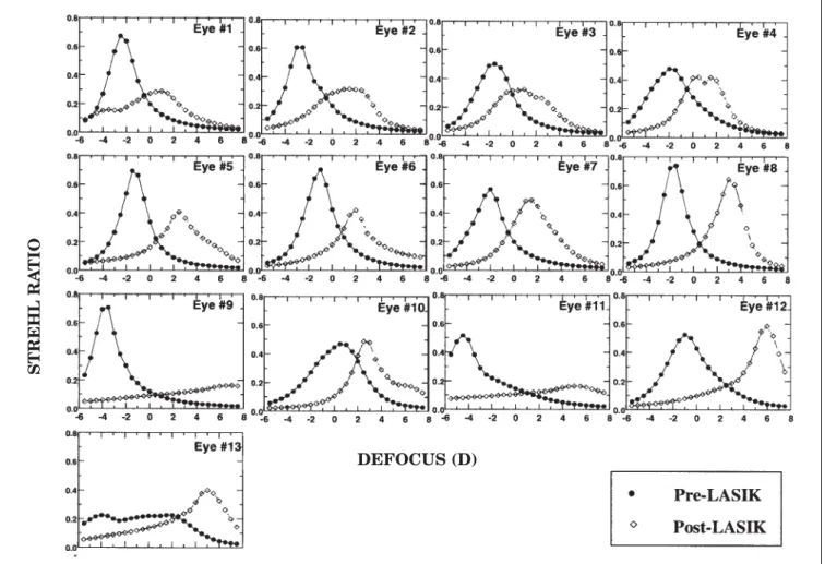
diffraction-limited modulation transfer function), it was possi-ble to obtain the refractive error from the wave aberration, in good agreement with the subjective refraction. We computed the Strehl ratio (truncating the volume beyond 80 c/deg, since those high fre-quencies are not relevant to the visual system) as a function of defocus and estimated the best image quality (in terms of Strehl ratio) achieved with sphero-cylindrical correction. Figure 5 shows the through-focus Strehl ratio for all eyes, before (black circles) and after (open diamonds) hyperopic LASIK. Defocus (D) is relative to the Z2
0term in the Zernike
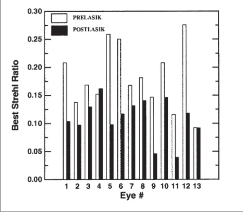
polynomial expansion. A negative sign is indicative of a myopic shift and a positive sign is indicative of a hyperopic shift. The maximum value of these curves represents the best Strehl ratio (or Strehl ratio at best focus). Figure 6 represents best Strehl ratio for preoperative and postoperative eyes, for 6.5-mm-diameter pupils (with best defocus and astigmatism correction). This graph is directly Figure 5. Strehl ratio as a function of defocus (relative to Z0
2= 0) for all eyes in this study. Black circles indicate preoperative values and white
squares indicate postoperative values. Pupil size was 6.5 mm.
STREHL
RA
TIO
comparable with Figure 2, in which best-corrected RMS wavefront error was used as a metric (cancel-ing defocus and astigmatism terms in the Zernike polynomial expansion). On average, there was a decrease in best-corrected image quality (or decrease in Strehl ratio) by a factor of 1.84 ± 0.76 for a 6.5-mm pupil (1.74 ± 1.04 for a 3-mm pupil). Figure 5 also shows the defocus shifts caused by the interactions of aberrations and defocus. This can be assessed by a metric such as Strehl ratio, but not RMS. We found a consistent myopic shift of the best image quality in preoperative eyes (by -1.41 ± 0.95 D) and a hyperopic shift in postoperative eyes (by 1.95 ± 0.95 D). This relatively large effect of the spherical aberration is consistent with the positive spherical aberration that is higher in emmetropes and myopes than in hyperopes, and the induced negative spherical aberration with surgery. The depth-of-field (defined as the dioptric range for which the Strehl ratio is at least 80% of the maxi-mum Strehl ratio) also increases after surgery (from 1.09 to 1.63 D) consistent with the increase of opti-cal aberrations.46Given that the best focus location
is not relevant for those eyes with large depth-of-field (eyes #9, 11 and 13), these were not included in the computation. When the astigmatism terms are not cancelled (before and after surgery), there is not
a significant degradation of retinal image quality (ratio preoperative/postoperative = 1.04 ± 0.52), indicating that the surgery was successful at cor-recting cylinder. Also, the average depth-of-field decreased from 2.15 ± 1.00 D preoperatively (enlarged by the effect of astigmatism) to 1.60 ± 0.77 D postoperatively. The tendency toward a myopic shift preoperatively (-0.90 ± 1.67 D) and a hyperopic shift postoperatively (1.80 ± 1.49 D) was not affected by the presence of astigmatism.
This analysis based on Strehl ratio yields a simi-lar conclusion in terms of degradation induced by LASIK for hyperopia on overall best-corrected optical quality than that obtained using RMS wave-front error. However, the through-focus analysis using Strehl ratio provides a better understanding of the interactions of the aberrations (natural or induced) with the residual defocus, and intersubject variability of out-of-focus optical performance. Interestingly, hyperopic eyes seem to benefit from the positive spherical aberration, which tends to reduce the hyperopic defocus, while after surgery the induced negative spherical aberration tends to produce a hyperopic shift of the best focus that reduces the effect of the surgery in the refraction.
All conclusions stated in this article apply to the standard application of hyperopic ablation profiles. Figure 6. Best Strehl ratio (computed as the
maxima of the curves in Figure 5) before and after hyperopic LASIK. Pupil size was 6.5 mm.
It would be interesting to investigate how these effects are corrected in newer generations of ablation algorithms.
REFERENCES
1. Strang N, Schmid K, Carney L. Hyperopia is predominantly axial in nature. Curr Eye Res 1998;17:380-383.
2. Grosvenor T, Goss DA. Clinical Management of Myopia. Boston, MA: Butterworth-Heinemann; 1999.
3. Budak K, Khater T, Friedman NJ, Holladay JT, Koch DD. Evaluation of relationships among refractive and topo-graphic parameters. J Cataract Refract Surg 1999;25: 814-820.
4. Carney L, Mainstone J, Henderson B. Corneal topography and myopia. A cross-sectional study. Invest Ophthalmol Vis Sci 1997;38:311-320.
5. Carkeet A, Luo H, Tong L, Saw SM, Tan DT. Refractive error and monochromatic aberrations in Singaporean children. Vision Res 2002;42:1809-1824.
6. Mainstone J, Carney L, Anderson C, Clem PM, Stephensen AL, Wilson MD. Corneal shape in hyperopia. Clin Exp Optom 1998;81:131-137.
7. Argento C, Cosentino M. Laser in situ keratomileusis for hyperopia. J Cataract Refract Surg 1998;24:1050-1058. 8. Choi R, Wilson S. Hyperopic laser in situ keratomileusis:
primary and secondary treatments are safe and effective. Cornea 2001;20:388-393.
9. Zadok D, Maskaleris G, Montes M, Shah S, Garcia V, Chayet A. Hyperopic laser in situ keratomileusis with the Nidek EC-5000 excimer laser. Ophthalmology 2000;107:1132-1137. 10. Salz J, Stevens C. LASIK correction of spherical hyperopia, hyperopic astigmatism, and mixed astigmatism with the LADARVision excimer laser system. Ophthalmology 2002;109:1647-1657.
11. Dierick H, Missotten L. Corneal ablation profiles for correc-tion of hyperopia with the excimer laser. J Refract Surg 1996;12:767-773.
12. Seiler T, Kaemmerer M, Mierdel P, Krinke H-E. Ocular opti-cal aberrations after photorefractive keratectomy for myopia and myopic astigmatism. Arch Ophthalmol 2000;118:17-21. 13. Moreno-Barriuso E, Merayo-Lloves J, Marcos S, Navarro R, Llorente L, Barbero S. Ocular aberrations before and after myopic corneal refractive surgery: LASIK-induced changes measured with Laser Ray Tracing. Invest Ophthalmol Vis Sci 2001;42:1396-1403.
14. Marcos S, Barbero B, Llorente L, Merayo-Lloves J. Optical response to LASIK for myopia from total and corneal aber-ration measurements. Invest Ophthalmol Vis Sci 2001; 42:3349-3356.
15. Martinez C, Applegate R, Klyce S, McDonald MB, Medina JP, Howland HC. Effects of pupillary dilation on corneal optical aberrations after photorefractive keratectomy. Arch Ophthalmol 1998;116:1053-1062.
16. Oshika T, Klyce SD, Applegate RA, Howland HC. Comparison of corneal wavefront aberrations after photore-fractive keratectomy and laser in situ keratomileusis. Am J Ophthalmol 1999;127:1-7.
17. Oliver K, Hemenger R, Corbett M, O’Brart DP, Verma S, Marshall J, Tomlinson A. Corneal optical aberrations induced by photorefractive keratectomy. J Refract Surg 1997;13:246-254.
18. Mrochen M, Krueger R, Bueeler M, Seiler T. Aberration-sensing and wavefront-guided laser in situ keratomileusis: management of decentered ablation. J Refract Surg 2002;18:418-429.
19. Oliver K, O'Brart D, Stephenson C, Hemenger RP, Applegate RA, Tomlinson A, Marshall J. Anterior corneal optical aberrations induced by photorefractive keratectomy
for hyperopia. J Refract Surg 2001;17:406-413.
20. Wang L, Koch D. Anterior corneal optical aberrations induced by laser in situ keratomileusis for hyperopia. J Cataract Refract Surg 2003;29:1702-1708.
21. Chen C, Izadshenas A, Rana M, Azar D. Corneal aspherici-ty after hyperopic laser in situ keratomileusis. J Cataract Refract Surg 2002;28:1539-1545.
22. Ma L, Atchison DA, Albietz J, Lenton LM, McLennan SG. Wavefront aberrations following LASIK and lensectomy cor-rections for hypermetropia. J Refract Surg, in press. 23. Navarro R, Losada MA. Aberrations and relative efficiency
of light pencils in the living human eye. Optom Vis Sci 1997;74:540-547.
24. Moreno-Barriuso E, Marcos S, Navarro R, Burns SA. Comparing laser ray tracing, Spatially Resolved Refractometer and Hartmann-Shack sensor to measure the ocular wavefront aberration. Optom Vis Sci 2001;78: 152-156.
25. Marcos S, Díaz-Santana L, Llorente LCD. Ocular aberra-tions with ray tracing and Shack-Hartmann wavefront sen-sors: does polarization play a role? J Opt Soc Am A 2002;19:1063-1072.
26. Marcos S. Aberrations and visual performance following standard laser vision correction. J Refract Surg 2001; 17:596-601.
27. Llorente L, Diaz-Santana L, Lara-Saucedo D, Marcos S. Aberrations of the human eye in visible and near infrared illumination. Optom Vis Sci 2003;80:26-35.
28. American National Standard Institute. American National Standard for the safe use of lasers, Standard Z-136.1-1993. Orlando, FL: The Laser Institute of America, 1993. 29. Thibos LN, Applegate RA, Schwiegerling JT, Webb RH,
VSIA Standards Taskforce Members. Standards for report-ing the optical aberrations of eyes. Vision Science and Its Applications, OSA Trends in Optics & Photonics 2000; 35:110-130.
30. Barbero S, Marcos S, Merayo-Lloves J, Moreno-Barriuso E. Validation of the estimation of corneal aberrations from videokeratography in keratoconus. J Refract Surg 2002; 18:263-270.
31. Barbero S, Marcos S, Merayo-Lloves JM. Total and corneal aberrations in an unilateral aphakic subject. J Cataract Refract Surg 2002;28:1594-1600.
32. Gatinel D, Hoang-Xuan T, Azar D. Determination of corneal asphericity after myopia surgery with the excimer laser: a mathematical model. Invest Ophthalmol Vis Sci 2001;42:1736-1742.
33. Anera R, Jimenez J, Jimenez del Barco L, Hita E. Changes in corneal asphericity after laser refractive surgery, includ-ing reflection losses and non normal incidence upon the anterior cornea. Opt Lett 2003;28:417-419.
34. Marcos S, Cano D, Barbero S. The increase of corneal asphericity after standard myopic LASIK surgery is not inherent to the Munnerlyn algorithm. J Refract Surg 2003;19:592-596.
35. Mrochen M, Seiler T. Influence of corneal curvature on cal-culation of ablation patterns used in photorefractive laser surgery. J Refract Surg 2001;17:S584-S587.
36. Berret R, Kemmner D, Oltrup T, Bende T, Jean B. Influence of the reflection on the ablation rate during excimer laser ablation and its compensation. ARVO E-abstract 2593: 2003; accessed www.arvo.org.
37. Roberts C, Dupps W. Corneal biomechanics and their role in corneal ablative procedures. In: MacRae S, Krueger R, Applegate R, eds. Customized Corneal Ablation: The Quest for Super Vision. Thorofare, NJ: Slack Inc; 2001.
38. MacRae S. Excimer laser design and elliptical transition zones. J Cataract Refract Surg 1999;25:1191-1197.
39. Artal P, Berrio E, Guirao A, Piers P. Contribution of the cornea and internal surfaces to the change of ocular
aberrations with age. J Opt Soc Am A 2002;19:137-143. 40. Marcos S, Barbero S, Llorente L. The sources of optical
aber-rations in myopic eyes. 2002 Annual Meeting Abstract and Program Planner. Association for Research in Vision and Ophthalmology. Abstract 1510: 2002; accessed www.arvo.org.
41. Llorente L, Barbero S, Cano D, Dorronsoro C, Marcos S. Myopic versus hyperopic eyes: axial length, corneal shape and optical aberrations. Journal of Vision 2004;4:288-298. 42. Marcos S. Are changes in ocular aberrations with age a
sig-nificant problem for refractive surgery? J Refract Surg 2002;18:572-578.
43. Seitz B, Torres F, Langenbucher A, Behrens A, Suarez E. Posterior corneal curvature changes after myopic laser in situ keratomileusis. Ophthalmology 2001;108:666-673. 44. Applegate R, Thibos L, Williams D. Converting wavefront
aberration to metrics predictive of visual performance. ARVO E-abstract 2124: 2003; accessed www.arvo.org. 45. Guirao A, Williams D. A method to predict refractive errors
from wave aberration data. Optom Vis Sci 2003;80:36-42. 46. Marcos S, Moreno E, Navarro R. The depth-of-field of the
human eye from objective and subjective measurements. Vis Res 1999;39:2039-2049.