ARTICLE
Human immunodeficiency virus (HIV) invades the central nervous system (CNS) soon after the initial systemic infection. Three pathways have been proposed whereby the virus breaches the blood-brain barrier and enters CNS parenchymal tissue: (i) HIV may be carried into the brain by infected macrophages-monocytes and/or CD4+ T lymphocytes (Trojan horse effect); (ii) cell-free virus may pass from the vascular compartment into brain tissue between cerebral microvascular endothelial cells (CMVECs) or by transmigration (transcytosis) through CMVECs; and (iii) following direct HIV infection of the specialised endothelial cells of the blood/brain barrier or CMVECs, newly formed HIV viral particles may be released into the cerebral compartment.1,2 Once established within the CNS compartment, the virus infects various cell populations, primarily perivascular macrophages and microglia, and activates several cell types including macrophages, microglia and astrocytes.2,3 The consequent inflammatory response induces neuronal apoptotic pathways causing neuronal death and attrition. Apoptosis may be activated by viral glycoproteins including gp120, tat, nef, vpr and gp41, or a variety of host-derived mediators including inflammatory cytokines, free radicals and excitatory amino acids.4 There is some evidence suggesting that HIV may also inhibit neural progenitor cell proliferation, negatively affecting de novo neurogenesis.5 The consequence of the above is the development of neurological and neurocognitive manifestations.s Classically, HIV affects the immature brain causing static or progressive encephalopathy. Progressive encephalopathy has been defined by the Centers for Diseases Control (CDC), and is characterised by microcephaly, failure to attain or loss of neurodevelopmental milestones, or loss of intellectual ability,
and acquired symmetrical motor defects.6 The prevalence of HIV encephalopathy varies from 13% to 35%, and up to 50% of children with full-blown AIDS (CDC clinical category C disease) have evidence of this complication.7 HIV encephalopathy may be the first AIDS-defining illness in as many as 18% of HIV-infected children.7 Encephalopathy may become static, plateau or progressively deteriorate.8 Without highly active antiretroviral therapy (HAART) the median survival of children who progress is less than 24 months.9 Aetiological agents other than HIV may cause neurological and/ or neurocognitive deficits in HIV-infected children. Because of generalised immunodeficiency, HIV-infected children are predisposed to CNS opportunistic infections including acute bacterial meningitis, tuberculous meningitis, cytomegalovirus co-infection, Epstein-Barr virus-associated primary lymphoma, and in older children cryptococcal meningitis.10-14 HIV-infected patients are at risk for thromboembolic strokes caused by disturbances in blood coagulation via antiphospholipid antibodies or reduced protein S concentration, and ischaemic strokes due to infections, coagulopathies or HIV-associated vasculopathy.15 Following initiation of antiretroviral therapy (HAART) an immune reconstitution inflammatory response to several CNS pathogens including John Cunningham (JC) virus, a polyomavirus, may occur resulting in CNS deterioration.16 Neurological manifestations may be complex in their presentation due to multiple pathologies.17 However, isolated neurocognitive deficits may occur, particularly in the presence of advanced HIV disease.18
Studies have reported on the frequency of CNS manifestations in HIV-infected children living in resource-constrained
Lara Smith,
MB ChB, DCH (SA), FCPaed (SA), MMed (Paed)
Colleen Adnams,
MB ChB, FCPaed (SA), BSc (Hons)
Brian Eley,
MB ChB, FCPaed (SA), BSc (Hons)
Red Cross War Memorial Children’s Hospital and School of Child and Adolescent Health, University of Cape Town
Neurological and neurocognitive
function of HIV-infected children
commenced on antiretroviral therapy
S
A
Jo
ea
lth
S
A
Jo
ur
nal
of Child
H
ea
lth
S
A
Jo
ur
nal
of Child
H
ea
lth
Aim: To describe neurological and neurocognitive deficits in HIV-infected children and the short-term effect of highly active
antiretroviral therapy (HAART) on the observed deficits
Methods: In this prospective study, 39 children (15 females) were evaluated before the start of HAART and 30 reassessed 6 months later. The subjects were evaluated with a range of cognitive tests used in everyday clinical practice.
Results: At enrolment, the mean (±SD) age was 60±46 months, 17 (44%) and 22 (56%) had Centers for Disease Control (CDC) clinical category B and C disease respectively, and 36 (92%) had severe immunosuppression. At the start of HAART no child had cranial nerve or cerebellar dysfunction, but 13/29 (33.3%) had evidence of motor dysfunction. By 6 months 1 child had developed cerebellar dysfunction, but there was no statistically significant change in the frequency of motor dysfunction. Mean baseline performances on cognitive testing were generally subnormal. Between 33% and 81% of the children recorded subnormal intelligence quotients on various cognitive tests. Mean performances did not change significantly after 6 months of HAART.
ARTICLE
settings. These studies generally documented significantly higher prevalences of neurological and cognitive deficits in HIV-infected children compared with those who serorevert or in uninfected controls.19-22 An exception to this is described in a Ugandan study that showed similar frequencies of neurological and cognitive deficits in small groups of HIV-infected, HIV-exposed but unHIV-infected, and uninfected children of schoolgoing age.23 Neurological and cognitive deficits have been documented in up to 80% of HIV-infected children.22 The risk for neurodevelopmental delay may be greatest in those who have documented HIV infection at birth, compared with children who seroconvert after birth.21 In the present study we describe the neurological and neurocognitive deficits in a group of children with HIV infection in Cape Town, and assess the response of these deficits after 6 months on HAART.
Methods
This prospective study was completed on the first group of HIV-infected children enrolled on the antiretroviral treatment programme at Red Cross Children’s Hospital, Cape Town, between February 2002 and December 2003. Children were started on HAART based on criteria derived from the Paediatric European Network for the Treatment of AIDS as previously described.24 The present study describes the neurological and neurocognitive effects in these children at two time points, before and after 6 months of HAART. This study overlapped with the establishment of a donor-funded antiretroviral treatment programme at the hospital during August 2002. The initial impact of this treatment programme was previously described.24,25 The Research Ethics Committee of the University of Cape Town approved the study (reference number 290/2002). Written informed consent was obtained from a parent or legal guardian prior to study entry.
Participants
A convenience sample of 40 ambulant children attending the outpatient HIV service were considered for inclusion. All children met criteria for starting HAART.24 All were commenced on a triple combination regimen comprising stavudine and lamivudine plus either ritonavir (18 children) or efavirenz (21 children). Children were excluded from the study if they had pre-existing neurocognitive disabilities due to causes other than HIV, previously documented neonatal hypoxic ischaemic encephalopathy, or were hospitalised at the time of neurocognitive testing. One child with a pre-existing genetic disorder was excluded. A further 2 children had possible non-HIV-related risk factors for neurodevelopmental delay; one child with premature birth, and the other with low Apgar scores and a history of prenatal alcohol exposure but no history of hypoxic ischaemic encephalopathy or features of fetal alcohol syndrome. Both these children were included. Thirty-nine children (15 females, 24 males) therefore participated in the study.
Demographic and social data
At baseline, the mean age of the participants (± standard deviation, SD)was 60±46 months. The primary caregiver was the mother in 29 cases (74.4%) and another family member in 8 (20.5%), 1 child was in foster care, and 1 child had an unrelated legal guardian. Twenty-nine (74.4%) of caregivers were unemployed, 7 (17.4%) were employed, 2 (5.1%) were receiving a pension and one child was institutionalised in foster care. However, 62% (24) of the primary caregivers
were receiving a social grant. At enrolment, 10 (25.6%) of the caregivers had achieved an education level ≥ grade 12, 22 (56.4%) had completed grade 7 - 11, 5 (12.8%) had been educated to a level < grade 7, and the educational status of 2 was unknown. Fourteen children (35.9%) were >6 years old and therefore eligible for school attendance. Of these 7 were attending school and had started at the appropriate age, 3 had never attended school because of financial reasons or poor health/recurrent infections, and 4 had either delayed school entry or failed a year. Whether or not the remaining 25 were attending preschool was not documented. The predominant languages spoken were Xhosa by 34 (87.2%) children, Xhosa at home and English at school by 2 (5.1%), Afrikaans by 1 (2.6%), Zulu and Xhosa by 1 (2.6%), and Sotho and Xhosa by 1 (2.6%).
The severity of the children’s HIV infection was determined using the CDC clinical and immunological classification for children.6 Weight-for-age and height-for-age z-scores were calculated using EpiInfo 2000, version 1.0, Division of Surveillance and Epidemiology, CDC, Atlanta, Georgia. After baseline neurological and cognitive assessment, the children were commenced on standard three-drug triple combination antiretroviral therapy, comprising two nucleoside reverse transcriptase inhibitors (NRTIs) plus either a non-nucleoside reverse transcriptase inhibitor (NNRTI) or a protease inhibitor (PI). Response to HAART was determined by comparing 6-month CD4 percentage, HIV-1 RNA concentration (viral load) and growth parameters with corresponding baseline measurements. Viral loads were expressed in log10 values.
Outcome measures
Neurological examination was completed at enrolment and
after 6 months. This covered cranial nerves, cerebellar function and motor system, which included evaluation of reflexes, power, tone and gait.
Cognitive testing was completed at baseline and after 6
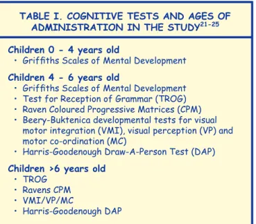
months using a battery of standardised tests covering the age range 0 - 13 years. Tests were administered according to their applicability to three age ranges; <4 years, 4 - 6 years and >6 years (Table I). Four general tests of intelligence and one motor test were administered.
General tests of intelligence
Griffiths Mental Development Scales26 measure progression
of development in six areas: locomotor, personal-social, hearing and speech, eye and hand co-ordination, performance, and practical reasoning. The first five scales assess development from infancy to age 8 years, and the last scale from 3 years to 8 years. Each subscale is expressed as a quotient of performance mental age as a percentage of chronological age. The general quotient (GQ) is a composite of all the subscales. Subscales can be used alone, with subscale means ranging from 99.79 to 100.46 and standard deviations from 15.58 to 17.43. Norms for the Griffiths Scales are available for some South African population groups. The Griffiths Scales were administered to all children <6 years of age (N=26) (see Table I).
Test of the Reception of Grammar (TROG)27 consists of
Raven Progressive Coloured Matrices (Raven CPM)28 is a cross-cultural test that assesses non-verbal problem solving based on matching components of patterns. It is normed for children and has been used widely in cognitive research on developing countries. Test scores are reported as percentile scores. For interpretation, above the 95th centile is intellectually superior; above the 75th centile is above average; between the 25th and 75th centiles is intellectually average; below the 25th centile is below average; and at or below the 5th centile represents intellectual impairment.
The Harris-Goodenough-Draw-A-Person Test (DAP),29
adapted from the Goodenough Draw-a-Man Test originally developed as a scaled measure of non-verbal intelligence, was included in this study to represent a general screen for maturity of cognitive and non-verbal function in children. In this context, the test provides a useful tool with which to identify children who have cognitive, motor or visual-spatial delays in maturation. The Harris-Goodenough Draw-a-Person score was converted to a general intellectual function (IF) score by dividing the score obtained in months by the chronological age in months.24
Motor test
Beery-Buktenica Developmental Test of Visual-Motor
Integration30 is a pencil and paper test that measures the
extent to which individuals can integrate their visual and motor abilities. This test, extensively used in clinical practice in South Africa, includes visual motor integration (VMI) as well as visual perception (VP) and motor co-ordination (MC) components. Scores were converted to percentiles using test manual norms.30
All tests were performed in the patient’s first language, using an interpreter where appropriate. All test materials were available in Xhosa, Afrikaans and English, translated from the English originals.26-30 Standardised charts were used to convert TROG and Raven CPM percentiles into intelligence quotient (IQ) scores.27,28 An IQ, GQ or intellectual function (IF) score was considered sub-normal if <75.
During the study period, apart from HAART, the participants received no other specific interventions such as physical, occupational or speech therapy, because of lengthy waiting lists.
Statistical analysis
The data were entered into a Microsoft Excel spreadsheet and analysed using standard statistical methods in SPSS Version 10.0, SPSS Inc., Chicago, USA. The Lilliefors test for normality was applied to all sets of data. Baseline and 6-month results were analysed using the paired t-test for normally distributed data, or the Wilcoxon signed ranks sum test for non-normally distributed data. Pearson’s correlation coefficient was used to correlate neurocognitive results with either CD4% or log10
viral load. A p-value of <0.05 was regarded as statistically significant.
Results
Participant attrition
Thirty (76.9%) children completed the 6-month assessment. The reasons for non-completion were: loss to follow-up (2), discontinuation of HAART because of poor adherence (2), died (3), and medically unfit or hospitalised at the time of the 6-month evaluation (2).
HIV status, nutrition and response to
HAART
At the start of HAART 17 (44%) and 22 (56%) children had CDC clinical category B and C disease respectively. According to the CDC immunological classification, 3 (8%) were moderately immunosuppressed (immune category 2) and 36 (92%) severely immunosuppressed (immune category 3). The initial CD4% (mean ± SD) was 9.3±4.7% and the initial log10
viral load 5.59±0.75. Weight- and height-for-age z-scores were –2.52±1.29 and –3.05±1.28, respectively. At the start of HAART, 27 (69.2%) and 29 (74.3%) were underweight and stunted, respectively. Twenty children (51.3%) were started on an NNTRI-based regimen and the remaining 19 children (48.7%) on a PI-based regimen. The change in CD4%, log10 viral load
and nutritional status during the first 6 months of HAART in those children with paired samples was statistically significant and is described in Table II. After 6 months 16/22 (72.7%) achieved a viral load <400 copies/ml.
Neurological outcome
At the time of initial examiniation one child had a past history of seizures, i.e. had experienced a single generalised seizure
TAble I. COGNITIVe TeSTS AND AGeS Of ADMINISTRATION IN THe STuDy21-25
Children 0 - 4 years old
• Griffiths Scales of Mental Development Children 4 - 6 years old
• Griffiths Scales of Mental Development • Test for Reception of Grammar (TROG) • Raven Coloured Progressive Matrices (CPM) • Beery-Buktenica developmental tests for visual
motor integration (VMI), visual perception (VP) and motor co-ordination (MC)
• Harris-Goodenough Draw-A-Person Test (DAP) Children >6 years old
• TROG • Ravens CPM • VMI/VP/MC
• Harris-Goodenough DAP
TAble II. CHANGe IN CD4 COuNT, HIV VIRAl lOAD AND NuTRITIONAl STATuS DuRING THe
fIRST 6 MONTHS Of HAART
Parameter baseline(mean ± SD) 6 months(mean ± SD) N p
CD4% 8.92±4.47 15.99±6.30 30 <0.0001
Log10 viral
load 5.48±0.79 3.15±0.91 22 <0.0001
Weight-for-age z-score
–2.39±1.18 –1.64±1.07 30 0.0002
Height-for-age z- score
ARTICLE
related to fever. At the start of HAART no child had cranial nerve or cerebellar dysfunction. None of the children had neuroradiological investigations done before enrolment. Five weeks after commencing HAART one child developed cerebellar signs related to progressive multifocal leucoencephalopathy. Although he responded to glucocorticosteroids, he never fully recovered and residual cerebellar ataxia persisted.16 The findings on baseline and 6-month motor system examinations are documented in Table III. All motor deficits documented on clinical examination were symmetrical, consistent with HIV encephalopathy.
Neurocognitive assessment
A summary of GQs at baseline and 6 months for the Griffiths Mental Development Scales is set out in Table IV. Reasons for non-completion have been described. All baseline and post-HAART subscales were in the low normal, borderline normal or below normal range. Lower performance (in the mild disability range) was observed both at baseline and post HAART in locomotor, hearing and speech and motor performance subscale domains. There was no statistical
difference in scores after 6 months of HAART.
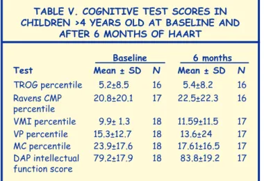
For children >4 years, the baseline and post-HAART test scores for TROG, Raven CPM, Beery-Buktenica VMI, VP, MC and Harris-Goodenough DAP are presented in Table V. As with the Griffiths Scales, all scores in this age group fell in the below average or below normal range, with specific deficits noted in language (TROG = 5th percentile) and visual perception (VP between 15th and 13th percentile) for age. The scores for tests administered remained similar at baseline and post-HAART; changes were not statistically significant.
The percentages of children at baseline with IQ, GQ or IF scores below 75 are set out recorded in Table VI.
Correlations between neurocognitive measures and either CD4 percentage or viral load were not statistically significant.
Discussion
In this pilot study, a small number of HIV-infected children were screened for neurological and cognitive deficits. The neurocognitive tools used are employed in everyday paediatric neurodevelopmental practice in South Africa. They evaluated non-verbal (Griffiths scales of mental development, Ravens CPM and Harris-Goodenough DAP) and verbal (TROG) cognition, and visual perception (Beery-Buktenica tests). The tests were selected to cover the age range 0 - 13 years, i.e. the age range of HIV-infected children who attend the infectious diseases service at Red Cross Children’s Hospital. Furthermore, the assessment tools were translated into the three dominant languages of the Western Cape and administered in the first language of the participants.
TAble III. MOTOR SySTeM exAMINATION AT bASelINe AND AfTeR 6 MONTHS Of HAART
Assessment
frequency at baseline (%) (N=39)
frequency at 6 months (%) (N=30) Normal motor
examination 26 (66.7) 19 (63.3)
Hyperreflexia 9 (23.1) 9 (30)
Hyperreflexia and
hypertonia 1 (2.6) 1 (3.3)
Hyperreflexia and
abnormal gait 1 (2.6) 1 (3.3)
Hyperreflexia and
clonus 1 (2.6) 0
Hyperreflexia,
hyper-tonia and reduced power 1 (2.6) 0
TAble IV. GRIffITHS MeNTAl DeVelOPMeNT SCAleS GeNeRAl quOTIeNTS AND Sub-quOTIeNTS AT bASelINe AND AfTeR 6
MONTHS Of HAART
Griffiths Mental Develop-ment Scales test results
baseline (mean ± SD) (N=26)
6 months (mean ± SD) (N=17)
General quotient (GQ) 71.6±16.3 72.7±14.8
Locomotor sub-quotient 67.3±18.6 68.1±16.3
Personal-social
sub-quotient 80.5±25.1 82.8±24.6
Hearing & speech
sub-quotient 69.1±16.8 70.7±16.0
Eye & hand co-ordination
sub-quotient 74.3±15.9 74.9±12.9
Performance sub-quotient 69±17.7 69.8±18.8
Practical reasoning
sub-quotient 70.8±16.5 71.5±16.2
TAble V. COGNITIVe TeST SCOReS IN CHIlDReN >4 yeARS OlD AT bASelINe AND
AfTeR 6 MONTHS Of HAART
baseline 6 months
Test Mean ± SD N Mean ± SD N
TROG percentile 5.2±8.5 16 5.4±8.2 16
Ravens CMP
percentile 20.8±20.1 17 22.5±22.3 16
VMI percentile 9.9± 1.3 18 11.59±11.5 17
VP percentile 15.3±12.7 18 13.6±24 17
MC percentile 23.9±17.6 18 17.61±16.5 17
DAP intellectual
function score 79.2±17.9 18 83.8±19.2 17
TAble VI. PeRCeNTAGeS Of CHIlDReN AT bASelINe wITH Sub-NORMAl INTellIGeNCe quOTIeNT (Iq), GeNeRAl quOTIeNT (Gq) OR
INTelleCTuAl fuNCTION (If) SCORe
No. of
children tested Percentage (N) <75
Griffiths (GQ) 26 62% (16) TROG (IQ) 16 81% (13) Ravens CMP (IQ) 17 59% (10)
ARTICLE
Notwithstanding the study limitations discussed below, our findings in a South African population are consistent with other studies in developing countries which describe a significant rate of early neurocognitive and motor developmental disabilities in HIV-infected children.19-22 Experience from these settings suggests that HIV is a major determinant of neurocognitive dysfunction.19-22 Notably, in this study language deficits in all age groups were greatest, as evidenced by the Griffiths Mental Development Scales and TROG scores, which were in the mild disability range. This could be anticipated, as language is the most representative developmental domain of higher order cognitive function and would therefore be expected to be the most sensitive to, and affected by, the CNS challenges described in HIV infection.
The study had several limitations. Despite careful selection of children and the exclusion of those with pre-existing deficits due to causes other than HIV, the socio-demographic and nutritional profile of the participants suggests that factors such as limited maternal education, poor household intellectual stimulation and malnutrition may themselves have impacted negatively on neurocognitive development. Furthermore, maternal emotional status, which could have influenced development, was not evaulated. While the spectrum of motor defects documented in the patient group is consistent with HIV encephalopathy, the absence of an HIV-uninfected control group for comparison limits our ability to estimate the relative contribution of HIV to neurocognitive dysfunction. However, a crude comparison of neurocognitive scores of HIV-infected children in the present study and scores of uninfected children from low socio-economic communities in the Western Cape who were enrolled in other studies showed that verbal and non-verbal intelligence performances of HIV-infected children were lower, and comparable to performance levels of children with mild developmental disability.31,32
The small, non-random sample in this study limits our ability to generalise our findings of prevalence and range of neurological and cognitive deficits to other groups of HIV-infected children in South Africa. Despite these weaknesses, the research findings strengthen the hypothesis that HIV-infected children in South Africa are vulnerable to neurological and cognitive problems and that they should be identified and actively managed.
Antiretroviral therapy may prevent CNS manifestations of HIV infection and reverse the process in children with established HIV encephalopathy (although not completely, as there are residual features of arrested HIV encephalopathy).33,34 The age of starting HAART is an important determinant of neurological and neurocognitive outcome. When HAART is commenced in HIV-infected children during early infancy, the prevalence of CNS effects may be reduced to less than 2%.33 In the present study, the neurological and cognitive deficits observed in the children remained static after 6 months of HAART, i.e. there was neither spontaneous resolution nor deterioration of the children’s function. Non-reversal after only 6 months of HAART was not completely unanticipated. Although HAART causes decline in cerebrospinal fluid HIV viral load, reversal of neurological and cognitive deficits caused
by HIV is unpredictable and arrested encephalopathy with persistent deficits are frequent.34 Because encephalopathy was not classified at baseline we were unable to determine whether some children with the progressive type were beginning to develop arrested HIV encephalopathy after 6 months of HAART. Furthermore, the children were exposed to unchanged socio-demographic factors throughout the study period, which may have contributed to non-improvement of CNS deficits. Documentation of outcomes over a longer time period would better elucidate neurodevelopmental trajectory and response to HAART and in this study group could be achieved by longitudinal follow-up, specifically to the age of regular school attendance. One factor that may affect the long-term clinical response to HAART is the variable penetration of the CNS compartment by antiretroviral agents, which should be evaluated in studies of longer duration.
In South Africa, there is currently limited potential to alter poverty-related parameters that impact on the intellectual development of children. However, increased intellectual stimulation is possible in resource-limited settings including South Africa through optimal utilisation of existing resources, such as early child-care facilities and toy libraries. These potential interventions should be explored in the context of paediatric HIV infection.
In conclusion, because of delayed introduction of HAART and a continued low coverage rate among HIV-infected children in South Africa, neurological and cognitive deficits are likely to be highly prevalent. Early, persisting receptive and expressive language delay in children is a well-documented predictor of poor long-term educational and life achievement outcomes. This has significant implications for children with HIV infection, their families, communities and society in general. A shift in national treatment guidelines towards earlier introduction of HAART, and a commitment to early intervention with child development programmes, should lead to reduced prevalence of CNS deficits and improved neurocognitive outcomes.
Acknowledgements
Dr T Fish, Statistical Department, University of Stellenbosch Business School, provided statistical support and Dr D Michaels, School of Child and Adolescent Health, University of Cape Town, invaluable comments.
References
1. Kramer-Hämmerle S, Rothenaigner I, Wolff H, Bell JE, Brack-Werner R. Cells of the central nervous system as targets and reservoirs of the
human immunodeficiency virus. Virus Research 2005; 111: 194-213.
2. Banks WA, Ercal N, Price TO. The blood-brain barrier in neuroAIDS.
Curr HIV Res 2006; 4: 259-266.
3. Dunfee R, Thomas ER, Gorry PR, Wang J, Ancuta P, Gabuzda D.
Mechanisms of HIV 1 neurotropism. Curr HIV Res 2006; 4: 267-278.
4. Kaul M, Zheng J, Gendelman HE, Lipton SA. HIV-1 infection and
AIDS: consequences for the central nervous system. Cell Death and
Differentiation 2005; 12: 878-892
5. Okamoto S, Kang Y, Brechtel CW, et al. HIV/gp120 decreases adult
neural progenitor cell proliferation via checkpoint kinase-mediated
cell-cycle withdrawal and G1 arrest. Cell Stem Cell 2007; 1: 230-236.
6. Centers for Disease Control and Prevention, Division of HIV/AIDS.
The prevalence and
extent of deficits
did not change
significantly in
response to
short-term HAART.
S
A
Jo
ur
H
ea
lth
S
A
Jo
ur
nal
of Child
H
ea
lth
S
A
Jo
ur
nal
of Child
H
ea
ARTICLE
1994 revised classification system for human immunodeficiency virus
infection in children less than 13 years of age. MMWR 1994; 43 (no.
RR-12): 1-10.
7. Van Rie A, Harrington PR, Dow A, Robertson K. Neurologic and neurodevelopmental manifestations of pediatric HIV/AIDS: a global
perspective. Eur J Pediatr Neurol 2007; 11: 1-9.
8. Brouwers P, Walters P, Civitello L. Central nervous system manifestations
and assessment. In: Pizzo PA, Wilfert CM, eds. Pediatric AIDS. 3rd ed.
Baltimore, Md: Williams & Williams, 1998: 293-308.
9. Lobato MN, Caldwell MB, Ng P, Oxtoby MJ. Encephalopathy in children with perinatally acquired human immunodeficiency virus infection.
Pediatric spectrum of disease consortium. J Pediatr 1995; 126: 710-715.
10. Molyneux EM, Tembo M, Kayira K, et al. The effect of HIV infection on
paediatric bacterial meningitis in Blantyre, Malawi. Arch Dis Child 2003;
88: 1112-1118.
11. van der Weert EM, Hartgers NM, Schaaf HS, et al. Comparison
of diagnostic criteria of tuberculous meningitis in human
immunodeficiency virus-infected and uninfected children. Pediatr Infect
Dis J 2006; 25: 65-69.
12. Centers for Disease Control and Prevention. Treating opportunistic infections among HIV-exposed and infected children: recommendations from CDC, the National Institutes of Health, and the Infectious
Diseases Society of America. MMWR 2004; 53(No. RR-14): inclusive
page numbers.
13. Bigger RJ, Frisch M, Goedert JJ, et al. Risk of cancer in children with
AIDS. J Am Med Assoc 2000; 284: 205-209.
14. McCarthy KM, Morgan J, Wannemuehler KA, et al. Population-based
surveillance for cryptococcosis in an antiretroviral-naïve South African
province with a high HIV seroprevalence. AIDS 2006; 20: 2199-2206.
15. Connor M. Stroke in patients with human immunodeficiency virus
infection. J Neurol Neurosurg Psychiatry 2007; 78: 1291.
16. Nuttall JJC, Wilmshurst JM, Yeats J, et al. Progressive multifocal
leukoencephalopathy after initiation of highly active antiretroviral therapy in a child with advanced human immunodeficiency virus infection: A case of immune reconstitution inflammatory syndrome.
Pediatr Infect Dis J 2004; 23: 683-685.
17. Wilmshurst JM, Burgess J, Hartley P, Eley B. Specific neurologic complications of human immunodeficiency virus type 1 (HIV-1)
infection in children. J Child Neurol 2006; 21: 788-794.
18. Smith R, Malee K, Leighty R, et al. Effects of perinatal HIV infection
and associated risk factors on cognitive development among young
children. Pediatrics 2006; 117: 851-862.
19. Msellati P, Lepage P, Hitimana D, Van Goethem C, Van der Pierre P, Dabis F. Neurodevelopmental testing of children born to human immunodeficiency virus type 1 seropositive and seronegative mothers:
a prospective cohort study in Kigali, Rwanda. Pediatrics 1993; 92:
843-848.
20. Sanmaneechai O, Puthanakit T, Louthrenoo O, Sirisanthana V. Growth, developmental, and behavioural outcomes of HIV-affected preschool
children in Thailand. J Med Assoc Thai 2005; 88: 1873-1879.
21. McGrath N, Fawzi WW, Bellinger D, et al. The timing of
mother-to-child transmission of human immunodeficiency virus infection and the
neurodevelopment of children in Tanzania. Pediatr Infect Dis J 2006; 25:
47-52.
22. Tahan TT, Bruck I, Burger M, Cruz CR. Neurological profile and neurodevelopment of 88 children infected with HIV and 84 seroreverter
children followed up from 1995 to 2002. Br J Infect Dis 2006; 10:
322-326.
23. Bagenda D, Nassali A, Kalyesubula I, et al. Health, neurologic, and
cognitive status of HIV-infected, long-surviving, and
antiretroviral-naive Ugandan children. Pediatrics 2006; 117: 729-740.
24. Eley B, Nuttall J, Davies M, et al. Initial experiences of a public sector
antiretroviral treatment programme for HIV-infected children and their
infected parents. S Afr Med J 2004; 94: 643-646.
25. Eley B, Davies M, Apolles P, et al. Antiretroviral treatment for children.
S Afr Med J 2006; 96: 988-993.
26. Griffiths R. Griffiths Mental Development Scales. ARICD, Bucks, UK,
1984.
27. Bishop DVM. T.R.O.G. Test for the Reception of Grammar. Oxford: Medical
Research Council, 1989.
28. Raven, JC. Raven Coloured Matrices. Oxford: Oxford Psychologists Press,
1947.
29. Harris DB. Children’s Drawings as Measures of Intellectual Maturity: A
Revision and Extension of the Goodenough Draw-a-Man Test. New York: Harcourt, Brace & World, 1963.
30. Beery KE. Revised Administration, Scoring and Teaching Manual for the
Developmental Test of Visual-Motor Integration. Cleveland: Modern Curriculum Press, 1982.
31. Adnams CM, Kodituwakku PW, Hay A, Molteno C, Viljoen D, May PA. Patterns of cognitive-motor development in children with fetal alcohol
syndrome from a community in South Africa. Alcohol. Clin Exp Res
2001; 25: 557-562.
32. May PA, Brooke L, Gossage JP, et al. Epidemiology of fetal alcohol
syndrome in a South African community. Am J Public Health 2000; 90:
1905-1912.
33. Shanbhag MC, Rutstein RM, Zaoutis T, Zhao H, Chao D, Radcliffe J. Neurocognitive functioning in pediatric human immunodeficiency
virus infection. Arch Pediatr Adolesc Med 2005; 159: 651-656.
34. Chiriboga CA, Fleishman S, Champion S, Gaye-Robinson L, Abrams EJ. Incidence and prevalence of HIV encephalopathy in children with HIV infection receiving highly active anti-retroviral therapy (HAART).