Achiraya Teyateeti, M.D.*, Pornpirom Laojunun, M.Sc.*, Porntip Iampongpaiboon, M.Sc.*, Nucharee Poon-iad, M.Sc. *, Sunanta Rojwatkarnjana, M.D.***, Titiporn Jiranantanakorn, M.D.****, Ajalaya Teyateeti, M.D. *
* Department of Radiology, Faculty of Medicine Siriraj Hospital, Mahidol University, ** Radiation Oncology Unit, Chulabhorn Hospital, ***Diagnostic Radiology and Nuclear Medicine Unit, National Cancer Institute, Thailand.
Outcome of External Beam Radiation in
Differentiated Thyroid Cancer: A 10-year Experience
Correspondence to: Ajalaya Teyateeti E-mail: [email protected]
Received 5 June 2017 Revised 6 July 2017 Accepted 11 July 2017 doi:10.14456/smj.2017.50
ABSTRACT
Objective: Role of external beam radiation (EBRT) in differentiated thyroid cancer (DTC) is controversial. Outcomes
of EBRT are also not uniform among studies due to variation in practices. Therefore, this study was elicited to evaluate the outcome of EBRT in DTC in context of our institutional practice.
Methods: Medical records of DTC patients who received EBRT to neck with or without mediastinum from January
2004 to December 2013 were retrospectively reviewed. All patients underwent total thyroidectomy (TT) or completion of thyroidectomy. EBRT could be given immediate after TT as adjuvant treatment or given at time of recurrence or RAI refractory as salvage treatment. Radioactive iodine (RAI) treatment could be given before or after EBRT or even be omitted. The primary end-points were locoregional failure-free survival (LRFS) and overall survival (OS).
Results: Among 47 patients, most of them were female (70%), age 45 years or over (83%), papillary thyroid cancer
(75%) and stage III-IV (62%). There were adjuvant EBRT in 8 patients and salvage EBRT in 39 patients. At time of EBRT, there were high risk histology, gross residual tumor, positive surgical margin, lymph node metastasis, distant metastasis and RAI refractory in 21%, 62%, 72%, 66%, 47% and 59% of patients, respectively. Radiation techniques were conventional radiotherapy, three-dimensional conformal radiotherapy (3D-CRT) and intensity modulated radiotherapy (IMRT) in 14, 19 and 14 patients with median EBRT dose of 50, 60 and 66 Gy (30-70 Gy), respectively. At median follow-up time of 26.6 months (1-82), LRFS at 2 and 4 year were 74% and 63%, respectively. The 2- and 4-year OS were 62% and 42%, respectively. On multivariate analysis, age ≥ 45 years tended to associate with worse OS (p=0.058). EBRT dose ≤ 50 Gy also had a trend to be related with both worse LRFS (p=0.069) and OS (p=0.079).
Conclusion: EBRT might be given either as adjuvant or salvage treatment, particularly in case of gross residual
tumor and RAI refractory. Escalation of EBRT dose to over 50 Gy with advanced EBRT technique such as IMRT might improve the LRFS and OS.
Keywords: External beam radiation; radiotherapy; differentiated thyroid cancer (Siriraj Med J 2017;69: 247-254)
INTRODUCTION
Treatment of differentiated thyroid cancer (DTC) generally comprises of total thyroidectomy (TT) with or without lymph node dissection (LND), radioactive iodine (RAI) treatment and Thyroid-Stimulating Hormone (TSH) suppression therapy.1,2 Role of external beam
radiation (EBRT) is still controversial due to lack of randomized control trials. According to previous studies
and in clinical practice, EBRT may be given in case of (1) gross residual tumor (2) pathological T4 or extrathyroidal extension (ETE), (3) positive surgical margin (SM), (4) presence of lymph node (LN) metastasis with or without extranodal extension (ENE) and (5) RAI refractory.1-24
(LRC) in patients with high risk factors such as gross residual disease, microscopic positive SM, ETE, and RAI refractory 7-20, whereas few studies reported that EBRT
increased toxicity without additional benefit over surgery alone.25-28 As a result, this study was elicited to evaluate
the result of EBRT in DTC in the context of institutional practice.
MATERIALS AND METHODS
Retrospective chart review of DTC patients who received EBRT at Division of Radiation Oncology, Siriraj Hospital during January 2004 to December 2013, were performed following approval of the ethical committee of the faculty. Inclusion criteria were patients with (1) age of 18 years old and over, (2) underwent or attempted TT or completion of thyroidectomy with or without LND, (3) histopathologically proven DTC; papillary (PTC) and follicular carcinoma (FTC) with variants e.g. classical, tall cell, columnar cell, diffuse sclerosing, hobnail, oncocytic (Hürthle cell) and poorly differentiated, (4) any risk stratification and stage of disease, (5) given EBRT to gross residual tumor and/or thyroid bed with or without neck and mediastinal LN and (6) completed EBRT course as planned. Exclusion criteria were (1) presence of other malignancies (2) histopathological diagnosis of anaplastic thyroid cancer and medullary thyroid cancer or mixed DTC with anaplastic thyroid cancer (3) cytological diagnosis of DTC from fine needle aspiration (FNA) without histopathological confirmation, or (4) previous EBRT to neck and/or mediastinal region.
All patients were referred for EBRT following TT or completion of thyroidectomy with or without gross residual tumor. RAI dose of 30–200 mci may be omitted, given before or given after EBRT. RAI treatment may be given repeatedly depending on RAI avidity and nuclear medicine physician discretion. EBRT were given in 2 ways; (1) adjuvant treatment - defined as immediate post-operative EBRT within 3 months without evidence of RAI refractory and (2) salvage treatment – defined as delayed EBRT to time of recurrence or RAI refractory.2
Radiation technique was varied based on availability at that time and physician preference – i.e. conventional radiotherapy, three-dimensional conformal radiotherapy (3D-CRT), intensity-modulated radiotherapy (IMRT) and volumetric arc radiotherapy (VMAT). Machines were Cobalt-60 or linear accelerator with or without electron enface. Typically, radiation oncologists prescribed dose of 66-70 Gy to gross residual tumor, 63-66 Gy to area of positive or closed SM, 59.4-63 Gy to area of surgical bed/tumor bed and positive LN region and 50-54 Gy to
disease. Dose constraints for normal tissue were referred to QUANTEC.29
The primary end-points of this study were locoregional failure-free survival (LRFS) and overall survival (OS). LRFS was defined from date of start radiation to date of recurrence or progression in irradiated region or date of last follow-up if no progression or recurrence in irradiated region. Recurrence or progression was defined by clinical examination and/or imaging with or without pathologic confirmation. OS was defined from date of start radiation to date of death or date of last follow-up alive. Secondary outcome was progression-free survival (PFS) which was defined from date of start radiation to date of recurrence or progression or date of death or date of last follow-up alive without progression or recurrence.
Survival analysis was performed by using Kaplan-Meier methods. Univariate analysis was also done by using Log-rank. Factors which had p-value < 0.1 from univariate analysis were proceeded to enter the multivariate analysis by Cox proportional-hazards model.
RESUTLS
There were 47 patients included in this study. All patients underwent surgical attempt of TT or completion of thyroidectomy prior to RAI treatment and EBRT. Twenty-nine patients (61.7%) underwent LND. Major histopathology was PTC (74.5%). High risk histology (HR) was found in 8 patients (17%). (Table 1)
There were 29 of 47 patients (61.7%) who experienced additional surgical procedures prior to EBRT. The majority of procedures were LND performed in 18 patients (38.3%). Median number of additional surgical procedures was 1 time (1-8 times). At time of EBRT, there was transformation of mixed papillary and follicular thyroid cancer and FTC to poorly differentiated thyroid cancer in 2 patients. Tumor and treatment characteristics at time of EBRT were shown in Table 2 and 3, respectively.
Of 47 patients, 41 patients received RAI treatment (87.3%) – before, after or both before and after EBRT. RAI treatment was not administrated in 5 patients with HR (10.6%) and sequence of RAI was unknown in 1 patient (2.1%).
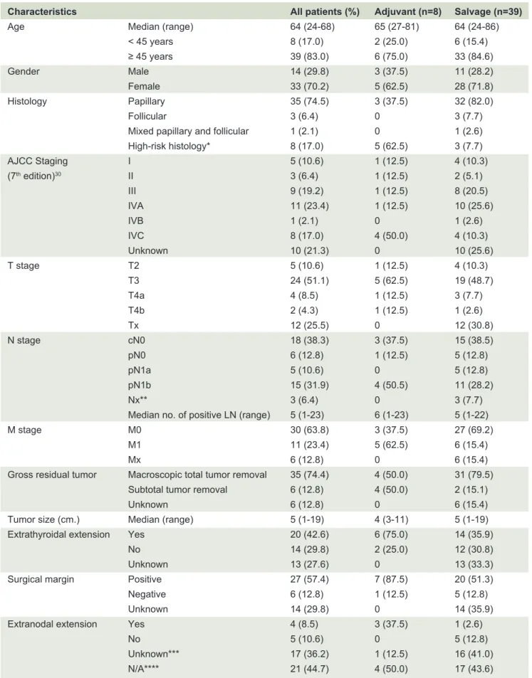
TABLE 1. Patient and tumor characteristics at time of diagnosis.
*High risk histology 8 patients – 4 poorly differentiated (8.5%), 2 insular (4.3%), 1 tall cell (2.1%) and 1 Hürthle cell (2.1%). **Patients underwent LN dissection but pathological reports were not available.
***Patients underwent cervical LN dissection, but pathological report did not mention extranodal extension.
****LN dissection was not performed (n=18) or LN dissection was performed but pathological reports were not available (n=3). Characteristics All patients (%) Adjuvant (n=8) Salvage (n=39)
Age Median (range) 64 (24-68) 65 (27-81) 64 (24-86)
< 45 years 8 (17.0) 2 (25.0) 6 (15.4)
≥ 45 years 39 (83.0) 6 (75.0) 33 (84.6)
Gender Male 14 (29.8) 3 (37.5) 11 (28.2)
Female 33 (70.2) 5 (62.5) 28 (71.8)
Histology Papillary 35 (74.5) 3 (37.5) 32 (82.0)
Follicular 3 (6.4) 0 3 (7.7)
Mixed papillary and follicular 1 (2.1) 0 1 (2.6)
High-risk histology* 8 (17.0) 5 (62.5) 3 (7.7)
AJCC Staging I 5 (10.6) 1 (12.5) 4 (10.3)
(7th edition)30 II 3 (6.4) 1 (12.5) 2 (5.1)
III 9 (19.2) 1 (12.5) 8 (20.5)
IVA 11 (23.4) 1 (12.5) 10 (25.6)
IVB 1 (2.1) 0 1 (2.6)
IVC 8 (17.0) 4 (50.0) 4 (10.3)
Unknown 10 (21.3) 0 10 (25.6)
T stage T2 5 (10.6) 1 (12.5) 4 (10.3)
T3 24 (51.1) 5 (62.5) 19 (48.7)
T4a 4 (8.5) 1 (12.5) 3 (7.7)
T4b 2 (4.3) 1 (12.5) 1 (2.6)
Tx 12 (25.5) 0 12 (30.8)
N stage cN0 18 (38.3) 3 (37.5) 15 (38.5)
pN0 6 (12.8) 1 (12.5) 5 (12.8)
pN1a 5 (10.6) 0 5 (12.8)
pN1b 15 (31.9) 4 (50.5) 11 (28.2)
Nx** 3 (6.4) 0 3 (7.7)
Median no. of positive LN (range) 5 (1-23) 6 (1-23) 5 (1-22)
M stage M0 30 (63.8) 3 (37.5) 27 (69.2)
M1 11 (23.4) 5 (62.5) 6 (15.4)
Mx 6 (12.8) 0 6 (15.4)
Gross residual tumor Macroscopic total tumor removal 35 (74.4) 4 (50.0) 31 (79.5)
Subtotal tumor removal 6 (12.8) 4 (50.0) 2 (15.1)
Unknown 6 (12.8) 0 6 (15.4)
Tumor size (cm.) Median (range) 5 (1-19) 4 (3-11) 5 (1-19)
Extrathyroidal extension Yes 20 (42.6) 6 (75.0) 14 (35.9)
No 14 (29.8) 2 (25.0) 12 (30.8)
Unknown 13 (27.6) 0 13 (33.3)
Surgical margin Positive 27 (57.4) 7 (87.5) 20 (51.3)
Negative 6 (12.8) 1 (12.5) 5 (12.8)
Unknown 14 (29.8) 0 14 (35.9)
Extranodal extension Yes 4 (8.5) 3 (37.5) 1 (2.6)
No 5 (10.6) 0 5 (12.8)
Unknown*** 17 (36.2) 1 (12.5) 16 (41.0)
TABLE 2. Tumor characteristics at time of external beam radiation.
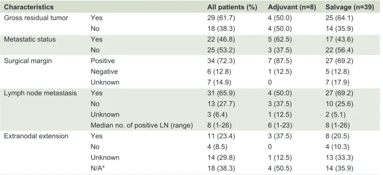
TABLE 3. Treatment characteristics at time of external beam radiation.
Characteristics All patients (%) Adjuvant (n=8) Salvage (n=39)
Gross residual tumor Yes 29 (61.7) 4 (50.0) 25 (64.1)
No 18 (38.3) 4 (50.0) 14 (35.9)
Metastatic status Yes 22 (46.8) 5 (62.5) 17 (43.6)
No 25 (53.2) 3 (37.5) 22 (56.4)
Surgical margin Positive 34 (72.3) 7 (87.5) 27 (69.2)
Negative 6 (12.8) 1 (12.5) 5 (12.8)
Unknown 7 (14.9) 0 7 (17.9)
Lymph node metastasis Yes 31 (65.9) 4 (50.0) 27 (69.2)
No 13 (27.7) 3 (37.5) 10 (25.6)
Unknown 3 (6.4) 1 (12.5) 2 (5.1)
Median no. of positive LN (range) 8 (1-26) 6 (1-23) 8 (1-26)
Extranodal extension Yes 11 (23.4) 3 (37.5) 8 (20.5)
No 4 (8.5) 0 4 (10.3)
Unknown 14 (29.8) 1 (12.5) 13 (33.3)
N/A* 18 (38.3) 4 (50.5) 14 (35.9)
*LN dissection was not performed or was performed but no evidence of cervical LN involvement determined by pathological report.
Characteristics Number (%) Adjuvant (n=8) Salvage (n=39) RAI treatment 41 (87.3) 4 (50.0) 37 (94.9)
Before EBRT 31 (66.0) 0 31 (19.5)
Median RAI dose (mCi) 450 (150-1300) 0 450 (150-1300)
Median number of courses (range) 3 (1-9) 0 3 (1-9)
Before and after EBRT 6 (12.8) 0 6 (15.4)
Mean RAI dose before EBRT (mCi) 367 (150-600) 0 367 (150-600)
Mean RAI dose after EBRT (mCi) 217 (150-450) 0 217 (150-450)
Mean total RAI dose (mCi) 583 (350-750) 0 583 (350-750)
Median number of courses before EBRT (range) 2 (1-4) 0 2 (1-4)
Median number of courses after EBRT (range) 2 (1-3) 0 2 (1-3)
Median total number of courses (range) 4 (2-5) 0 4 (2-5)
After EBRT 4 (8.5) 4 (8.5) 0
Median RAI dose (mCi) 450 (150-1300) 450 (150-1300) 0
Median number of courses (range) 3 (1-7) 3 (1-7) 0
No RAI treatment 5 (10.6) 4 (50.0) 1 (2.6)
Unknown 1 (2.1)* 0 1 (2.6)
External beam radiation
Technique
Conventional radiotherapy 14 (29.8) 2 (25.0) 12 (30.8)
3D-CRT 19 (40.4) 4 (50.0) 15 (38.4)
IMRT/VMAT 14 (29.8) 2 (25.0) 12 (30.8)
Dose
Median dose for all technique (Gy) 60 (30#-70) 55 60
Median dose for conventional radiotherapy (Gy) 50 (30#-66) 55+ 50
Median dose for 3D-CRT (Gy) 60 (30#-70) 50 60
Median dose for IMRT/VMAT (Gy) 66 (60-70) 66+ 66
*Unknown sequence of RAI treatment
At last follow-up, pattern of first failure or first progression were locoregional failure (LRF) in 8 patients (17%), distant metastatic failure in 11 patients (23.4%) and both locoregional and distant metastatic failure in 6 patients (12.8%). At median follow-up time of 26.6 months (range 1-82), the LRFS at 2 and 4 years were 74% and 63%, respectively. The OS at 2 and 4 years were 62% and 42% and PFS at 2 and 4 years were 45% and 23%, respectively.
The univariate analysis to predict factors which had effects on LRFS and OS were performed (Table 4). Factors which significantly had a negative impact on LRFS were positive LN and EBRT dose ≤ 50 Gy. Regarding
Fig 1. Kaplan-Meier curves of locoregional-free survival, progression-free survival and overall survival.
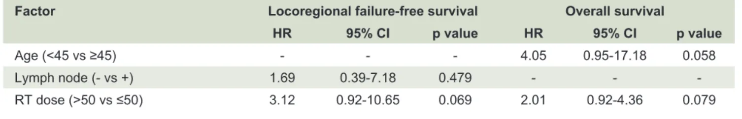
the OS, age ≥ 45 years and EBRT dose ≤ 50 Gy were related to worse outcome. Conventional radiotherapy as compared to IMRT tended to associate with decreased both LRFS and OS. According to Table 3, there was a correlation between higher radiation dose and more advanced radiation technique. Therefore, we decided to include only radiation dose in the multivariate analysis along with LN status for LRFS and age for OS. Multivariate analysis showed that none of factors had significant impact on LRFS and OS (Table 5). However, better OS tended to observe in patients < 45 years and radiation dose > 50 Gy.
TABLE 4. Univariate analysis prognostic factors of LRFS and OS.
Factor Locoregional failure-free survival Overall survival
HR 95% CI p value HR 95% CI p value Characteristics at time of diagnosis
Age (<45 vs ≥45) 2.04 0.45-9.19 0.500 4.26 1.01-18.02 0.032
Gender (F vs M) 0.71 0.22-2.26 0.390 0.48 0.20-1.19 0.169
Tumor size (≤4 vs >4) 1.63 0.48-5.61 0.238 1.78 0.73-4.38 0.546
Extrathyroidal extension (- vs +) 1.84 0.53-6.35 0.408 1.70 0.67-4.28 0.154
Characteristics at time of radiation
Adjuvant vs Salvage 0.47 0.06-3.67 0.724 0.93 0.35-2.45 0.609
Histology (PTC vs FTC)* - - - 3.06 0.87-10.77 0.142
High risk histology (- vs +)* - - - 1.89 0.83-4.30 0.441
Gross residual tumor (- vs +) 1.62 0.54-4.86 0.750 1.14 0.54-2.39 0.281
Metastasis (- vs +) 1.55 0.53-4.52 0.521 1.15 0.55-2.40 0.519
Surgical margin (- vs +) 1.86 0.24-14.55 0.763 1.14 0.34-3.86 0.967
Lymph node (- vs +) 2.49 0.67-9.21 0.048 1.22 0.52-2.84 0.495
RT technique 0.064 0.074
IMRT vs 2D 3.37 0.91-12.52 0.070 2.51 0.96-6.55 0.061
IMRT vs 3D 1.28 0.32-5.15 0.729 1.89 0.72-4.9 0.194
RT dose (>50 vs ≤50) 3.79 1.21-11.87 0.014 2.13 0.98-4.62 0.039
TABLE 5. Multivariate analysis prognostic factors of LRFS and OS.
Factor Locoregional failure-free survival Overall survival
HR 95% CI p value HR 95% CI p value
Age (<45 vs ≥45) - - - 4.05 0.95-17.18 0.058
Lymph node (- vs +) 1.69 0.39-7.18 0.479 - -
-RT dose (>50 vs ≤50) 3.12 0.92-10.65 0.069 2.01 0.92-4.36 0.079
Noticeably, low EBRT dose such as 30 Gy in 10 fractions was given to some friable or metastatic patients as a palliation. Inclusion of these patients in a group who received EBRT ≤ 50 Gy might result in worse outcome. To prove the effect of EBRT dose, we further analyzed impact of EBRT dose 50 Gy vs > 50 Gy and 50-54 Gy vs > 54 Gy on LRFS and OS. The results showed that lower EBRT doses were associated with significantly worse LRFS (p=0.013 and p< 0.001) and a trend toward worse OS (p=0.106 and p=0.160), respectively.
DISCUSSION
EBRT could be given either immediately after operation as adjuvant treatment or delayed until recurrence or RAI refractory as salvage treatment. Regarding adjuvant EBRT in our institute, the most common indications were HR and gross residual tumor. As we know that RAI avidity varied in HR, response to RAI treatment was consequently quite unpredictable. Additionally, RAI treatment might be inadequate for LRC in bulky disease and even worsen compressive symptoms due to edema. For these reasons, EBRT might be considered immediately after surgery to provide robust LRC whereas RAI treatment might be given later to provide systemic control.
Salvage EBRT was much more common in our practice since RAI treatment together with TSH suppression therapy was frequently used as an initial adjuvant treatment. We preferred this regimen owing to the fact that RAI treatment provided LRC and systemic control with minimal toxicity. Moreover, deferring EBRT until recurrence to avoid toxicity would not compromise ultimate LRC if total tumor removal could be achieved. There was also no significant difference in LRFS and OS among patients who received EBRT whether recurrence or not.29
The LRFS and OS in this study were inferior to other recent studies. Schwartz et al., found that 4-year LRFS and OS were 79% and 73%, respectively29 while
another study of non-anaplastic thyroid cancer (including medullary thyroid cancer) reported the 2- and 4-year LRFS of 86% and 72% and 2- and 4-year OS of 74% and
55%, respectively.30 We hypothesized that our worse
outcomes might be contributed by two factors; gross residual tumor and radiation dose.
Gross residual tumor at time of EBRT was a significant factor related to worse LRFS and OS.19,29,31 There was only
around 10% of patients who had gross residual tumor at time of EBRT in those studies compared to 62% of patients in our study. Therefore, worse LRFS and OS in our study were understandable. However, volume of gross residual tumors was another concern because it might affect the outcomes. Unfortunately, it was not evaluated in any of the aforementioned studies.
Although the median EBRT dose in our study, 60 Gy (30-70) was comparable to that of 60 Gy (38-72) of Schwartz et al.29, and 63 Gy (59.4-70) of Terezakis
et al.30, our treatment outcomes were inferior. We
presumed that it might have resulted from a relatively high proportion of patients receiving low dose EBRT. Esik et al., revealed that EBRT dose of 40-45 Gy or higher after macroscopic tumor removal provided significantly better LRFS. Additional studies of higher EBRT dose of 50-60 Gy with RAI also demonstrated improvement in LRFS.9,11 One third of patients in our study received
EBRT ≤ 50 Gy, consequently, lower LRFS and OS might be inevitable.
A trend of dose response effect was observed in a previous study.18 The univariate and multivariate analysis
in the present study also showed a trend toward worse LRFS and OS with RT dose ≤ 50 Gy. There is no evidence that increased radiation dose to certain dose level will lead to better outcomes. Nevertheless, study suggested that LRF after IMRT was least in irradiated area of 70 Gy and most of LRF were marginal failure outside irradiated area of 70 Gy or less.33 This finding implied that high
EBRT dose might provide better LRC.
higher than 3D-CRT.34 Dosimetric studies revealed that
IMRT provided higher EBRT dose, better dose target coverage and lower EBRT dose to critical structures such as spinal cord than 3D-CRT.34,35 The OS with IMRT was
also superior or comparable to 3D-CRT, even with the presence of more adverse features.34
In our study, around 30% of patients were treated with conventional technique compared to studies of Schwartz et al,29 and Terezakis et al,30 in which all patients
received EBRT with 3D-CRT or IMRT. Therefore, our worse treatment outcomes might be related to higher proportion of patients who received lower EBRT dose with conventional technique. Analysis of the effect of EBRT techniques in the current study found a trend toward worse LRFS and OS with conventional technique as well. However, this result might be an indirect effect of EBRT dose rather than the direct effect from EBRT technique itself.
Owing to relatively small population in this study, the effect of RAI on treatment outcomes, particularly for LRC might be confounded by EBRT. Even so, a much larger study from Brierley et al, showed that RAI was related to overall improvement in LRFS with possible cause-specific survival (CSS) in some patient subgroups.31
At our institute, we preferred to not give RAI treatment until there was evidence of refractory - e.g. non-avid lesion on post-treatment or diagnostic whole body scan. Accordingly, median number of courses and cumulative doses of RAI before EBRT in this study were much higher than Schwartz et al,29 (3 vs 1 time and
450 vs 150 mCi, respectively). Interestingly, even with higher RAI dose, distant failure was still common in our institute and both LRFS and OS remained inferior to that reported by Schwartz et al.29 Therefore, we postulated
that early salvage EBRT when locoregional recurrence or persistent disease tended to be RAI refractory probably provided better LRC and further reduced risk of distant metastasis.
According to previous studies, many patient and tumor characteristics were found to have effect on treatment outcomes. Ages of 60 years and older, FTC (as compared to PTC), HR, large tumor size, ETE, positive SM, LN metastasis and distant metastasis were associated with worse LRFS.18,19,29,31 Female, ages of 60 years and older,
FTC (as compared to PTC), HR, large tumor size and distant metastasis were related to lower CSS.19,29,31 Distant
metastasis and HR also resulted in inferior OS.29 However,
included subjects in this study were too few to demonstrate the effect of these aforementioned factors on LRFS and OS.
Another major limitation of our study was incomplete
follow-up data, especially for EBRT toxicities. As reported by Terezakis et al., common late toxicities were xerostomia, dysphagia and hoarseness.30 Severe late toxicities such
as esophageal stricture requiring dilatation, laryngeal stenosis or edema requiring tracheostomy and dysphagia requiring long-term tube feeding were also reported, particularly among patients treated with 60 Gy or more and toxicities were found less frequently with IMRT compared to 3D-CRT.29 This finding implied that using
IMRT in DTC not only enhances treatment outcomes through dose escalation, but also alleviated treatment toxicities.
According to the previous version of NCCN guidelines 36, adjuvant EBRT should be considered for
high risk features which are T4 with surgical evidence of gross ETE and age > 45 years old. However, adjuvant EBRT for this group of patients who have no gross residual tumor is currently no longer recommended by NCCN guidelines.1 Nonetheless, adjuvant EBRT in gross residual
tumor or inadequate RAI uptake and salvage EBRT still exist. As a result, further studies should be focused on 1) appropriate time to initiate salvage EBRT - whether early EBRT provides better outcome, and 2) whether integration of systemic treatment such as chemotherapy or targeted therapy to EBRT help in improving treatment outcome, particularly for high risk histology.
In conclusion, management of DTC with TT (with or without postoperative RAI) could be followed by EBRT as adjuvant or salvage treatment, particularly in case of gross residual tumor and RAI refractory. Escalation of EBRT dose to over 50 Gy with advanced EBRT technique such as IMRT might improve the LRFS and OS.
REFERENCES
1. NCCN Guidelines Version 1.2017
2. Haugen BR, Alexander EK, Bible KC, Doherty GM, Mandel SJ, Nikiforov YE, et al. 2015 American Thyroid Association Management Guidelines for Adult Patients with Thyroid Nodules and Differentiated Thyroid Cancer: The American Thyroid Association Guidelines Task Force on Thyroid Nodules and Differentiated Thyroid Cancer. Thyroid. 2016;26:1-133.
3. Weiss TE, Grigsby PW. Thyroid cancer. In: Halperin E, Perez C, Brady L, eds. Principles and practice of radiation oncology. 5th ed. Philadelphia: Lippincott Williams & Wilkins, a Wolters Kulwer business; 2008: 1055-75
4. Cooper DS, Doherty GM, Haugen BR, Kloos RT, Lee SL,
Mandel SJ, et al. Revised American Thyroid Association management guidelines for patients with thyroid nodules and differentiated thyroid cancer. Thyroid : official journal of the American Thyroid Association 2009;19:1167-214.
Perez C, Brady L, eds. Principles and practice of radiation oncology. 6th ed. Philadelphia: Lippincott Williams & Wilkins, a Wolters Kulwer business; 2013: 918-37.
7. Benker G, Olbricht T, Reinwein D, Reiners C, Sauerwein W, Krause U, et al. Survival rates in patients with differentiated thyroid carcinoma. Influence of postoperative external
radiotherapy. Cancer 1990;65:1517–1520.
8. Chung CT, Sagerman RH, Ryoo MC, King GA, Yu WS, Dalal
PS, et al. External irradiation for malignant thyroid tumors. Radiology 1980;136:753–756.
9. Farahati J, Reiners C, Stuschke M, Müller SP, Stüben G, Sauerwein W, et al. Differentiated thyroid cancer. Impact of adjuvant external radiotherapy in patients with perithyroidal tumor infiltration (stage pT4).Cancer 1996;77:172–180.
10. Harmer CL. External beam radiotherapy for thyroid cancer. Ann Radiol 1977;20:791–800.
11. Phlips P, Hanzen C, Andry G, Van Houtte P, Früuling J. Postoperative irradiation for thyroid cancer. Eur J Surg Oncol 1993;19:399–404.
12. Sheline GE, Galante M, Lindsay S. Radiation therapy in the control of persistent thyroid cancer. Am J Roentgenol Radium Ther Nucl Med 1966;97:923–930.
13. Simpson WJ, McKinney SE, Carruthers JS, Gospodarowicz MK, Sutcliffe SB, Panzarella T. Papillary and follicular thyroid cancer. Prognostic factors in 1,578 patients. Am J Med 1987;83:479–488.
14. Tsang RW1, Brierley JD, Simpson WJ, Panzarella T, Gospodarowicz MK, Sutcliffe SB. The effects of surgery, radioiodine, and external radiation therapy on the clinical outcome of patients with differentiated thyroid carcinoma. Cancer 1998;82:375–388.
15. Wu XL, Hu YH, Li QH, Guo JR, Sun D, Yan JH, et al. Value of postoperative radiotherapy for thyroid cancer. Head Neck Surg 1987;10:107–112.
16. O’Connell ME, A’Hern RP, Harmer CL. Results of external beam radiotherapy in differentiated thyroid carcinoma: A retrospective study from the Royal Marsden Hospital. Eur J Cancer 1994; 30A:733–739.
17. Esik O, Nemeth G, Eller J. Prophylactic external irradiation in differentiated thyroid cancer: A retrospective study over a 30-year observation period. Oncology 1994;51:372–379.
18. Ford D, Giridharan S, McConkey C, Hartley A, Brammer C, Watkinson JC, et al. External beam radiotherapy in the management of differentiated thyroid cancer. Clin Oncol 2003;15:337–341.
19. Meadows KM, Amdur RJ, Morris CG, Villaret DB, Mazzaferri EL, Mendenhall WM. External beam radiotherapy for differentiated thyroid cancer. Am J Otolaryngol 2006;27:24–28.
20. Kim TH, Yang DS, Jung KY, Kim CY, Choi MS. Value of external irradiation for locally advanced papillary thyroid cancer. Int J Radiat Oncol Biol Phys 2003;55:1006–1012.
21. Keum KC, Suh YG, Koom WS, Cho JH, Shim SJ, Lee CG,
et al. The role of postoperative external-beam radiotherapy in the management of patients with papillary thyroid cancer invading the trachea. Int J Radiat Oncol Biol Phys 2006;65: 474–480.
22. Hu A, Clark J, Payne RJ, Eski S, Walfish PG, Freeman JL.
Arch Otolaryngol Head Neck Surg 2007;133:644–649.
23. Brierley JD, Tsang RW. External-beam radiation therapy in the treatment of differentiated thyroid cancer. Semin Surg Oncol 1999;16:42–49.
24. Tubiana M, Haddad E, Schlumberger M, Hill C, Rougier P, Sarrazin D. External radiotherapy in thyroid cancers. Cancer 1985;55:2062-2071
25. Mazzaferri EL, Young RL. Papillary thyroid carcinoma: A 10 year follow-up report of the impact of therapy in 576 patients. Am J Med 1981;70:511–518.
26. Samaan NA, Schultz PN, Hickey RC, Goepfert H, Haynie TP, Johnston DA, et al. The results of various modalities of treatment of well differentiated thyroid carcinomas: A retrospective review of 1599 patients. J Clin Endocrinol Metab 1992;75:714–720.
27. Jensen MH, Davis RK, Derrick L. Thyroid cancer: A computer assisted review of 5287 cases. Otolaryngol Head Neck Surg 1990;102:51–65.
28. Lin JD, Tsang NM, Huang MJ, Weng HF. Results of external beam radiotherapy in patients with well differentiated thyroid carcinoma. Jpn J Clin Oncol. 1997;27:244-7.
29. Schwartz DL, Lobo MJ, Ang KK, Morrison WH, Rosenthal DI, Ahamad A, et al. Postoperative external beam radiotherapy for differentiated thyroid cancer: outcomes and morbidity with conformal treatment. Int J Radiat Oncol Biol Phys. 2009;74: 1083-91.
30. Terezakis SA, Lee KS, Ghossein RA, Rivera M, Tuttle RM, Wolden SL, et al. Role of external beam radiotherapy in patients with advanced or recurrent nonanaplastic thyroid cancer: Memorial Sloan-kettering Cancer Center experience. Int J Radiat Oncol Biol Phys. 2009;73:795-801.
31. Brierley J, Tsang R, Panzarella T, Bana N. Prognostic factors and the effect of treatment with radioactive iodine and external beam radiation on patients with differentiated thyroid cancer seen at a single institution over 40 years. Clin Endocrinol. 2005;63:418-27.
32. Esik O, Nemth G, Eller J. Prophylactic external irradiation in differentiated thyroid cancer: a retrospective study over a 30- year observational period. Oncology 1994;51:372–379
33. Shugard E, Chen J, Quivey JM, Glastonbury CM, Khanafshar E, Garsa AA, et al. Does radiation dose matter in thyroid cancer?: Patterns of local-regional failure in recurrent and metastatic well-differentiated thyroid cancers treated with dose-painted intensity-modulated radiation therapy. J Med Imaging Radiat Oncol. 2016;60:560-7.
34. Goffredo P, Robinson TJ, Youngwirth LM, Roman SA, Sosa JA. Intensity-modulated radiation therapy use for the localized treatment of thyroid cancer: Nationwide practice patterns and outcomes. Endocrine. 2016 Sep;53:761-73.
35. Nutting CM, Convery DJ, Cosgrove VP, Rowbottom C, Vini L, Harmer C, et al. Improvements in target coverage and reduced spinal cord irradiation using intensity-modulated radiotherapy (IMRT) in patients with carcinoma of the thyroid gland. Radiother Oncol 2001;60:173–180.