Original Article
Isolation of Small Number of Cryptosporidium parvum Oocyst
Using Immunochromatography
Elahe EBRAHIMZADE, *Parviz SHAYAN, Zeinab ASGHARI, Sedighe JAFARI, Zahra OMIDIAN
Department of Parasitology, Iranian Center for Ticks and Tick-borne Diseases, Faculty of Veterinary Medicine, Uni-versity of Tehran, Tehran, Iran
Received 12 Feb 2014
Accepted 10 Aug 2014 Abstract Background: Cryptosporidium parvum causes severe gastroenteritis in immunocom-promised human and new borne animals. The organism can be transmitted through water. Since small number of C. parvum is infectious, the aim of the present study was to develop a chromatography method for the isolation of C. parvum oocyst in samples with limited number of oocysts.
Methods: Antibody was prepared against whole antigen from C. parvum oocysts, the achieved Ab bound to the sepharose 4B and used for the isolation of oocysts. Antibody against P23 bound to the sepharose 4B, used also for the isolation of C. parvum oocyst. In comparison to these both methods, 2 traditional methods (Salt floatation and 55% sucrose floatation) were also performed.
Results: Both chromatography methods could bind oocysts with capacity depends on the column size. The isolated oocysts were free of bacteria. Our results showed that the traditional methods are useful for the isolation of oocysts from feces, in its smear stained with ziehl-nelsen, at least 3 oocyts are detectable in each microscopic field under 1000 X magnification. In contrast to the chromatography methods, the bacterial contamination was always observed in oocysts isolated with traditional methods.
Conclusion: Immunochromatography could be used for the successful isolation of C. parvum oocysts from the samples containing limited number of oocysts.
Keywords:
Cryptosporidium parvum
Oocyst
Chromatography Flotation
*Correspondence
Email:
Iranian Society of Parasitology http:// isp.tums.ac.ir
Iranian J Parasitol
Open access Journal at http:// ijpa.tums.ac.ir Tehran University of Medical
Introduction
ryptosporidium parvum is a coccidian protozoon that causes
gastrointes-tinal illness in
im-munocompromised human and newborn ani-mals (1). Billions oocysts of C. parvum can be released from the infected animals and can contaminate soil, food, water or surfaces (2). The oocysts can also remain viable for several months under a wide range of environmental stresses (1, 3). Unfortunately, there are no ef-fective methods of treating or preventing C. parvum infection in animals or human (1).
A large number of waterborne outbreaks of cryptosporidiosis have been reported world-wide (4, 5). Since water is the most important source of infection and the number of oocysts required for initiation of infection is relatively low, a sensitive method is required for the de-tection of C. parvum oocysts in samples (6, 7). Cryptosporidium spp. oocysts present in small numbers in the water sources (5). One of the methods for detection of small amount of this parasite in water sources is based on filtration and immune magnetic separation (IMS) fol-lowed by immunofluorescence assays (IFA) detection (8, 9). This method has limitation by some difficulties like need of equipment for immune fluorescence and high cost.
Manouchehri Naeini et al. and Mahmoudi et al. detected Cryptosporidium in recreational and surface waters using SSU rRNA-based PCR-RFLP or nested- PCR technique followed by filtering the samples through a membrane fil-ter (10, 11).
A number of conventional methods have been described for concentrating and isolating C. parvum oocysts from feces. These tech-niques include sucrose floatation (12), salt floatation (13), percoll or ficoll gradient cen-trifugation (14) and discontinuous sucrose gradient centrifugation in combination with percoll gradient isolation (15). Certain works (proteomics studies or cell culture) with Cryp-tosporidium require a large number of highly
purified oocysts (16). The isolated oocysts from abovementioned methods are always contaminated with bacteria that make them inappropriate for such studies. Moreover, they are not suitable for samples with small num-ber of oocysts.
The aim of this study was to develop a chro-matography column method based on the binding of rabbit anti C. parvum oocyst anti-body to the sepharose 4B for the isolation of the oocysts from samples with limited amount of oocysts.
Materials and Methods
Collection of C. parvum oocysts and ex-perimental infection
Fecal samples from naturally infected calves with C. parvum were collected; prepared smear was stained with modified Ziehl Neelsen method (17) and purified as described by Petri et al. (13). To confirm that the collected oo-cysts belong to C. parvum, DNA was extracted from oocysts using DNA extraction kit (MBST, Tehran, Iran) according to the manu-facturer’s instruction. DNA was amplified us-ing two primers (F1=5'AAGCTCGTAG-TTGGATTTCTG3' and R1=5'
TAAGGAACAA-CCTCCAATCTC 3') derived from 18SrRNA
gene of Cryptosporidium spp. The PCR product was purified using PCR-Purification Kit (MBST, Tehran, Iran) and amplified with C. parvum-specific primers (F2=5'
CATATT-ACTATTTTTTTTTTTAG 3'and R1) in a
semi-nested PCR reaction. 10×106oocysts were in-oculated orally in a 1-day old C. parvum sero-negative calf. C. parvum oocysts were then col-lected from the feces of the calf during the days 5 to 11 post inoculation. The isolated oocysts were treated in 10% sodium hypo-chlorite and subsequently washed three times in double distilled water and stored at 4°C un-til use. The experimental infection was
formed with consent given according to insti-tutional guidelines.
Antigen preparation from oocysts of C. parvum
C. parvum oocysts were isolated by method described by Winter et al. (18) with minor changes. Briefly, 50 ml of fecal sample were diluted with 3 volumes water and centrifuged at 3000xg for 10 min. This stage was repeated again. The pellet was suspended in 3 volumes ice-cold 1% (w/v) NaHCO3 solution, adding 1 volumes ice cold ether and centrifuged at 3000xg for 10 min. The supernatant was dis-carded and the pellet resuspended in 3 vol. ice-cold 1% (w/v) NaHCO3. The final pellet was resuspended in 40 ml ice-cold 55% (w/v) sucrose solution and 10 ml ice-cold H2O was layered on to the surface. After centrifugation at 3000xg for 20 min, oocysts were collected from the interface between layers. Purified oocysts were washed in PBS (pH=7.2). The isolated oocysts were passed through the membrane filter (Cellulose Nitrate Filter, pure size 3.0µm, No. of units: 100, biotech, Ger-man) (19). 1×10⁶ purified C. parvum oocyst in 300 µl PBS (pH=7.2) buffer were autoclaved by 121ºC for 20 min. The autoclaved oocysts were sonicated at 60% amplitude, and 0.5 cy-cles (Dr.Hielscher GmbH, Germany).
Immunization of rabbits with whole anti-gen of C. parvum oocysts
Two 4-6 months old rabbits (Pasteur Insti-tute, Karaj, Iran) were immunized subcutane-ously 3 times every two weeks with whole an-tigen of C. parvum. The first immunization was performed using emulsion of 250 µl antigen (500µg/ml) in PBS (pH=7.2) and 250 µl com-plete Freund’s adjuvant. The second and third immunizations were performed with emulsion of 250 µl antigen (500µg/ml) in PBS (pH=7.2) and 250 µl incomplete Freund’s adjuvant. Two days after the third immunization, serum was prepared from the immunized rabbits and maintained at -20ºC until use.
Preparation of serum IgG
The immune globulins precipitation was performed using saturated ammonium sul-phate at 4 ºC. Diluted serum was mixed with equal volume of saturated ammonium phate by slow addition of the ammonium sul-phate solution during gentle stirring for over-night. This material was subsequently centri-fuged at10000xg for 20 min, and washed twice with 50% saturated ammonium sulphate solu-tion. The precipitate was dissolved in distilled water and dialyzed against PBS (pH= 7.5) overnight at 4 °C.
Indirect fluorescent Antibody Test (IFAT)
The oocysts were stained with prepared anti-body against whole antigen of C. parvum by IFAT method. After isolation of C. Parvum oocysts from feces of experimentally infected calf, oocysts number was determined by hae-mocytometer under light microscope at 40X magnification. One hundred microliter of mix-ture containing 106 oocysts was centrifuged in a 1.5 ml sterile tube and the supernatant was removed. The oocyst pellet was washed twice in PBS (pH=7.5) and incubated for 45 min in diluted serum prepared from immunized rab-bit (1:200) or in negative serum (1:200). Sub-sequently, the oocysts were washed twice with PBS (pH=7.5) and incubated with fluorescein isothiocyanate (FITC)-conjugated goat anti-rabbit Ig (Dako, Denmark) (1:2000) for 45 min. After the oocysts were washed twice in PBS (pH= 7.5), they were analysed by fluores-cence microscope (Olympus, Germany).
Affinity chromatography
borate- saline buffer [H3BO3 (3.1gr), Na2B4O7 10 xH2O (4.75 gr), NaCl (2.2 gr), the final vol-ume was adjusted to 0.5 liter (pH=8.4)]. Se-pharose beads were settled and supernatant was removed. Twenty milligrams rabbit anti C. parvum oocysts antibody in 1 ml borate- saline buffer was added to the activated sepharose 4B and incubated under stirring condition at 4°C for 4 hours. Beads were washed 3 times with borate- saline buffer followed by 2 times with PBS (pH= 7.5). One molar glycine (pH=8) was used to block the unbounded sites on the beads. Different amount of C. par-vum oocysts (106, 5×10⁵, 4×10⁵, 3×10⁵, 2×10⁵, 105) in 1 ml borate buffer was added to 500 µl anti C.parvum oocyst antibody bounded se-pharose 4B beads and shacked for 30 min at RT. Subsequently the solution was transferred into the column, which was prepared with a 5 ml disposable syringe its outlet was blocked with nylon wool. The sepharose column was washed with PBS (pH=7.5) until all unbound C. parvum oocysts were exit completely by con-trolling under microscopic observation. Finally, oocysts were eluted with 500 µl of 2 M NaCl in multiple steps and each elution was col-lected in separate microtube. Elution was con-tinued until at least in the continuous three elutions no oocyst was observed. Alternatively, oocysts were eluted first with 2 M NaCl fol-lowed by elution with PBS (pH=7.5). All un-bound and eluted C. parvum oocysts were counted by haemocytometer.
The p23 gene of C. parvum and surface pro-tein of Theileria annulata (TaSp) were previ-ously cloned in pQE-32 vector at parasitology department of University of Tehran. The re-combinant p23 of C. parvum and recombinant TaSp were prepared and antibody against the-se two recombinant proteins was produced in rabbits (20, 21). As negative controls, the col-umn prepared with sepharose 4B or with rab-bit anti TaSp antibody was simultaneously used.
All steps of affinity chromatography using antibodies against these 2 recombinant pro-teins were carried out in parallel with antibody
against C. parvum oocysts. For this aim, 20 mg rabbit anti P23 Ig and 20 mg rabbit anti-TaSp Ig (Theileria annulata surface protein, as nega-tive control) were used.
Traditional methods
Stool samples from calf that was infected ex-perimentally with C. parvum was prepared by rectal examination, mixed to the same volume of potassium dichromate 3% and transferred to the parasitology department of Tehran Uni-versity. Fecal sample was washed 3 times and sieved through a series of metal meshes to re-move large debris. The resulting suspension was then centrifuged at 3000xg for 10 min. The two floatation procedures for isolation of C.parvum oocysts were performed. For this aim, salt floatation and 55% sucrose floatation techniques were performed. In salt floatation fatty materials was removed by suspending the pellet in 1/2 (V/V) ether-water and centri-fuged at 3000xg for 10 min. The resulting pel-let containing the oocysts was washed three times with water and treated with 10% sodium hypochlorite for 10 min. The pellet was sus-pended in 10 volumes of saturated NaCl solu-tion. The supernatant containing oocyst was removed and washed with 10 volumes water. Further washing was performed and final pel-let was diluted with PBS (pH=7.5).
inter-face between layers. Purified oocysts were washed in PBS (pH=7.5).
Results
The IFAT results showed that the oocysts had round morphology with 4 to 6 mic diame-ter exhibiting an apple-green fluorescence (Fig. 1).
The rabbit anti C. parvum oocysts antibody was bound to the activated sepharose 4B, and then transferred into the column and used for
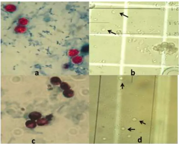
the oocysts separation. To determine the col-umn capacity for the binding of oocysts, dif-ferent amounts of oocysts (106, 5×10⁵, 4×10⁵, 3×10⁵, 2×10⁵, 1 × 10⁵) were passed through the column. All unbound and eluted oocysts were counted by haemocytometer. The capaci-ty of column was 50000 oocysts. The isolated oocysts were pure and without contamination with bacteria, which was determined by bacte-riological examinations performed in Depart-ment of Microbiology of Faculty Veterinary Medicine, University of Tehran (Fig. 2).
Fig. 1: a. Oocysts were analyzed with serum from rabbit immunized with whole antigen of C. parvum using the
IFAT method. B. Negative control with serum from rabbit before immunization
Fig. 2: a, b = Oocysts of Cryptosporidium before passing through chromatography column, a. Ziehl neelsen
stained oocyst under 1000 x magnification. b. unstained oocysts under 40 x nagnification. C,d = Oocysts of
Interestingly, the column prepared with anti-body against p23 of C. parvum had comparable oocysts retention to the column prepared with Ab against oocysts, since the capacity of the columns prepared with Ab against p23 of C. parvum was approximately 40000 oocysts.
As negative controls, the column prepared with sepharose 4B or with rabbit anti TaSp (Theileria annulata surface protein) antibody, which was previously produced in our depart-ment, bound to the activated sepharose 4B were simultaneously used. In contrast to the results obtained with anti C. parvum column no retention of oocysts could be observed by col-umns prepared with rabbit anti TaSp bound to the activated sepharose 4B or with sepharose 4B alone.
Moreover, salt floatation and 55% sucrose flotation techniques were also used for the isolation of C. parvum oocysts. In the salt floatation producer, approximately 50% C. parvum oocysts did not float on the superna-tant and retained in the pellet. Isolated C. par-vum oocysts showed high contamination with bacteria, according to the bacteriological ex-aminations performed by Department of Mi-crobiology of Faculty Veterinary Medicine, University of Tehran. C. parvum oocysts, which were isolated by 55% sucrose flotation, showed lower contamination with bacteria in comparison to the salt floatation method.
In addition, we used 55% sucrose flotation followed by Cellulose Nitrate membrane filtra-tion for isolafiltra-tion of C. parvum oocysts from feces. The isolated oocysts by filtration meth-od were pure and without contamination with bacteria, but more than 70% of C. parvum oo-cysts were missed through membrane filtra-tion.
Discussion
Cryptosporidium parvum is a zoonotic coccidian parasite, which plays an important role in the human and animal health management. One oocyst can also cause disease in immunosup-pressed individual (6, 7), which speaks for the
aggressiveness of this parasite. It is also known that an infected calf with C. parvum can shed billions of oocysts through the feces and can contaminate the soil, water, food and sur-face areas (2). The oocysts of C. parvum are very resistance with robust nature and no ef-fective treatment can be achieved by conven-tional drugs or practical disinfectants (22, 23). Diagnosis of cryptosporidiosis can be per-formed by serological methods or by detection of oocysts in the feces of the suspected ani-mals; the latter is more practicable by routine diagnosis. The detection of oocysts in the samples with limited number of oocysts is much more difficult.
One of the most used method for detection of Cryptosporidium species in samples with small number of oocysts is the immunomag-netic separation (IMS) (24- 26). The recovery efficiency of the IMS procedure for the isola-tion of Cryptosporidium oocysts was evaluated as 82.3% - 86.3% (24). This method has limi-tation by some difficulties like need of equip-ment for immunofluorescence and high cost.
In the present study, we developed an affin-ity chromatography method for the isolation and detection of C. parvum oocysts. For this aim, membrane filteration was used to achieve highly pure C. parvum oocyst. The isolated oo-cysts were pure and without contamination with bacteria, but large number (70%) of C. parvum oocysts were lost through the physical pore in the filter. As we showed previously (19), filtration could not isolate limited num-ber of C. parvum oocysts. In another approach, antibody prepared in rabbit against the pure oocysts obtained from filtration was used for isolation of C. parvum oocysts in immunochro-matography system. The antibody was bound to the sepharose 4B, transferred in to the col-umn and used for isolation of oocysts. The capacity of column was determined as 50000 oocysts.
of C.parvum oocyst was previously produced and analyzed at Parasitology Department of University of Tehran. Shayan et al. (28) showed that recombinant C. parvum P23 was an important target to screen the serum of pregnant dams and their calves for detection of Cryptosporidium specific antibody. Moreover, they showed that the specific IgY against re-combinant P23 provides protection against cryptosporidiosis in a mouse model (21). Pre-viously, we also developed an affinity chro-matography method based on antibody against p23 protein. The capacity of prepared column was 40000 oocysts. The isolated oo-cysts were pure and without contamination with bacteria (21). Our results showed that the affinity chromatography based on antibody against oocysts was comparable to the affinity chromatography based on antibody against p23. Since immunochromatography methods separated oocysts specifically, the use of men-tioned methods could be recommended for detection of oocysts in samples with limited amount of oocysts.
A number of methods have been described for isolating of C. parvum oocysts from feces. These include sucrose floatation (12), salt floatation (13, 29), percoll or ficoll gradient centrifugation (14, 30), and discontinuous su-crose gradient centrifugation in combination with percoll gradient isolation (31, 32). In this study, traditional methods such as salt and 55% sucrose floatation, which separate oo-cysts unspecifically, were also used. Our re-sults showed that beside loss of high number of oocysts, the isolated oocysts were always contaminated with high amount of bacteria. Therefore, we believe that such methods are not suitable for detection of oocysts in the samples with low number of oocysts. Further-more, due to the bacteria contamination, the isolated oocysts cannot be used for at least proteomic studies. However, Koompapong et al.(33) reported a detection system based on sucrose floatation with FA and they are the opinion that this method is efficient enough and economical compared to the IMS method.
Our results showed both immunochroma-tography methods could bound oocysts with capacity depend on the column size. Increase the size of column will increase its capacity.
Conclusion
The ice-cold 55% (w/v) sucrose solution followed by anti C. parvum oocyst antibody column-based chromatography or anti p23 antibody column-based chromatography could be recommended for isolation of C. parvum oocyst from bacterial contaminated samples like feces. These combination methods could give us highly pure oocysts for proteomics studies or cell culture. Furthermore, anti C. parvum oocyst antibody column-based chro-matography or anti p23 antibody column-based chromatography could be recommend-ed for the isolation of C. parvum oocysts from samples with small number of oocysts like drinking or recreational water.
Acknowledgements
We thank Iranian Higher Council of Sci-ences, Research and Technology and the Cen-tral Excellence for Immunopathology of Iran for the financial support according to the na-tional project “development of vaccine against cryptosporidiosis”. The authors declare that there is no conflict of interests.
References
1. Fayer R, Xia L. Cryptosporidium and cryptospor-idiosis. 2nd ed. CRC, New York; 2008. 2. Anderson BC. Patterns of shedding of
cryptos-poridial oocysts in Idaho calves. J Am Vet Med Assoc. 1981; 178 (9):982–4.
3. De Graaf DC, Vanopdenbosch E, Ortega-Mora LM, Abbassi H, Peeters JE. A review of the importance of cryptosporidiosis in farm an-imals. Int J Parasitol. 1999; 29(8):1269–87. 4. Fricker CR, Crabb JH. Water-borne
5. Smith HV, Rose JB. Waterborne cryptosporid-iosis. Parasitol Today. 1990; 6 (1):8–12.
6. Blewett DA, Wright SE, Casemore DP, Booth
NE, Jones CE. Infective dose size studies on
Cryptosporidium parvum using gnotobiotic lambs. Water Sci Technol. 1993; 27(3):61–4.
7. Pereira SJ, Ramirez NE, Xiao L, Ward LA. Pathogenesis of human and bovine Cryptosporid-ium parvum in gnotobiotic pigs. J Infect Dis. 2002; 186 (5):715–8.
8. Anonymous . Method 1622: Cryptosporidium in Water by Filtration/IMS/FA EPA-821-R-99-001. US Environmental Protection Agency, Washington, DC. 1999a
9. Anonymous. Method 1623: Cryptosporidium and
Giardia in Water by Filtration/IMS/FA EPA-821-R-99-006. US Environmental Protection Agency, Washington, DC. 1999b.
10. Manouchehri Naeini K, Asadi M and
Hashemzade Chaleshtori M. Detection and Molecular Characterization of Cryptosporidium
species in Recreational Waters of Chaharmahal va Bakhtiyari Province of Iran using nested-PCR-RFLP. Iran J Parasitol. 2011; 6(1):20-27. 11. Mahmoudi M, Ashrafi K, Abedinzadeh H,
Tahvildar-Bideruni F, Haghighi A, Bandehpour M, Taghipour Lailabadi N, Kazemi B. Devel-opment of sensitive detection of Cryptosporidium
and giardia from surface water in iran. Iran J Parasitol. 2011; 6(3):43-51.
12. McNabb SJ, Hensel DM, Welch DF, Heijbel H, Mckee GL, Istre GR. Comparison of Sedi-mentation and Flotation Techniques for Identi-fication of Cryptosporidium sp. Oocysts in a Large Outbreak of Human Diarrhea. J Clin Mi-crobiol. 1985; 22 (4):587-89.
13. Petry F, Robinson HA, McDonald V. Murine
infection model for maintenance and amplifica-tion of Cryptosporidium parvum oocysts. J Clin Microbiol. 1995; 33(7):1922–4.
14. Waldman E, Tzipori S, Forsyth JR. Separation of Cryptosporidium species oocysts from feces by using a percoll discontinuous density gradient. J Clin Microbiol. 1986; 23(1): 199-200.
15. Arrowood MJ, Sterling CR. Isolation of Cryptos-poridium Oocysts and Sporozoites Using Dis-continuous Sucrose and Isopycnic Percoll Gradients. J Parasitol. 1987; 73(2): 314-19. 16. Truong Q, Ferrari BC. Quantitative and
quali-tative comparisons of Cryptosporidium faecal pu-rification procedures for the isolation of
oo-cysts suitable for proteomic analysis. Int J Para-sitol. 2006; 36(7):811–9.
17. Henriksen SVA, Poglenz JF. Staining of Cryp-tosporidium by a modified ZiehlNeelsen tech-nique. Acta Vet Scand. 1981; 22: 594 -96. 18. Winter G, Andrew AG, Williams LK, Slade
MB. Characterization of a major sporozoite surface glycoprotein of Cryptosporidium parvum. Funct Integr Genomics. 2000; 1 (3): 207-17 19. Shayan P, Asghari Z, Ebrahimzadeh E,
Omid-ian Z, Zarghami F. Presenting an Appropriate Method for Isolation of Bacteria Free Cryptos-poridium parvum Oocysts. J VM. 2012; 8 ( 2): 75-90.
20. Sadr-Shirazi N , Shayan P, Eckert B, Ebrahim-zadeh E, Amininia N . Cloning, and Molecular Characterization of Polymorphic Iranian Iso-late Theileria annulata Surface Protein (Tasp). Iran J Parasitol: 2012; 7(2) :29-39.
21. Omidian Z, Ebrahimzadeh E, Shahbazi P, Asghari Z, Parviz Shayan. Application of re-combinant Cryptosporidium parvum P23 for isola-tion and prevenisola-tion. Parasitol Res.2013; DOI 10.1007/s00436-013-3648-0
22. Harp JA, Goff JP. Protection of calves with a vaccine against Cryptosporidium parvum. J Parasi-tol. 1995; 81(1):54–7.
23. Jenkins MC, O’Brien C, Trout J, Guidry A, Fayer R. Hyperimmune bovine colostrum spe-cific for recombinant Cryptosporidium parvum an-tigen confers partial protection against cryptos-poridiosis in immunosuppressed adult mice. Vaccine.1998; 17(19):2453–60.
24. Reynolds DT, Slade RB, Sykes NJ, Jonas A, Fricker CR. Detection of Cryptosporidium oo-cysts in water: techniques for generating precise recovery data. J Appl Microbiol.1999; 87(6): 804–13.
25. Deng MQ, Lam KM, Cliver DO. Immuno-magnetic separation of Cryptosporidium parvum
oocysts using MACS Micro Beads and high gradient separation columns. J Microbiol Methods. 2000; 40 (1) 11–17.
26. Fontaine M, Guillot E. An immunomagnetic separation–real-time PCR method for quantifi-cation of Cryptosporidium parvum in water sam-ples. J Microbiol Methods. 2003; 54(1) : 29– 36. 27. Boulter-Bitzer JI, Lee H, Trevors J T. Molecu-lar targets for detection and immunotherapy in
28. Shayan P, Ebrahimzadeh E, Mokhber-Dezfouli MR, Rahbari S. Recombinant Cryptos-poridiumparvum p23 as a target for the detection of Cryptosporidium-specific antibody in calf sera. Parasitol Res. 2008; 103:1207–11.
29. O'Brien CN, Jenkins MC. A Rapid Method for Producing Highly Purified Cryptosporidium par-vum oocysts. J Parasitol. 2007; 93 (2): 434-36.
30. Hijjawi NS, Meloni BP, Morgan UM,
Thomp-son RCA. Complete development and long-term maintenance of Cryptosporidium parvum hu-man and cattle genotypes in cell culture. Int J Parasitol. 2001; 31(10):1048–1055.
31. Arrowood MJ, Sterling CR . Isolation of Cryp-tosporidium oocysts and sporozoites using
dis-continuous sucrose and isopycnic Percoll gradi-ents. J Parasitol. 1987; 73 (2):314–19.
32. Truong Q. and Ferrari BC. Quantitative and qualitative comparisons of Cryptosporidium faecal purification procedures for the isolation of oo-cysts suitable for proteomic analysis. Int J Para-sitol.2006; 36 (7): 811-19.
33. Koompapong K, Sutthikornchai Ch and